Skip to main page content
U.S. flag

An official website of the United States government

Dot gov

The . gov means it’s official. Federal government websites often end in . gov or . mil. Before sharing sensitive information, make sure you’re on a federal government site VSports app下载. .

Https

The site is secure V体育官网. The https:// ensures that you are connecting to the official website and that any information you provide is encrypted and transmitted securely. .

. 2018 Jan 5;122(1):58-73.
doi: 10.1161/CIRCRESAHA.117.311307. Epub 2017 Nov 1.

"VSports注册入口" Mitochondrial Reactive Oxygen Species in Lipotoxic Hearts Induce Post-Translational Modifications of AKAP121, DRP1, and OPA1 That Promote Mitochondrial Fission

Affiliations

V体育安卓版 - Mitochondrial Reactive Oxygen Species in Lipotoxic Hearts Induce Post-Translational Modifications of AKAP121, DRP1, and OPA1 That Promote Mitochondrial Fission

Kensuke Tsushima et al. Circ Res. .

Abstract

Rationale: Cardiac lipotoxicity, characterized by increased uptake, oxidation, and accumulation of lipid intermediates, contributes to cardiac dysfunction in obesity and diabetes mellitus. However, mechanisms linking lipid overload and mitochondrial dysfunction are incompletely understood VSports手机版. .

Objective: To elucidate the mechanisms for mitochondrial adaptations to lipid overload in postnatal hearts in vivo. V体育安卓版.

Methods and results: Using a transgenic mouse model of cardiac lipotoxicity overexpressing ACSL1 (long-chain acyl-CoA synthetase 1) in cardiomyocytes, we show that modestly increased myocardial fatty acid uptake leads to mitochondrial structural remodeling with significant reduction in minimum diameter. This is associated with increased palmitoyl-carnitine oxidation and increased reactive oxygen species (ROS) generation in isolated mitochondria. Mitochondrial morphological changes and elevated ROS generation are also observed in palmitate-treated neonatal rat ventricular cardiomyocytes. Palmitate exposure to neonatal rat ventricular cardiomyocytes initially activates mitochondrial respiration, coupled with increased mitochondrial polarization and ATP synthesis. However, long-term exposure to palmitate (>8 hours) enhances ROS generation, which is accompanied by loss of the mitochondrial reticulum and a pattern suggesting increased mitochondrial fission. Mechanistically, lipid-induced changes in mitochondrial redox status increased mitochondrial fission by increased ubiquitination of AKAP121 (A-kinase anchor protein 121) leading to reduced phosphorylation of DRP1 (dynamin-related protein 1) at Ser637 and altered proteolytic processing of OPA1 (optic atrophy 1) V体育ios版. Scavenging mitochondrial ROS restored mitochondrial morphology in vivo and in vitro. .

Conclusions: Our results reveal a molecular mechanism by which lipid overload-induced mitochondrial ROS generation causes mitochondrial dysfunction by inducing post-translational modifications of mitochondrial proteins that regulate mitochondrial dynamics. These findings provide a novel mechanism for mitochondrial dysfunction in lipotoxic cardiomyopathy VSports最新版本. .

Keywords: heart failure; metabolism; mitochondrial dynamics; oxidative stress; reactive oxygen species. V体育平台登录.


Conflict of interest statement

Disclosures: The authors have declared no conflict of interest exists.

Figures (VSports)

Figure 1
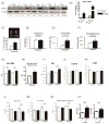
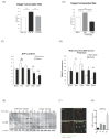
Figure 1. ACStg mice develop mild LVH with modest systolic dysfunction
(A) Representative western blot of Acsl1 protein expression in wild-type (WT) and Acsl1 transgenic mice (ACStg) from birth to 12-weeks of age. Numbers beneath each lane represents ACSL1 densitometry. The antibody recognizes both endogenous ACSL1 and the transgene. (B) mRNA expression of Acsl1 was quantified by RT-PCR at the age of P0 and 12-weeks. * P<0.05, ** P<0.01 vs. WT. (C) Representative PET image and quantification of 1-C palmitate biodistribution in 12-wk-old WT and ACStg hearts; n =10-12. * P <0.05 (D-F) Cardiac triacylglycerol (D), ceramide (E), or diacylglycerol (F) content in 12-week-old WT and ACStg hearts; n =5. * P<0.05 vs. WT. (G) Quantification of ventricular weight (VW) vs body weight (BW) ratio. n =4 in each group. (H) Myocyte cross-sectional areas estimated from WGA-stained cross sections obtained from 24-wk-old ACStg mice and age-matched controls (n=2 hearts per genotype). See Online Fig.I for representative image. (I-K) Echocardiographic analysis. Interventricular septal thickness (I), Left ventricle end-diastolic volume (LVEDV) (J), LV ejection fraction (LVEF) (K), at 12 and 24-weeks of age. n= 5 at 12-weeks of age and n = 4 at 24-weeks of age. (L-M) Cardiac catheterization. Max dP/dt (L) and Min dP/dt (M) and arterial blood pressure (N) in ACStg hearts at 12 and 24-weeks of age. n =5 at 12-weeks of age and n =4 at 24-weeks of age. (O) BNP and Acta1 mRNA expression were quantified by RT-PCR at 12-weeks of age. n =4, ** P < 0.01. All data are mean±sem.
Figure 2
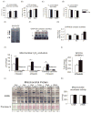
Figure 2. Mitochondrial respiratory function and ROS production in isolated mitochondria from ACStg hearts
(A,B) Maximal ADP-stimulated mitochondrial oxygen consumption (A) and ATP synthesis rates (B) in mitochondria isolated from 12- and 24-week-old WT and ACStg hearts using palmitoyl-carnitine as a substrate; n =4-5. * P<0.05 vs. WT. (C,D) Maximal ADP-stimulated mitochondrial oxygen consumption in saponin-permeabilized cardiac fibers of 12- and 24-week-old WT and ACStg hearts using pyruvate (C) or glutamate (D) as a substrate; n =6. (E-G) Electrophoretic separation of OXPHOS complexes by blue-native PAGE (E), representative images of in-gel activities of complexes I, IV, and V (F), and quantification of OXPHOS complex activities (G), measured in 12-week-old WT and ACStg hearts; n =4. *P<0.05 vs. WT. (H) H2O2 production with succinate as a substrate in mitochondria isolated from 12- or 24- week-old WT and ACStg hearts, or with palmitoyl-carnitine as a substrate in mitochondria from 12-week-old WT and ACStg hearts, in the absence or presence of rotenone (Rot); n =3 at 12wks, n =6 at 24wks. *P<0.05 vs. WT. (I) Oxidation of DCFDA in whole tissue extracts of 24-week-old WT and ACStg hearts; n =5-6. *P<0.05 vs. WT. (J) Western blot for 4HNE protein adducts in mitochondrial protein isolated from WT and ACStg hearts. Numbers beneath each lane represent densitometry of 4HNE immunoreactivity of all bands in that lane. (K) Aconitase activity measured in mitochondria isolated from 12- or 24-week-old WT and ACStg hearts; n =3 at 12weeks, n =6 at 24weeks. All data are mean ± sem.
Figure 3
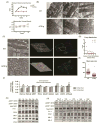
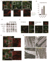
Figure 3. Mitochondrial fragmentation in ACStg hearts
(A) Postnatal mitochondrial enlargement was attenuated in ACStg hearts. Stereologic quantification of mitochondrial minimum diameter and volume density was performed at the ages as indicated in 2-D electron micrographs (EM) presented in Online Fig.III A. ; n =3-4. * P<0.05, ** P<0.01, *** P<0.001. (B) Representative electron micrographs of longitudinal sections of WT and ACStg cardiomyocytes at the age of 8weeks, 3 hearts per genotype. Scale bars indicate 200nM. (C-E) Representative electron tomographic micrographs and corresponding 3D models of the mitochondrial network. Scale bars indicate 100nM (C). Number of mitochondrial outer membranes transitioned on a straight line trajectory between M-lines of two neighbouring myofilament bundles, counted in 22 (WT) and 26 (ACStg) electron tomograms (3 hearts per genotype) (D). Short axis diameter was measured in 132 (WT) and 290 (ACStg) mitochondria of 22 (WT) and 26 (ACStg) electron tomograms from 3 hearts per genotype; ** P <0.01(E). (F) Myocardial gene expression in 12- and 24-week-old WT and ACStg mice normalized to 16S RNA transcript levels. Values represent fold change in mRNA transcript levels relative to WT, which was assigned as 1 (dashed line), n=8 *; P<0.05 vs WT. (G) Western blot of whole heart lysates probed with antibodies as indicated at postnatal ages as shown. d=day, w=week. See also Online Fig.IV A. Each sample at 0d, 1w and 3w was isolated more than 3 times and immunoblotted separately, and each sample at day1, 2w and 8w was repeated twice. A representative western blot is shown. (H) Western blot of whole heart lysates from WT and ACStg mice probed with antibodies as indicated and harvested at the ages shown. See also Online Fig.IV B. All data are mean ± sem. Each sample at day0, 1wk and 3wk was isolated more than 3times and immunoblotted separately, and each sample at 2wk and 8wk was repeated twice. A representative western blot is shown.
Figure 4
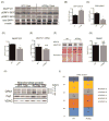
Figure 4. Differential post-translational modifications of DRP1 and OPA1 in ACStg mice
(A-D) Decreased protein content of AKAP121, and altered phosphorylation profile of DRP1. Whole heart lysates were subjected to western blot analysis (A) and densitometric quantification of DRP1pS616 (B), DRP1pS637 (C) and AKAP121 (D), n =4. ** P <0.01, * P <0.05. (E) AKAP121 mRNA expression of 12-week-old WT and ACStg mice was determined by quantitative RT-PCR (n=4) (F) AKAP121 degradation is mediated by the Ubiquitin-Proteasome pathway in ACStg hearts. WT or ACStg mice were injected intraperitoneally twice with DMSO or MG132 (18hrs and 6hrs before euthanasia). Whole-heart lysates were subjected to western blot analysis. (G) Siah2 gene expression in 12-week-old WT and ACStg mouse hearts was determined by realtime-PCR. Values are normalized to GAPDH expression. n=4. (H and I) Increased OPA1 cleavage in ACStg hearts. Mitochondrial fractions were prepared from 12-week-old WT or ACStg hearts and subjected to western blot analysis. Each lane shows the sample from different animals. The top 2 bands correspond to non-cleaved isoforms of OPA1 (long-form) and the bottom 3 bands correspond to cleaved isoforms of OPA1 (short-form) (H), quantitative densitometric analysis of OPA1 isoforms (I), n =4. * P<0.05 vs. WT. All data are mean ± sem.
Figure 5
Figure 5. Mitochondrial metabolism and ROS production in rat neonatal cardiomyocytes following increasing duration of free fatty acids exposure
(A) Basal oxygen consumption rate (OCR) (assayed by the Seahorse XF24 system) in NRVCs incubated with BSA alone, palmitate-BSA (500μM) or oleate-BSA (500μM). ** P <0.01. (B) Time course analysis of OCR of NRVCs treated with palmitate-BSA. Increased basal OCR after short-term exposure to palmitate (3hr) and reduced basal OCR after long-term exposure (15hr) of palmitate. **P <0.01. See also Online Fig.V. (C) Time course analysis of ATP content in NRVCs after palmitate or oleate treatment. **P <0.01 vs BSA # P <0.05, ## P<0.01. (D) Time course analysis of mitochondrial membrane potential after palmitate or oleate treatment. Cardiomyocytes were stained with TMRM and Hoechst at indicated times and fluorescence intensity was assayed with a plate reader. The ratio of TMRM/Hoechst fluorescence are shown. n = 4, * P <0.05 vs BSA, # P <0.05. (E) Western blot for 4HNE protein adducts in NRVCs after palmitate or oleate treatment. (F and G) CellROX green staining in NRVCs after palmitate treatment. CellROX Green is a DNA dye, and upon oxidation, it binds to DNA. Representative confocal image (F) and fluorescence intensity was quantified (G). ** P <0.01 vs BSA. All data are mean±sem. Scale bars indicate 20μm.
Figure 6
Figure 6. DRP1 mediates mitochondrial fission after lipid overload
(A) Rat neonatal cardiomyocytes were stimulated with growth medium with or without 500μM palmitate or oleate. 12hrs after stimulation, cells were fixed with 4% paraformaldehyde and immunostained with α-actinin (green), Tom20 (red) and DAPI (blue). Scale bars indicate 20μm. (B) Quantification of mitochondrial fragmentation presented in Figure 6A. More than 100 cells were counted to determine the percentage (%) of cells with fragmented mitochondria. n=3, **: P<0.01. (C) Increased phosphorylation of DRP1 at Ser616 after palmitate treatment. NRVCs were treated in growth medium with 500μM palmitate and cell lysates were harvested at the indicated times in hr. (D) NRVCs were treated in growth medium with or without 500μM palmitate and subjected to immunohistochemistry for DRP1 (green) and Tom20 (red). Note that DRP1 is co-localized with Tom20 after palmitate treatment. Scale bars indicate 20μm. (E) NRVCs were infected with AdGFP or Ad DRP1K38E and were treated in growth medium with 500μM palmitate. NRVCs were subjected to immunohistochemistry for α-actinin (green) and Tom20 (red). See also Online Fig.VII A. Scale bars indicate 20μm. (F) Representative electron micrographs of longitudinal heart sections obtained from WT, DRP1+/-, ACStg and ACStg × DRP1+/-. DRP1 knockdown partially rescued mitochondrial morphology in ACStg hearts. See also Online Fig.VII B-E.
Figure 7
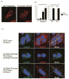
Figure 7. Mitochondrial fusion in L6 myoblasts was impaired by palmitate exposure
(A) L6 myoblasts transfected with mitoRFP were incubated in growth medium with or without 500μM palmitate for 5h, and representative images are shown. (B) Cells were evaluated to quantify tubular or fragmented mitochondrial networks, and cells with fragmented mitochondria were expressed as percentage of all viewed cells, n=4 and 80 cells counted per group. ** P < 0.01 vs BSA. All data are mean±sem. (C) L6 myoblasts were transfected either with mitoRFP or mitoGFP, co-plated (25,000 cells each) on cover slips for 24h, and then cell fusion was induced with polyethylene glycol (PEG). Cells were fused for 8h in regular growth medium with no palmitate (top row), 8h in growth medium with 500μM palmitate, following 3h pre-incubation with 500μM palmitate prior to PEG fusion (middle row), or 8h in the regular growth medium with no palmitate, following 3h pre-incubation with 500μM palmitate prior to PEG fusion (bottom row).
Figure 8
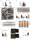
Figure 8. Mitochondrial superoxide dismutase (SOD2) overexpression partially rescued abnormal mitochondrial dynamics in ACStg mice
(A,B) Enhanced ROS production in ACStg hearts was rescued by SOD2 overexpression. H2O2 production was determined in isolated mitochondria from 12-week-old WT, SOD2tg, ACStg and SOD2 × ACS double tg hearts in the absence or presence of rotenone (A). Mitochondrial fractions were prepared from 12-week-old WT, SOD2tg, ACStg and SOD2 × ACS double transgenic hearts and subjected to western blot for 4HNE and quantified by densitometry (B), n =3-4 in each group. ** P <0.01. See also Online Fig.VIII A (C) SOD2 overexpression partially rescued fragmented mitochondria in ACStg hearts. Representative electron micrographs from WT, SOD2tg, ACStg and SOD2 × ACS double tg mice. Bar indicates 1μm. See also Online Fig.VIII B. (D-H) SOD2 overexpression reversed altered phosphorylation pattern of Drp1 and processing of OPA in ACStg hearts. Mitochondrial fractions were prepared from 12-week-old WT, SOD2tg, ACStg and SOD2 × ACS double tg hearts and subjected to western blot analysis for AKAP121 and total and phosphorylated Drp1at Ser637 and Ser616 and the distribution of OPA1 isoforms were quantified (F-H). n =3-4 in each group. * P <0.05, ** P <0.01 vs WT, # P <0.05 vs ACStg. (I,J) MnTBAP prevented palmitate-induced mitochondrial fragmentation in NRVCs. NRVCs were treated with or without 400μM MnTBAP, a SOD mimetic and peroxynitrite scavenger, and then stimulated with or without growth medium containing 500μM Palmitate. After 12h of stimulation, cells were fixed and stained with Tom20 antibody. Representative image from each group. (I) and quantification of the ratio of fragmented cells (J), n =3 in each group. ** P <0.01. Scale bars indicate 20μm.

Comment in

  • What You Eat Affects Your Shape.
    Murphy E, Glancy B, Steenbergen C. Murphy E, et al. Circ Res. 2018 Jan 5;122(1):8-10. doi: 10.1161/CIRCRESAHA.117.312335. Circ Res. 2018. PMID: 29301836 Free PMC article. No abstract available.

V体育2025版 - References

    1. Doenst T, Nguyen TD, Abel ED. Cardiac metabolism in heart failure: implications beyond ATP production. Circ Res. 2013;113:709–24. - PMC - PubMed
    1. de Jong H, Neal AC, Coleman RA, Lewin TM. Ontogeny of mRNA expression and activity of long-chain acyl-CoA synthetase (ACSL) isoforms in Mus musculus heart. Biochim Biophys Acta. 2007;1771:75–82. - PMC - PubMed
    1. Lopaschuk GD, Collins-Nakai RL, Itoi T. Developmental changes in energy substrate use by the heart. Cardiovasc Res. 1992;26:1172–80. - "V体育平台登录" PubMed
    1. Lopaschuk GD, Witters LA, Itoi T, Barr R, Barr A. Acetyl-CoA carboxylase involvement in the rapid maturation of fatty acid oxidation in the newborn rabbit heart. J Biol Chem. 1994;269:25871–8. - PubMed
    1. Kolwicz SC, Jr, Purohit S, Tian R. Cardiac metabolism and its interactions with contraction, growth, and survival of cardiomyocytes. Circ Res. 2013;113:603–16. - PMC - PubMed

Publication types (VSports注册入口)

MeSH terms