Skip to main page content
U.S. flag

An official website of the United States government

Dot gov

The . gov means it’s official. Federal government websites often end in VSports app下载. gov or . mil. Before sharing sensitive information, make sure you’re on a federal government site. .

Https

The site is secure. The https:// ensures that you are connecting to the official website and that any information you provide is encrypted and transmitted securely V体育官网. .

. 2013 Jul 1;191(1):488-99.
doi: 10.4049/jimmunol.1300657. Epub 2013 May 24.

Thymic damage, impaired negative selection, and development of chronic graft-versus-host disease caused by donor CD4+ and CD8+ T cells

Affiliations

V体育平台登录 - Thymic damage, impaired negative selection, and development of chronic graft-versus-host disease caused by donor CD4+ and CD8+ T cells

Tao Wu et al. J Immunol. .

Abstract

Prevention of chronic graft-versus-host disease (cGVHD) remains a major challenge in allogeneic hematopoietic cell transplantation (HCT) owing to limited understanding of cGVHD pathogenesis and lack of appropriate animal models VSports手机版. In this study, we report that, in classical acute GVHD models with C57BL/6 donors and MHC-mismatched BALB/c recipients and with C3H. SW donors and MHC-matched C57BL/6 recipients, GVHD recipients surviving for >60 d after HCT developed cGVHD characterized by cutaneous fibrosis, tissue damage in the salivary gland, and the presence of serum autoantibodies. Donor CD8(+) T cells were more potent than CD4(+) T cells for inducing cGVHD. The recipient thymus and de novo-generated, donor-derived CD4(+) T cells were required for induction of cGVHD by donor CD8(+) T cells but not by donor CD4(+) T cells. Donor CD8(+) T cells preferentially damaged recipient medullary thymic epithelial cells and impaired negative selection, resulting in production of autoreactive CD4(+) T cells that perpetuated damage to the thymus and augmented the development of cGVHD. Short-term anti-CD4 mAb treatment early after HCT enabled recovery from thymic damage and prevented cGVHD. These results demonstrate that donor CD8(+) T cells cause cGVHD solely through thymic-dependent mechanisms, whereas CD4(+) T cells can cause cGVHD through either thymic-dependent or independent mechanisms. .


Conflict of interest statement (VSports在线直播)

All authors declare no conflict of interest

Figures

Fig. 1
Fig. 1. Low-dose of donor spleen cells induced cGVHD
Lethally irradiated BALB/c recipients were transplanted with titrated numbers of spleen cells (5-1.25 ×10⁶) and TBCD-BM cells (2.5×10⁶) from C57BL/6 donors. Control recipients were given TBCD-BM cells alone. Recipients were monitored for clinical GVHD including bodyweight changes, diarrhea, hair-loss, and survival (†, indicating death of all recipients in a group). 15 and 60 days after HCT, recipient serum samples were tested for presence of autoantibodies by staining donor-type Rag-2-/- C57BL/6 skin and salivary gland tissues. 60 days after HCT, recipients were measured for percentage and yield of CD4+CD8+ thymocytes. A-D: Percentage of bodyweight changes, percentage of recipients without diarrhea, clinical cutaneous GVHD score, and percentage of survival. Each group contained 12 recipients combined from three replicate experiments. E: Representative photomicrographs of GVHD recipient serum autoantibody staining of Rag-2-/- skin and salivary gland tissues. DAPI staining is shown in blue, and autoantibody staining is shown in green. One representative result is shown from 8 samples evaluated in each group. F: One representative of CD4+CD8+ thymocytes staining pattern is shown of 8 recipients examined in each group. The mean ± SE of yield of CD4+CD8+ thymocytes (N=8).
Fig. 2
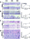
Fig. 2. Kinetic changes of GVHD target tissue pathology
GVHD target tissues of colon (A), jejunum (B), skin (C) and salivary gland (D) were harvested and processed for evaluation of histopathology on days 15, 30, and 60 days after HCT from recipients given donor TBCD-BM alone or additional 1.25 ×10⁶ spleen cells. Arrows indicate the following changes in GVHD recipients as compared to control recipients: infiltration and loss of crypts in the colon; blunting and loss of crypts in the jejunum; expansion of dermis and loss of fat tissues in the skin; infiltration and destruction of secretory follicles in the salivary gland. One representative photomicrograph and mean ± SE of histopathology scores are shown for 6 recipients in each group.
Fig. 3
Fig. 3. High dose of donor CD4+ T cells induced more severe aGVHD but low-dose of donor CD8+ T cells induced more severe cGVHD
Lethally irradiated BALB/c recipients were injected with titrated numbers (5, 0.5, and 0.1 ×10⁶) of sorted donor CD4+ or CD8+ T cells and TBCD-BM cells from C57BL/6 donors. Recipients given TBCD-BM alone were used as controls. Recipients were monitored for bodyweight change, hair-loss, diarrhea, and survival (†, indicating death of all recipients in a group). There were 8 recipients in each group, combined from two replicate experiments. A: Recipients given 5 ×10⁶ donor CD4+ T cells developed diarrhea, and all died before development of hair-loss; in contrast, recipients given 5 ×10⁶ CD8+ T cells showed little diarrhea, and most survived for more than 60 days, but all developed severe hair-loss. B: Recipients given 0.5 ×10⁶ donor CD4+ T cells developed diarrhea and hair-loss, although most survived for more than 60 days. In contrast, recipients given 0.5 ×10⁶ CD8+ T cells showed severe hair-loss without diarrhea, and most survived for more than 60 days. C: Recipients given 0.1 ×10⁶ donor CD4+ T cells showed mild weight-loss with little hair-loss. In comparison, recipients given 0.1 ×10⁶ CD8+ T cells showed more severe weight-loss and hair-loss 40 days after HCT (P<0.01).
Fig. 4
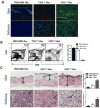
Fig. 4. Recipients given 0.1 ×10⁶ CD8+ but not CD4+ T cells developed autoantibody production, thymic damage, and tissue damage
Recipients from Fig. 3C were measured for autoantibodies in serum, percentage and yield of CD4+CD8+ thymocytes, and histopathology in skin and salivary gland at 60 days after HCT. A: Serum autoantibody staining of donor-type Rag-2-/- C57BL/6 skin and salivary gland tissues. DAPI staining is shown in blue. Autoantibody staining is shown in green. One representative photomicrograph is shown of 6 recipients in each group. B: Representative flow cytometry patterns of CD4+CD8+ thymocytes and mean ± SE of yield of CD4+CD8+ thymocytes of 6 recipients in each group. C: Representative photomicrograph and histopathology scores of 6 recipients examined in each group. Arrows indicate the following changes in GVHD recipients as compared to control recipients: expansion of dermis and loss of fat tissues in the skin; infiltration and destruction of secretory follicles in the salivary gland.
Fig. 5
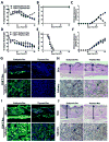
Fig. 5. Donor CD8+ but not CD4+ T cell induction of cGVHD required recipient thymus
Lethally irradiated, euthymic and thymectomized BALB/c recipients were transplanted with 0.5 ×10⁶ CD4+ T cells from CD8+ T deficient or CD8+ T cells from CD4+ T cell-deficient C57BL/6 and TBCD-BM cells from wild-type C57BL/6 mice. The use of CD4+ or CD8+ T cell-deficient donors avoided contamination of CD4+ or CD8+ T cells in injected CD8+ or CD4+ T cells. Recipients were monitored for bodyweight change, diarrhea, hair-loss, and survival after HCT. Data are combined from two replicate experiments (N=8). At day 60 after HCT, recipient sera were tested for autoantibodies. Recipient skin and salivary gland tissues were used for histopathology. A & D: Percentage bodyweight changes. Compared with euthymic recipients, thymectomized recipients given donor CD8+ T cells showed bodyweight recovery 45 days after HCT (P<0.01), but no difference was observed between thymectomized or euthymic recipients given donor CD4+ T cells. B & E: Percentage of mice without diarrhea. Recipients given donor CD8+ T cells showed no diarrhea, but recipients given donor CD4+ T cells all showed diarrhea. C & F: Hair-loss. Thymectomized recipients that were transplanted with donor CD8+ T cells showed transient weak hair-loss and then re-growth of hair at approximately 45 days after HCT, while euthymic recipients showed worsening hair-loss (P<0.01). Both thymectomized and euthymic recipients given CD4+ T cells showed severe hair-loss. G & I: Serum autoantibody staining of donor-type Rag-2-/- C57BL/6 skin and salivary gland tissues. DAPI staining is shown in blue, and autoantibody staining is shown in green. G shows recipients injected with donor CD8+ T cells. I shows recipients injected with donor CD4+ T cells. Left panels show euthymic recipients, and right-panels show thymectomized recipients. One representative photograph of 6 recipients examined in each group. H & J: Histopathology of skin and salivary gland of euthymic and thymectomized recipients given donor CD8+ or CD4+ T cells. Arrows indicate the following changes in GVHD recipients as compared to GVHD-free recipients: expansion of dermis and loss of fat tissues in the skin; infiltration and destruction of salivary follicles in the salivary gland. A representative photomicrograph from 1 of 6 recipients in each group is shown.
Fig. 6
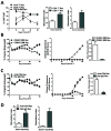
Fig. 6. Low-dose donor CD8+ T cells was more potent than CD4+ T cells for inducing thymic damage and development of cGVHD
A: Lethally irradiated BALB/c recipients were transplanted with 0.1 ×10⁶ CD4+ or CD8+ T cells in addition to TBCD-BM from donor C57BL/6 mice. Recipients given TBCD-BM alone was used as controls. Percentage of CD4+CD8+ thymocytes was measured on 15, 30, 45, and 60 days after HCT. Mean ± SE of 4 recipients at each time point from each group were combined from two replicate experiments. At 60 days after HCT, de novo-generated donor-derived CD4+ T cells (0.2 ×10⁶) from recipients given donor CD4+ or CD8+ T cells were stimulated with recipient- or donor-derived CD11c+ DCs (0.1 ×10⁶). Proliferation of donor CD4+ T cells was measured with 3H-TdR incorporation. Mean ± SE of the stimulation index was combined from 4 replicate experiments. B: Lethally irradiated BALB/c recipients were transplanted with donor CD8+ T cells (0.5 ×10⁶) with TBCD-BM cells (2.5 ×10⁶) from CD4+ T- or CD8+ T-deficient C57BL/6 donors. Recipients were monitored for bodyweight changes and hair-loss. Thymocyte yield was also compared at 60 days after HCT. As compared to recipients given CD8+ T-deficient BM cells, recipients given CD4+ T-deficient BM cells showed bodyweight increase beginning at approximately 45 days after HCT (P<0.01). There were 8 recipients in each group, combined from two replicate experiments. C: Lethally irradiated recipients were transplanted with donor CD8+ T cells (0.5 ×10⁶) and TBCD-BM cells from wild-type C57BL/6 donors. On days 15 and 30 days after HCT, the recipients were injected with anti-CD4 mAb or control rat-IgG (500μg/mouse). Recipients were monitored for bodyweight changes and hair-loss. At 60 days after HCT, total thymocyte yield was compared. Data were combined from 8 recipients in each group from two replicate experiments. As compared with recipients treated with rat-IgG, recipients treated with anti-CD4 mAb showed bodyweight increase at approximately 45 days after HCT (P<0.01). There were 8 recipients in each group, combined from two replicate experiments. D. De novo-generated CD4+ T cell proliferation in response to recipient- or donor-derived CD11c+ DCs at 60 days after HCT. Mean ± SE of the stimulation index was combined from 4 replicate experiments.
Fig. 7
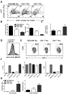
Fig. 7. Low numbers of donor CD8+ T cells preferentially damaged mTEC in the recipient thymus and caused defective thymic negative selection
Lethally irradiated BALB/c recipients were transplanted with 0.1 ×10⁶ CD4+ or CD8+ T cells and TBCD-BM cells from C57BL/6 donors. Recipients given TBCD-BM cells alone were used as controls. At 15 days after HCT, recipient thymus was measured for the percentage and yield of mTEC, percentage and yield of thymic CD11c+ DCs, and expression of MHC II and CCR9. At 30 days after HCT, recipient spleen cells were measured for MMTV-mediated clonal deletion of Vβ3, Vβ4, and Vβ5. In addition, flow cytometry-sorted de novo-generated donor-derived CD4+ T cells were measured for donor- and recipient-reactivity. A. Gated thymic epithelial cells are shown in UEA-1 (mTEC marker) versus Ly51 (cTEC marker). A representative flow cytometry pattern and mean ± SE of percentage and yield of mTEC is shown from1 of 4 replicate experiments. In each experiment, 6 thymi were combined from each group in order to obtain a sufficient number of thymic epithelial cells. B. Percentage and yield of mTEC, Mean ± SE, N=4. C. Total yield of donor CD11c+ DCs in the thymus, Mean ± SE, N=6. D. Expression levels of MHC II by CD11c+ DCs. E. Percentage of CCR9+CD11c+ DCs among total CD11c+ DCs. A representative flow cytometry pattern is shown from 1 of 6 samples. F. Percentage changes of Vβ3, Vβ4, and Vβ5 among de novo-generated donor-derived CD4+ T cells. Mean ± SE (N=6) is shown. G. Recipient- and donor-reactivity of de novo-generated donor CD4+ T cells. Flow cytometry-sorted, de novo-generated, donor-derived CD4+ T cells (0.2 ×10⁶) were stimulated with donor- or recipient-type DCs (0.1×10⁶). T cell proliferation was measured with 3H-TDR incorporation. Mean ± SE of the stimulation index from 4 replicate experiments is shown.
Fig. 8
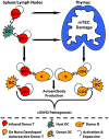
Fig. 8. Diagram of chronic GVHD Pathophysiology

VSports注册入口 - References

    1. Martin PJ. Donor CD8 cells prevent allogeneic marrow graft rejection in mice: potential implications for marrow transplantation in humans. J Exp Med. 1993;178:703–712. - PMC - PubMed
    1. Martin PJ, Rowley SD, Anasetti C, Chauncey TR, Gooley T, Petersdorf EW, van Burik JA, Flowers ME, Storb R, Appelbaum FR, Hansen JA. A phase I-II clinical trial to evaluate removal of CD4 cells and partial depletion of CD8 cells from donor marrow for HLA-mismatched unrelated recipients. Blood. 1999;94:2192–2199. - PubMed (VSports手机版)
    1. Ferrara JL, Levine JE, Reddy P, Holler E. Graft-versus-host disease. Lancet. 2009;373:1550–1561. - PMC - PubMed
    1. Welniak LA, Blazar BR, Murphy WJ. Immunobiology of allogeneic hematopoietic stem cell transplantation. Annu Rev Immunol. 2007;25:139–170. - PubMed
    1. Shlomchik WD, Lee SJ, Couriel D, Pavletic SZ. Transplantation’s greatest challenges: advances in chronic graft-versus-host disease. Biol Blood Marrow Transplant. 2007;13:2–10. - PubMed

Publication types

V体育2025版 - MeSH terms

Grants and funding (VSports注册入口)