<font draggable="BNjloT"></font><var lang="RLMiJw3"><style lang="L7hFw94O"></style></var> V体育平台登录 - Skip to main page content
U.S. flag

An official website of the United States government

Dot gov

The VSports app下载. gov means it’s official. Federal government websites often end in . gov or . mil. Before sharing sensitive information, make sure you’re on a federal government site. .

Https

The site is secure. The https:// ensures that you are connecting to the official website and that any information you provide is encrypted and transmitted securely. V体育官网.

. 2013 May;141(5):585-600.
doi: 10.1085/jgp.201210861.

"VSports在线直播" TMEM16F (Anoctamin 6), an anion channel of delayed Ca(2+) activation

Affiliations

TMEM16F (Anoctamin 6), an anion channel of delayed Ca(2+) activation

Søren Grubb et al. J Gen Physiol. 2013 May.

Abstract

Members of the TMEM16 (Anoctamin) family of membrane proteins have been shown to be essential constituents of the Ca(2+)-activated Cl(-) channel (CaCC) in many cell types. In this study, we have investigated the electrophysiological properties of mouse TMEM16F. Heterologous expression of TMEM16F in HEK293 cells resulted in plasma membrane localization and an outwardly rectifying ICl,Ca that was activated with a delay of several minutes. Furthermore, a significant Na(+) current was activated, and the two permeabilities were correlated according to PNa = 0. 3 PCl. The current showed an EC50 of 100 µM intracellular free Ca(2+) concentration and an Eisenman type 1 anion selectivity sequence of PSCN > PI > PBr > PCl > PAsp. The mTMEM16F-associated ICl,Ca was abolished in one mutant of the putative pore region (R592E) but retained in two other mutants (K616E and R636E). The mutant K616E had a lower relative permeability to iodide, and the mutant R636E had an altered anion selectivity sequence (PSCN = PI = PBr = PCl > PAsp). Our data provide evidence that TMEM16F constitutes a Ca(2+)-activated anion channel or a pore-forming subunit of an anion channel with properties distinct from TMEM16A. VSports手机版.


"VSports在线直播" Figures

Figure 1.
Figure 1.
TMEM16 expression in EATCs. (A) PCR for all known mouse TMEM16 genes in EATCs. (B) Western blots showing expression of TMEM16F, TMEM16FA, and TMEM16FK protein in EATCs. Presented blots are representative of three blots made.
Figure 2.
Figure 2.
Subcellular localization experiments. mTMEM16F-GFP expressed in HEK293 cells. The images were taken using an epifluorescence microscope. mTMEM16F-GFP was visualized with anti-GFP and Alexa Fluor 488. Anti-Hsp47 and Alexa Fluor 568 were used to label the ER, whereas the plasma membrane was stained using rhodamine-conjugated WGA. The nucleus was stained with DAPI.
Figure 3.
Figure 3.
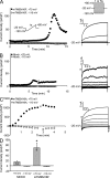
ICl,Ca densities in mock-, mTMEM16F-, and mTMEM16A-transfected HEK293 cells. (A) Typical current development recorded by a ramp protocol run every 15 s (left; see inset) and representative current-voltage relationships (right) of mTMEM16F (250 µM free [Ca2+]i). Inward currents were initially inactivated similar to those in C; however, this inactivation is not visible at the given scale. (B and C) Mock (250 µM free [Ca2+]i; B) and mTMEM16A (0.07 µM free [Ca2+]i; C). “a” indicates where the step protocols were recorded. (D) Mean peak ICa,Cl current density when activated by 250 µM free [Ca2+]i of mock and mTMEM16F, measured at 70 mV and −110 mV, respectively. Data are presented as mean ± SE of 14 mock- and 8 mTMEM16F-transfected cells and tested by one-way ANOVA: *, P < 0.05.
Figure 4.
Figure 4.
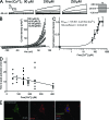
Ca2+ activation kinetics of mTMEM16F. (A) Typical current-voltage relationships of mTMEM16F at max current with 90, 190, or 250 µM free [Ca2+]i. (B) The mean mTMEM16F I-V relation at maximal activation with the indicated free [Ca2+]i, generated from ramp protocol sweeps (from −120 to 80 mV). Data are presented as mean ± SE of three to nine replicates. (C) Hill plot of normalized mTMEM16F ICl,Ca densities at 70 mV. Data are presented as mean ± SE of 3–12 replicates. (D) Ca2+ dependence of mTMEM16F versus time from whole start of whole-cell recording to peak ICa,Cl activation. Linear regression is included to illustrate the decline in the time to peak activation with increasing free [Ca2+]i and is given by f(x) = −0.04x + 14.11. (E) mTMEM16F-GFP expressed in HEK293 cells. Immediately before fixation, the cells were exposed to ionomycin for 20 min in a solution containing 500 µM free Ca2+. The images were taken using an epifluorescence microscope. mTMEM16F-GFP was visualized with anti-GFP and Alexa Fluor 488, whereas the plasma membrane was stained using rhodamine-conjugated WGA. The nucleus was stained with DAPI.
Figure 5.
Figure 5.
Anion selectivity of the mTMEM16F-induced current. (A and B) Representative anion selectivity current traces recorded in whole-cell configuration after stimulation with 250 µM [Ca2+]i of mock (A)- and mTMEM16F-transfected (B) HEK293 cells. (C and D) Mean relative anion permeabilities (PX/PCl) for mock (C)- and mTMEM16F-transfected (D) HEK293 cells. PX/PCl are presented as a mean of seven to nine cells and are tested by one way ANOVA; “*” indicates significant (P < 0.05) difference from PCl, and “†” indicates significant difference (P < 0.05) between the relevant anion permeabilities when tested with paired t test. Only experiments in which the anion permeabilities were paired were included in the statistical test.
Figure 6.
Figure 6.
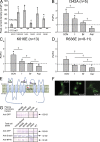
Effect of single amino acid mutations on whole-cell current and anion selectivity. (A) Mean peak ICa,Cl current density when activated by 250 µM free [Ca2+]i of mock, mTMEM16F, and single amino acid mutants in the putative pore site of mTMEM16F (the control mutation I342A and the pore region mutants R592E, K616E, and R636E) measured at 70 mV and −110 mV, respectively. Data are presented as mean ± SE; you can read the amount of replicates inside the 110-mV bar graphs; “*” and “†” indicate significant (P < 0.05, when tested with one-way ANOVA) difference from mock and mTMEM16F, respectively. (B–D) Anion permeabilities recorded in whole-cell mode after simulation with 250 µM free [Ca2+]i for I342A (B)-, K616E (C)-, and R636E-transfected (D) HEK293 cells. Data are presented as mean ± SE; the number of replicates are indicated in the figures; “*” indicates significant (P < 0.05) difference from PCl, and “†” indicates significant difference (P < 0.05) between the relevant anion permeabilities, when tested with paired t test. Only experiments in which the anion permeabilities were paired were included in the statistical test. (E) Putative topology of the TMEM16F channel with the mutated amino acids’ approximate positions. (F) Immunofluorescence images of localization of TMEM16F mutants expressed in HEK293 cells. The images were taken using an epifluorescence microscope. GFP was enhanced by anti-GFP and Alexa Fluor 488 (green). (G) Presence of mTMEM16F-GFP in the plasma membrane fraction and total cell lysate of HEK293 cells expressing either GFP-tagged WT mTMEM16F or one of the mutants I342A, R592E, K616E, and R636E. Plasma membrane proteins were isolated using a biotinylation assay. β-Actin and NPTII were used as controls for loading and transfection efficiency, respectively.
Figure 7.
Figure 7.
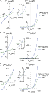
Instantaneous (IInst) and near steady-state (ISt-St) current-voltage relationships of HEK293 cells in whole-cell patch-clamp configuration. Green symbols show I-V relationships obtained with NMDG-Cl in bath. Blue symbols show I-V relationships obtained with NaCl in bath. IInst was recorded immediately after the voltage step from the holding potential, Vhold = 0 mV, to the new Vm. ISt-St was recorded toward the end of the 1-s voltage pulse. Thus, rather than estimating the PNa/PCl of each cell from a reversal potential obtained by interpolation, we could take advantage of IInst-Vm data points covering a broader range about Vrev. Current-voltage relationships are shown for the following (all permeabilities in units of 10−14 cm3/s/pF). (A) HEK293 cells expressing TMEM16F-WT (with NMDG-Cl in bath, PCl = 12.9 ± 0.6; with NaCl in bath, PCl = 31.7 ± 1.0 and PNa = 9.7 ± 0.5). (B) HEK293 cells expressing the neutral mutant TMEM16F-I342A (with NMDG-Cl in bath, PCl = 19.8 ± 0.8; with NaCl in bath, PCl = 93.8 ± 4.2 and PNa = 13.3 ± 3.6). (C) HEK293 cells expressing the anion sequence modifying TMEM16F-R636E mutant (with NMDG-Cl in bath, PCl = 16.9 ± 1.5; with NaCl in bath, PCl = 22.9 ± 1.7 and PNa = 8.0 ± 1.4). It is noted that for all three ISt-St/Vm relationships obtained with NMDG-Cl in bath that Vrev ≈ ECl, whereas the reversal potentials with NaCl in bath are significantly displaced to the right of ECl, which is emphasized by the insets of expanded axis scales.
Figure 8.
Figure 8.
Relationship between PCl and PNa of HEK293 cells expressing WT and engineered TMEM16F constructs. The permeabilities were obtained as described in the legend of Fig. 7. For expressing permeability in the conventional units of cm/s, the values given in Table 2 have been recalculated using the empirical approximation that 106 pF corresponds to a membrane area of 1 cm2. The linear fit to all data is given by PNa = 0.334 × PCl (±SD = 0.028), R2 = 0.903, and n = 15.

VSports最新版本 - References

    1. Almaça J., Tian Y., Aldehni F., Ousingsawat J., Kongsuphol P., Rock J.R., Harfe B.D., Schreiber R., Kunzelmann K. 2009. TMEM16 proteins produce volume-regulated chloride currents that are reduced in mice lacking TMEM16A. J. Biol. Chem. 284:28571–28578 10.1074/jbc.M109.010074 - DOI - PMC - PubMed
    1. André S., Boukhaddaoui H., Campo B., Al-Jumaily M., Mayeux V., Greuet D., Valmier J., Scamps F. 2003. Axotomy-induced expression of calcium-activated chloride current in subpopulations of mouse dorsal root ganglion neurons. J. Neurophysiol. 90:3764–3773 10.1152/jn.00449.2003 - DOI - PubMed
    1. Angermann J.E., Sanguinetti A.R., Kenyon J.L., Leblanc N., Greenwood I.A. 2006. Mechanism of the inhibition of Ca2+-activated Cl− currents by phosphorylation in pulmonary arterial smooth muscle cells. J. Gen. Physiol. 128:73–87 10.1085/jgp.200609507 - DOI - PMC - PubMed
    1. Bader C.R., Bertrand D., Schwartz E.A. 1982. Voltage-activated and calcium-activated currents studied in solitary rod inner segments from the salamander retina. J. Physiol. 331:253–284 - PMC - PubMed
    1. Barish M.E. 1983. A transient calcium-dependent chloride current in the immature Xenopus oocyte. J. Physiol. 342:309–325 - PMC - PubMed

Publication types

"VSports手机版" MeSH terms