VSports - Skip to main page content
U.S. flag

An official website of the United States government

Dot gov

The . gov means it’s official. Federal government websites often end in VSports app下载. gov or . mil. Before sharing sensitive information, make sure you’re on a federal government site. .

Https

The site is secure. The https:// ensures that you are connecting to the official website and that any information you provide is encrypted and transmitted securely. V体育官网.

. 2020 Apr 1;34(7-8):526-543.
doi: 10.1101/gad.334219.119. Epub 2020 Feb 20.

VSports - MDM2 and MDMX promote ferroptosis by PPARα-mediated lipid remodeling

Affiliations

MDM2 and MDMX promote ferroptosis by PPARα-mediated lipid remodeling

Divya Venkatesh et al. Genes Dev. .

Abstract

MDM2 and MDMX, negative regulators of the tumor suppressor p53, can work separately and as a heteromeric complex to restrain p53's functions. MDM2 also has pro-oncogenic roles in cells, tissues, and animals that are independent of p53. There is less information available about p53-independent roles of MDMX or the MDM2-MDMX complex. We found that MDM2 and MDMX facilitate ferroptosis in cells with or without p53 VSports手机版. Using small molecules, RNA interference reagents, and mutant forms of MDMX, we found that MDM2 and MDMX, likely working in part as a complex, normally facilitate ferroptotic death. We observed that MDM2 and MDMX alter the lipid profile of cells to favor ferroptosis. Inhibition of MDM2 or MDMX leads to increased levels of FSP1 protein and a consequent increase in the levels of coenzyme Q10, an endogenous lipophilic antioxidant. This suggests that MDM2 and MDMX normally prevent cells from mounting an adequate defense against lipid peroxidation and thereby promote ferroptosis. Moreover, we found that PPARα activity is essential for MDM2 and MDMX to promote ferroptosis, suggesting that the MDM2-MDMX complex regulates lipids through altering PPARα activity. These findings reveal the complexity of cellular responses to MDM2 and MDMX and suggest that MDM2-MDMX inhibition might be useful for preventing degenerative diseases involving ferroptosis. Furthermore, they suggest that MDM2/MDMX amplification may predict sensitivity of some cancers to ferroptosis inducers. .

Keywords: CoQ10; FSP1; MDM2; MDMX; PPARα; cancer; ferroptosis; lipid metabolism; p53-independent V体育安卓版. .


Figures (VSports)

Figure 1.
Figure 1.
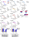
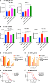
p53 is not required for suppression of ferroptosis caused by inhibition of MDM2. (A) Varying responses of HT-1080, SK-Hep1, HCT116, and H1299 cells to erastin treatment. (B,C) Dose response of wild-type and p53 KO cells to erastin in HT-1080-derived cells (B) and SK-Hep1-derived cells (C). (D,E) Inhibition of erastin-induced HT-1080 cell death by Fer-1 (D) and DFO (E). (FK) Effect of MDM2 antagonists (nutlin and MEL23) on the dose response of two different cell lines to erastin treatment. (F) Cartoon of known structure and mechanism of action of nutlin (Vassilev et al. 2004). (G,H) HT-1080-derived cells (G) and SK-Hep1-derived cells (H) treated with the combination of nutlin and erastin. (I) Cartoon of known structure and mechanism of action of MEL23 (Herman et al. 2011). (J,K) HT-1080-derived cells (J) and SK-Hep1-derived cells (K) treated with the combination of MEL23 and erastin. (L,M) Suppression of ferroptosis by the knockdown of MDM2 in two HT-1080-derived p53 KO clones. The top panel shows the viability of cells treated with a lethal dose of IKE when transfected with either the siRNA against Luciferase or one of two different siRNAs against MDM2. The bottom panel shows the corresponding decrease in the protein levels of MDM2 upon RNA interference against Luciferase (L) or MDM2 (1 and 2). The transfection was done using 15 nM of siRNA and the cells were treated with IKE 24 h after transfection. Cells in AK were treated with drugs for 24 h. Cells in L and M were treated with drugs for 18 h. The data in AE, G,H, J, and K represent the mean ± SE for two out of four independent experiments. The viability data in L and M represent the mean ± SE for four independent experiments. The viability data have been measured using ATP-based CellTiter-Glo reagent and have been normalized to the DMSO control.
Figure 2.
Figure 2.
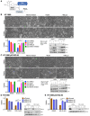
MDMX promotes sensitivity to ferroptosis independently of p53. (A) Cartoon of the known structure and mechanism of action of the MDMX inhibitor (Berkson et al. 2005; Wang and Yan 2011). (B,C) Effect of MDM2 and MDMX antagonists (MEL23, nutlin, and MDMX inhibitor) on the lethality of erastin in HT-1080-derived cells. This effect was analyzed using microscopic images taken at 10× magnification (top panel) and cell viability (bottom left panel). (Bottom right panel) The corresponding MDM2/X protein levels were also measured. (D,E) Suppression of ferroptosis by the knockdown of MDMX in HT-1080-derived cells. The left panel shows the viability of cells treated with a lethal dose of IKE when transfected with either the scrambled siRNA negative control or one of two different siRNAs against MDMX. The right panel shows the corresponding decrease in the protein levels of MDMX upon RNA interference against the control (N) or MDMX (1 and 2). The transfection was done using 15 nM of siRNA and the cells were treated with IKE 24 h after transfection. Cells in B and C were treated with drugs for 16 h. Cells in D and E were treated with drugs for 18 h. The viability data in B and C represent the mean ± SE for two independent experiments. The viability data in D and E represent the mean ± SE for three independent experiments. The viability data have been measured using ATP-based CellTiter-Glo reagent and have been normalized to the DMSO control.
Figure 3.
Figure 3.
Wild-type MDMX is more effective at sensitizing cells to ferroptosis than MDM2 binding-deficient mutants of MDMX. (AD) Effect of overexpression of MDMX variants (WT and C463A) on the lethality of IKE in p53 KO clones derived from two different cells. The transfection was done using 300 µg of plasmid of each variant in HT-1080-derived cells (A) and SK-Hep1-derived cells (B). The transfection was done using a dose-curve (0–300 µg) of each variant in HT-1080-derived cells (C) and SK-Hep1-derived cells (D). The right panels of A and B and the bottom panels of C and D show the corresponding expression levels of the MDMX variants. The cells were treated for 18 h with IKE (5 µM) 24 h after transfection. The viability data in AD represent the mean ± SE for three independent experiments. The viability after drug treatment has been measured using ATP-based CellTiter-Glo reagent and is normalized to the viability of the DMSO control under each transfection condition, respectively.
Figure 4.
Figure 4.
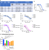
The MDM2–X complex can promote ferroptosis in patient-derived glioblastoma models and rat brain slices. (AF) The MDM2–X complex mediates the sensitivity of four different patient-derived glioblastoma models to RSL3. (A) RSL3's EC50, MDM2, and p53 status for the glioblastoma models. Dose response of the glioblastoma models to RSL3 (B), staurosporine (STS) (C), and doxorubicin (D). MEL23 is able to inhibit the sensitivity of MDM2 wild-type glioblastoma models (E) and MDM2-amplified glioblastoma models (F) to RSL3 treatment. (G) MEL23 is able to suppress the mutant Huntingtin (mHTT) protein-induced neurodegeneration of rat striatal medium spiny neurons in a brain slice model of Huntington's disease. Rat corticostriatal brain slice explants were cotransfected with the first exon of the mHTT (Q73), and YFP transfection was used as a control. Brain slices were treated with either DMSO, a positive control mixture of 50 µM KW-6002 and 30 µM of SP600125, Fer-1 as a second positive control, or a three-point concentration response curve for MEL23. Cells in AF were treated with drugs for 24 h. The data in AF represent the mean ± SE for two out of four independent experiments. The data in G represent the mean ± SE from one of two representative experiments, with at least 24 samples assessed per condition. The viability data have been measured using ATP-based CellTiter-Glo reagent and have been normalized to the DMSO control.
Figure 5.
Figure 5.
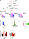
MDM2 and MDMX control a central checkpoint of ferroptosis. (A) Schematic depicting the previously established mechanisms of various FINs (Stockwell et al. 2017). (B) Inhibitory effect of MEL23 on the response of HT-1080-derived p53 KO cells to various classes of FINs: RSL3, FIN56, and FINO2. (C) Assessment of reduced glutathione levels in HT-1080-derived p53 KO cells treated with erastin with or without MEL23 or MDMX inhibitor. (D) Lipid ROS levels in HT-1080-derived p53 KO cells were measured by flow cytometry using C-11 Bodipy. The cells were treated with IKE in combination with either ferrostatin-1 or MDM2/X antagonists. The left panel represents the histogram showing the green fluorescence and the right panel represents the quantification of the normalized fluorescence. (E) The levels of labile (ferrous) iron in HT-1080-derived cells were measured using the FIP-1 probe. The cells were treated with erastin, either alone or in combination with DFO or MEL23. Cells in B were treated with drugs for 24 h. Cells in CE were treated with erastin/IKE (10 µM) for 6.5 h. The data in B represent the mean ± SE for two out of four independent experiments. The data in C represent the mean ± SE for three biological replicates. The data in the right panel of D represent the mean ± SE for three independent experiments. The data in E is from one representative experiment out of three independent experiments. The data in E show the mean ± SE obtained from analyzing at least 100 different cells (from a minimum of five different fields) for each treatment condition. The viability data have been measured using ATP-based CellTiter-Glo reagent and have been normalized to the DMSO control.
Figure 6.
Figure 6.
MDM2 and MDMX regulate lipid metabolism to favor ferroptosis. (A,B) Signal intensities of lipids (A) and triacyl glycerides (TAGs) (B) in HT-1080-derived p53 KO cells were measured by LC-MS and analyzed as significantly altered by ANOVA. The cells were treated with erastin in combination with MDM2 and MDMX antagonists. (CF) The cell death due to erastin was assessed when PPARα activity was modulated using an agonist, pirinixic acid, or an antagonist, GW6471, in two different cell lines. Pirinixic acid blocks the lethal dose of erastin in HT-1080-derived p53 KO cells (C) and SK-Hep1-derived p53 KO cells (D). GW6471 enhances the dose response to erastin in HT-1080-derived p53 KO cells (E) and SK-Hep1-derived p53 KO cells (F). Cells in A and B were treated with erastin (10 µM) for 6.5 h. Cells in C and D were treated with erastin and pirinixic acid for 16 h. Cells in E and F were treated with erastin for 24 h and with GW6471 for 48 h. The data in A and B represent the mean ± SE for three biological replicates. The data in C and D represent the mean ± SE for three or two of three independent experiments, respectively. The data in E and F represent the mean ± SE for two out of four independent experiments. The viability data have been measured using ATP-based CellTiter-Glo reagent and have been normalized to the DMSO control.
Figure 7.
Figure 7.
PPARα activity plays a key role in facilitating the abilities of MDM2 and MDMX to dampen the antioxidant responses of cells and promote ferroptosis. (A,B) Effect of PPARα antagonist GW6471 on the ability of MEL23, MDMX inhibitor, and Fer-1 to block ferroptosis in SK-HEP1-derived p53 KO cells. (B, left panel) The relative cell death due to erastin when in combination with these inhibitors and GW6471. (B, right panel) The effect of MEL23 and MDMX inhibitor on the protein levels of MDM2 and MDMX is unaffected by cotreatment with GW6471. (A) Visualization of the aforementioned changes in cell viability at 10× magnification. (C) PPARα antagonist GW6471 suppresses the ability of siMDMX and siMDM2 to block ferroptosis in SK-HEP1-derived p53 KO cells. Two different siRNAs (#1 and #2) were used against each protein and compared against a scrambled siRNA negative control (siCtrl). The right panel shows the corresponding decrease in the protein levels of MDM2 and MDMX upon RNA interference. The transfection was done using 20 nM of siRNA in media with GW4671 and the cells were treated with drugs 24 h after transfection. (D) MEL23 and MDMX inhibitor up-regulate the mRNA levels of some known PPARα downstream targets in SK-HEP1-derived p53 KO cells. (E) Immunoprecipitation using PPARα antibody in SK-HEP1-derived p53 KO cells shows binding to MDM2 and MDMX even under treatment with MDM2–X antagonists. (F) Ratio of reduced to oxidized CoQ10 levels in HT-1080-derived p53 KO cells treated with erastin in combination with MDM2 and MDMX antagonists. (G) PPARα activity modulates the levels and extent of increase of FSP1 protein in cells treated with the MDM2/X antagonists in HT-1080 p53 KO cells. (H) Summary schematic depicting the hypothesized role of MDM2 and MDMX in ferroptosis. Cells in AC, E, and G were treated with erastin/MEL23/MDMX inhibitor for 16 h and also with GW6471 for 40 h, as shown in AC and G. Cells in D and F were treated with erastin (10 µM) for 6.5 h. The data in B and C represent the mean ± SE for three or two of three independent experiments, respectively. The data in D and F represent the mean ± SE for three biological replicates. The viability data have been measured using ATP-based CellTiter-Glo reagent and have been normalized to either the DMSO control or the GW6471 control, respectively, in B, and to each transfection's respective control that is not treated with erastin in C.

References

    1. Afshinnia F, Rajendiran TM, Soni T, Byun J, Wernisch S, Sas KM, Hawkins J, Bellovich K, Gipson D, Michailidis G, et al. 2018. Impaired β-oxidation and altered complex lipid fatty acid partitioning with advancing CKD. J Am Soc Nephrol 29: 295–306. 10.1681/ASN.2017030350 - DOI (V体育ios版) - PMC - PubMed
    1. Alarcon-Vargas D, Ronai Z. 2002. p53-Mdm2—the affair that never ends. Carcinogenesis 23: 541–547. 10.1093/carcin/23.4.541 - DOI (VSports手机版) - PubMed
    1. Arena G, Cissé MY, Pyrdziak S, Chatre L, Riscal R, Fuentes M, Arnold JJ, Kastner M, Gayte L, Bertrand-Gaday C, et al. 2018. Mitochondrial MDM2 regulates respiratory complex I activity independently of p53. Mol Cell 69: 594–609.e8. 10.1016/j.molcel.2018.01.023 - DOI - PMC - PubMed
    1. Aron AT, Loehr MO, Bogena J, Chang CJ. 2016. An endoperoxide reactivity-based FRET probe for ratiometric fluorescence imaging of labile iron pools in living cells. J Am Chem Soc 138: 14338–14346. 10.1021/jacs.6b08016 - "V体育官网入口" DOI - PMC - PubMed
    1. Atatreh N, Ghattas MA, Bardaweel SK, Al Rawashdeh S, Al Sorkhy M. 2018. Identification of new inhibitors of Mdm2–p53 interaction via pharmacophore and structure-based virtual screening. Drug Des Devel Ther 12: 3741–3752. 10.2147/DDDT.S182444 - "VSports注册入口" DOI - PMC - PubMed

Publication types

MeSH terms

"V体育安卓版" Substances