<sup dir="RKsqE28L"></sup> "VSports在线直播" Skip to main page content
U.S. flag

An official website of the United States government

Dot gov

The . gov means it’s official VSports app下载. Federal government websites often end in . gov or . mil. Before sharing sensitive information, make sure you’re on a federal government site. .

Https

The site is secure V体育官网. The https:// ensures that you are connecting to the official website and that any information you provide is encrypted and transmitted securely. .

. 2019 Jul 16;51(1):90-103.e3.
doi: 10.1016/j.immuni.2019.06.003. Epub 2019 Jul 2.

T Cell Recruitment to the Intestinal Stem Cell Compartment Drives Immune-Mediated Intestinal Damage after Allogeneic Transplantation

Affiliations

T Cell Recruitment to the Intestinal Stem Cell Compartment Drives Immune-Mediated Intestinal Damage after Allogeneic Transplantation

Ya-Yuan Fu et al. Immunity. .

Abstract

The key sites within the gastrointestinal (GI) tract where T cells mediate effector responses and the impact of these responses on intestinal stem cells (ISCs) remain unclear. Using experimental bone marrow transplantation to model immune-mediated GI damage and 3D imaging to analyze T cell localization, we found that the ISC compartment is the primary intestinal site targeted by T cells after transplantation. Recruitment to the crypt base region resulted in direct T cell engagement with the stem cell compartment and loss of crypt base columnar ISCs, which expressed both MHC classes I and II VSports手机版. Vasculature expressing the adhesion molecule MAdCAM-1 clustered near the crypt base, preferentially regulating crypt compartment invasion and ISC reduction without affecting T cell migration to villi. These findings indicate that allogeneic T cells rapidly access the stem cell niche after transplantation, and this targeted recruitment to the stem cell compartment results in ISC loss during immune-mediated GI damage. .

Keywords: BMT; GVHD; ISCs; LPAM; MAdCAM-1; Paneth cells; allogeneic bone marrow transplantation; beta7 integrin; graft versus host disease; imaging of immunity; intestinal stem cells; mucosal immunology; transplantation. V体育安卓版.


Figures

Figure 1.
Figure 1.. 3-D imaging approach provides accurate tissue volume and cell localization.
(A) 3-D images of scanned volume and processed tissue volume in ileum. (B) 3-D reconstruction of villi and crypts from full-thickness BALB/c ileum; red, CD3+ T cells; white, nuclei. Note that villus tissue volume is larger than crypt tissue volume: mean villus Z-depth is approximately 250 μm; mean crypt Z-depth is approximately 100 μm. (C) Quantification of CD3+ T cell number per full-thickness 3-D field in BALB/c ileum. (D) Quantification of CD3+ T cell density in BALB/c ileum per full-thickness 3-D field; n = 17 (villus region) and n = 16 (crypt region) independent 3-D views (with 4–5 independent 3-D views per mouse and 4 mice per group). Bar graphs represent mean and SEM; **p < 0.01. Data combined from two independent experiments.
Figure 2.
Figure 2.. T cell density within the intestinal mucosa during immune-mediated GI damage is greatest in the crypt base region where the stem cell compartment is located.
Full-thickness SI (ileum) was imaged by performing immunofluorescent confocal microscopy on clarified whole-mount intestinal tissue. 3-D images were divided into upper and lower halves of the villus and crypt regions for quantification of T cell density after B6-into-BALB/c allogeneic BMT. Transplantation into irradiated recipients was performed with TCD marrow +/− 1 × 10 6 purified donor T cells, and T cells were identified by anti-CD3 immunofluorescence. (A) Representative images of full-thickness SI tissue divided into upper and lower halves of the villus and crypt regions; red, CD3+ T cells; white, nuclei. Green dots outline the scanned volume dimensions: 581.25 × 581.25 × 123 μm 3 (both in upper and lower villus regions); 581.25 × 581.25 × 62.5 μm 3 (both in upper and lower crypt regions). (B) 3-D projections of CD3+ T cells present at different ileal depths in normal untransplanted BALB/c controls and in BALB/c recipient mice four days after BMT; scale bars, 100 μm. (C) Quantification of CD3+ T cell densities four days after BMT; n = 21 (no BMT), n =18 (TCD BMT), and n = 13 (BMT + T) independent views in upper and lower villus regions; n = 20 (no BMT), n = 27 (TCD BMT), and n = 17 (BMT + T) independent views in upper and lower crypt regions. Data represent 3–6 independent 3-D views per mouse and 4–6 mice per group, combined from two independent experiments. (D) 3-D projections of CD3+ T cells present at different ileal depths in normal untransplanted BALB/c controls and in BALB/c recipient mice seven days after BMT; scale bars, 100 μm. (E) Quantification of CD3+ T cell densities seven days after BMT. n = 21 (no BMT), n =24 (TCD BMT), and n = 21 (BMT + T) independent views in upper and lower villus regions; n = 20 (no BMT), n = 23 (TCD BMT), and n = 21 (BMT + T) independent views in upper and lower crypt regions. Data represent 3–6 independent 3-D views per mouse and 4–6 mice per group combined from two independent experiments. Bar graphs represent mean and SEM; **p < 0.01; ***p < 0.001.
Figure 3.
Figure 3.. Crypt injury and loss of intestinal stem cells occur early after allogeneic bone marrow transplantation.
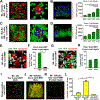
(A and B) 2-D optical slices from immunofluorescent 3-D imaging of Olfm4+ ISCs (GFP staining, green) and nuclei (DAPI staining, blue) in SI (ileum) from Olfm4-GFP mice after staining and scanning of intact clarified whole-mount ileal tissue. (A) Transverse view of Olfm4+ ISCs; scale bar, 100 μm. (B) 2-D optical slices from 3-D imaging of the ISC compartment with staining for both Olfm4+ ISCs and Paneth cells (lysozyme staining, red). Upper panel shows a high magnification view of the ISC compartment at transverse orientation. Lower panel shows a high magnification view of the ISC compartment with a viewing orientation from the serosal intestinal surface; scale bars, 20 μm. (C-E) Whole-mount staining of MHC (white) along with lysozyme to identify Paneth cells (red) and GFP to identify ISCs (green) in SI from Olfm4-GFP mice. (C) 2-D optical slice from 3-D imaging of MHC class I (white) with Olfm4+ ISCs and lysozyme+ Paneth cells at the crypt base; red arrows indicate lysozyme+ Paneth cells; green arrows indicate Olfm4+ ISCs; scale bars, 20 μm. (D) 2-D optical slice from 3-D imaging of MHC class II (white) with Olfm4+ ISCs and lysozyme+ Paneth cells at the crypt base; red arrows indicate lysozyme+ Paneth cells; green arrows indicate Olfm4+ ISCs; scale bars, 20 μm. (E) Upper panel, quantification of cell frequency with MHC class I signal detected on Olfm4+ ISCs and lysozyme+ Paneth cells; n = 6 independent 3-D views from three mice. Lower panel, quantification of the cell frequency with MHC class II signal detected on Olfm4+ ISCs and lysozyme+ Paneth cells; n = 6 independent 3-D views from three mice. (F-I) 3-D imaging and quantification of the SI (ileum) ISC compartment after B6-into-BALB/c allogeneic BMT with 1 × 10 6 donor T cells and assessment of lysozyme+ Paneth cells and adjacent CBC ISCs. (F) 3-D projections of lysozyme+ Paneth cells (red) indicating crypt base architecture in SI from normal untransplanted BALB/c mice and from mice four days after BMT; scale bars, 100 μm. (G) High magnification 2-D images of BALB/c crypt bases shown in (F) from a normal untransplanted mouse (left panel) and four days after BMT (middle and right panels), with lysozyme staining in red and DAPI nuclear signal in blue; red arrows indicate lysozyme+ Paneth cells, and light blue (cyan) arrows indicate nuclei from lysozyme CBC cells; scale bars, 25 μm. (H) 3-D projections of DAPI nuclear signal from the crypt bases shown in (G); dark blue nuclei indicate Paneth cells; light blue (cyan) nuclei indicate CBC cells; scale bars, 25 μm. (I) Quantification of Paneth cells and CBC cells from 3-D images. Data combined from two independent experiments; n = 13 independent views per group (with 3–4 independent 3-D views per mouse and four mice per group). Graphs indicate mean and SEM; ***p < 0.001.
Figure 4.
Figure 4.. Allogeneic donor T cells preferentially invade the stem cell compartment within the intestines.
(A-H) Whole-mount 3-D imaging of SI (ileum) after B6-into-BALB/c allogeneic BMT with 1 × 10 6 GFP+ purified donor T cells. (A) GFP+ donor T cell infiltration of the crypt base region in relation to lysozyme+ Paneth cells four days after BMT. Left panel, low magnification image shows donor T cells in relation to several crypts; scale bar, 100 μm. Right panel, close-up image shows GFP+ donor T cells in contact with a damaged crypt base lacking CBC cells. Green, GFP+ donor T cells; red, lysozyme+ Paneth cells; blue, nuclei of crypt base epithelium; scale bar, 25 μm. (B) Image and quantification of GFP+ donor T cell infiltration in full-thickness SI, greatest in lower crypt region four days after BMT. Image shows 3-D projection of GFP+ donor T cells (green), with 2-D nuclear background (blue) indicating the surrounding intestinal structure in transverse orientation; I, upper villus; II, lower villus; III, upper crypt; IV, lower crypt; scale bar, 100 μm. Graph shows quantification of donor T cell density in the different regions; n = 12 (upper and lower villus) and n = 10 (upper and lower crypt) independent views per region from 5–7 independent 3-D views per mouse and two mice per group; ***p < 0.001. (C) GFP+ donor T cell infiltration of the crypt base region in relation to lysozyme+ Paneth cells seven days after BMT. Left panel, low magnification image of the entire field shows donor T cells in relation to several crypts; scale bar, 100 μm. Right panel, high magnification image shows GFP+ donor cells in contact with a damaged crypt base. Green, GFP+ donor T cells; red, lysozyme+ Paneth cells; blue, nuclei of crypt base epithelium; scale bar, 25 μm. (D) Image and quantification of GFP+ donor T cell infiltration in full-thickness SI, greatest in lower crypt region seven days after BMT. Image shows 3-D projection of GFP+ donor T cells (green), with 2-D nuclear background (blue) indicating the surrounding intestinal structure in transverse orientation; I, upper villus; II, lower villus; III, upper crypt; IV, lower crypt; scale bar, 100 μm. Graph shows quantification of donor T cell density in the different regions; n = 12 independent views per region from 3–9 independent 3-D views per mouse and two mice per group. Data are mean and SEM; lower crypt region vs. other regions; ***p < 0.001. (E) High magnification 3-D projection of GFP+ donor T cells (green) with anti-CD4 (white) and anti-lysozyme (red) immunostaining shows CD4+GFP+ donor T cells infiltrating the crypt base compartment four days after BMT; scale bar, 50 μm. (F) CD4+ and CD4 frequencies in GFP+ donor T cells in the lower crypt region four days after BMT; n = 16 independent 3-D views from 4–8 views per mouse and four mice per group. Data are mean and SEM; ***p < 0.001. (G) High magnification 3-D projection of GFP+ donor T cells (green) with anti-CD4 (white) and anti-lysozyme (red) immunostaining shows both CD4+GFP+ and CD4GFP+ (i.e. CD8) donor T cells infiltrating the crypt base compartment seven days after BMT; scale bar, 50 μm. (H) CD4+ and CD4 frequencies in GFP+ donor T cells in the lower crypt region seven days after BMT; n = 16 independent 3-D views from 8 views per mouse and two mice per group. (I-L) 3-D images and quantification of proliferating GFP+ donor T cells (yellow) after B6-into-B6 syngeneic BMT or B6-into-BALB/c allogeneic BMT with 1 × 10 6 GFP+ purified donor T cells; dimensions of 3-D fields: 303.34 × 303.34 × 99 μm 3. (I) 3-D projection of double positive Ki67+GFP+ cells in MLN four days after syngeneic BMT. (J) 3-D projection of double positive Ki67+GFP+ cells in MLN four days after allogeneic BMT. (K) 3-D projection of double positive Ki67+GFP+ cells in SI crypt region four days after allogeneic BMT. (L) Quantification of Ki67+GFP+ cells in MLNs and SI crypt region four days after syngeneic (syn) or allogeneic (allo) BMT; ***p < 0.001.
Figure 5.
Figure 5.. Donor T cells invaded the ISC compartment adjacent to or replacing CBC ISCs after unirradiated BMT.
SI crypt base images of CBC stem cell nuclei next to Paneth cells and infiltrating T cells, day 7 after B6-into-unirradiated-BDF1 allogeneic BMT with 30 × 10 6 GFP+ purified donor T cells. DAPI+ nuclei are shown in blue, lysozyme+ Paneth cells are shown in red, GFP+ donor T cells are shown in green. Left panels show 2-D slices of nuclei and anti-lysozyme signal, with T cell nuclei highlighted by green arrowheads (left upper panels) and T cell shape demonstrated with GFP signal (left lower panels). Right panels show 3-D projections of the entire crypt base with ISC nuclei highlighted in light blue. (A) Images of the crypt base region with an invading GFP+ donor T cell adjacent to a stem cell and directly interacting with it. Yellow arrows indicate the nucleus of the ISC surrounded by the donor T cell; scale bars, 25 μm. (B) Images of the crypt base region with a GFP+ donor T cell between several Paneth cells in a position that would normally be occupied by an ISC, which appears to be absent; scale bars, 20 μm.
Figure 6.
Figure 6.. MAdCAM-1+ vessels primarily localize to the crypt regions of the intestinal mucosa.
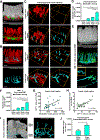
(A-D) Vessel painting and 3-D immunofluorescent imaging of ileum in normal (untransplanted) mice. (A) 3-D imaging of small intestine vasculature (red) after vessel painting in Olfm4-GFP mice shows greater vascular density in the villus region compared to the crypt region; white, nuclei (DAPI); green, ISCs (anti-GFP); scale bars, 100 μm. (B) 3-D immunofluorescent imaging of MAdCAM-1 staining in light blue (cyan), with MAdCAM-1vasculature shown in red, reveals that MAdCAM-1+ vessels are predominantly located around the crypt compartment; scale bars, 150 μm. (C) Representative 3-D imaging of MAdCAM-1vasculature (red) and MAdCAM-1+ vasculature (light blue) in different regions within ileum of BALB/c mice. Green dots outline the scanned volume dimensions: 303.34 × 303.34 × 105 μm 3 (both in upper and lower villus regions); 303.34 × 303.34 × 42 μm 3 (both in upper and lower crypt regions). (D) Quantification of MAdCAM-1 density in different regions as shown in (C); n = 18 (upper and lower villus regions) and n = 23 (upper and lower crypt regions) independent views per region combined from 3–7 independent 3-D views per mouse and five mice per group from two independent experiments. (E-H) Analyses of ileum four days after B6-into-BALB/c BMT with 1 × 10 6 GFP+ donor T cells. (E) 3-D imaging of MAdCAM-1+ vessels (light blue) and donor T cells (green) in recipient SI post-transplant. Upper and middle panels: upper panel provides architectural orientation from DAPI nuclear staining (white); in the middle panel, GFP+ donor T cells appear to localize near MAdCAM-1+ vessels; scale bars, 150 μm. Lower panel: close-up 2-D image of a MAdCAM-1+ vessel in the crypt base region from the 3-D projection image shown in middle panel. Green arrows indicate transendothelial migration of 4 GFP+ donor T cells. A MAdCAM-1+ vessel encircles the donor T cells (inset) while they are migrating out of the vessel to parenchymal areas; scale bar, 50 μm. (F) Quantification of MAdCAM-1 density in different regions four days after BMT as shown in (E); n = 11 (upper and lower villus regions) and n = 28 (upper and lower crypt regions) independent views per region combined from 2–6 independent 3-D views per mouse and 3–5 mice per group from two independent experiments. (G) Comparison of GFP+ donor T cells and MAdCAM-1 density in the lower crypt region; n = 28 independent 3-D views combined from two independent experiments. (H) Comparison of severely damaged crypts (<5 CBCs per crypt) and GFP+ donor T cells in the lower crypt region; n = 29 independent 3-D views combined from two independent experiments. (I) 3-D imaging of MAdCAM-1+ vessels in human duodenal biopsy specimen after clinical allogeneic hematopoietic transplantation; light blue, anti-MAdCAM-1; white, nuclei (DAPI); scale bar, 250 μm. (J) Quantification of patient duodenal MAdCAM-1 density in the villus and crypt regions after transplantation as shown in (I); n = 3 independent 3-D views. Bar graphs represent mean and SEM; lower crypt region vs. other regions; *p < 0.05; ***p < 0.001.
Figure 7.
Figure 7.. β7 integrin and MAdCAM-1 regulate T cell recruitment to the stem cell compartment and loss of intestinal stem cells during immune-mediated GI damage.
(A and B) Full-thickness SI tissue (ileum) was divided into upper and lower halves of the villus and crypt regions for quantification of T cell density after B6-into-BALB/c allogeneic BMT. BMT was performed with TCD marrow and 1 × 10 6 purified WT or β7−/− T cells. T cells were identified by anti-CD3 immunofluorescence. β7 deficiency specifically affected T cell density in the crypt compartment, not in the villi. (A) Representative 3-D images of CD3+ T cells in ileal crypt and villus compartments; scale bars, 100 μm. (B) Quantification of CD3+ T cell densities; n = 24 independent views from six mice per group and two independent experiments. (C-F) BMT recipients were treated with 150 μg anti-MAdCAM-1 or isotype antibodies on days 2 and 3 after BMT, then 3-D imaging was performed in the ileum on day 4 post-transplant. B6-into-BALB/c BMT was performed with TCD marrow and 1 × 10 6 purified GFP+ donor T cells. Treatment with anti-MAdCAM-1 neutralizing antibodies specifically reduced donor T cell infiltration in the crypt region without altering donor T cell migration to the villi and prevented loss of ISCs post-transplant. (C) Representative 3-D images of GFP+ donor T cells in ileal crypt and villus compartments; scale bars, 100 μm. (D) Quantification of GFP+ donor T cell densities; n = 24 (isotype) and n = 23 (anti-MAdCAM-1) independent views in upper and lower villus regions; n = 32 (isotype) and n = 33 (anti-MAdCAM-1) independent views in upper and lower crypt regions from 6–12 independent 3-D views per mouse and 3–4 mice per group from two independent experiments. (E) Representative nuclear images of CBC cells (light blue) and Paneth cells (dark blue) after treatment with anti-MAdCAM-1 or isotype; scale bars, 25 μm. (F) Quantification of CBC cells and Paneth cells from 3-D images after treatment with anti-MAdCAM-1 or isotype antibody. n = 9 independent views per group from 2–3 independent 3-D views per mouse and 4 mice per group from two independent experiments. Bar graphs represent mean and SEM; *p < 0.05; **p < 0.01.

References

    1. Adams DH, and Eksteen B (2006). Aberrant homing of mucosal T cells and extra-intestinal manifestations of inflammatory bowel disease. Nat Rev Immunol 6, 244–251. - PubMed
    1. Agudo J, Park ES, Rose SA, Alibo E, Sweeney R, Dhainaut M, Kobayashi KS, Sachidanandam R, Baccarini A, Merad M, and Brown BD (2018). Quiescent Tissue Stem Cells Evade Immune Surveillance. Immunity 48, 271–285 e275. - "V体育2025版" PMC - PubMed
    1. Akcora D, Huynh D, Lightowler S, Germann M, Robine S, de May JR, Pollard JW, Stanley ER, Malaterre J, and Ramsay RG (2013). The CSF-1 receptor fashions the intestinal stem cell niche. Stem Cell Res 10, 203–212. - PMC - PubMed
    1. Asquith M, and Powrie F (2010). An innately dangerous balancing act: intestinal homeostasis, inflammation, and colitis-associated cancer. J Exp Med 207, 1573–1577. - PMC - PubMed
    1. Barker N (2014). Adult intestinal stem cells: critical drivers of epithelial homeostasis and regeneration. Nat Rev Mol Cell Biol 15, 19–33. - PubMed

Publication types

MeSH terms

LinkOut - more resources (V体育官网入口)