Skip to main page content
U.S. flag

An official website of the United States government

Dot gov

The . gov means it’s official. Federal government websites often end in . gov or . mil. Before sharing sensitive information, make sure you’re on a federal government site. VSports app下载.

Https

The site is secure V体育官网. The https:// ensures that you are connecting to the official website and that any information you provide is encrypted and transmitted securely. .

. 2019 May 22;10(1):2263.
doi: 10.1038/s41467-019-10018-1.

Killer-like receptors and GPR56 progressive expression defines cytokine production of human CD4+ memory T cells

Affiliations

Killer-like receptors and GPR56 progressive expression defines cytokine production of human CD4+ memory T cells

Kim-Long Truong et al. Nat Commun. .

Abstract (VSports手机版)

All memory T cells mount an accelerated response on antigen reencounter, but significant functional heterogeneity is present within the respective memory T-cell subsets as defined by CCR7 and CD45RA expression, thereby warranting further stratification. Here we show that several surface markers, including KLRB1, KLRG1, GPR56, and KLRF1, help define low, high, or exhausted cytokine producers within human peripheral and intrahepatic CD4+ memory T-cell populations VSports手机版. Highest simultaneous production of TNF and IFN-γ is observed in KLRB1+KLRG1+GPR56+ CD4 T cells. By contrast, KLRF1 expression is associated with T-cell exhaustion and reduced TNF/IFN-γ production. Lastly, TCRβ repertoire analysis and in vitro differentiation support a regulated, progressive expression for these markers during CD4+ memory T-cell differentiation. Our results thus help refine the classification of human memory T cells to provide insights on inflammatory disease progression and immunotherapy development. .


"V体育安卓版" Conflict of interest statement

The authors declare no competing interests.

VSports app下载 - Figures
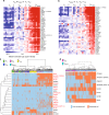
Fig. 1
Fig. 1
Specific mRNA expression profile and heterogeneity of CD4+ TEM and TEMRA cells. a, b Heatmaps of genes with significantly increased expression in a TEM/TEMRA compared to TN/TCM cells and b TEMRA compared with TN/TCM/TEM cells identified by an intersection analysis. Gene expression was determined in human CD4+ TN, TCM, TEM, and TEMRA cells sorted from peripheral blood of healthy subjects (n = 3–8, each consisting of 1–3 pooled sorted samples). The scale of both heatmaps is identical. c Single-cell profiling of TEM- and TEMRA-specific gene expression shown as unsupervised hierarchical cluster analysis of 16 gene candidates (red) and additional genes (black) in single blood CD4+ TEMRA (n = 226), TEM (n = 199), TCM (n = 186), TN (n = 94), and Treg cells (n = 178) from four healthy donors. d Unsupervised hierarchical cluster analysis of single-cell gene expression results from identified natural killer cell-associated markers in blood CD4+ TEM (n = 260) and TEMRA (n = 276) cells of five healthy individuals. Classification as expressing and non-expressing cells based on individually defined limit of detection (LoD) Ct values. Data are provided with the Source Data file
Fig. 2
Fig. 2
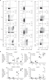
Heterogeneous surface expression of killer-like receptors and GPR56 in CD4+ T cells. a Exemplary dot plots of KLRB1, KLRG1, KLRF1, GPR56, TIGIT, and PD-1 surface expression in gated CD4+ TN, TCM, TEM, and TEMRA cells from blood of healthy donors. b Comparison of KLRB1-, KLRG1-, KLRF1-, and GPR56-positive cell frequencies obtained either from single-cell gene expression analysis (mRNA, n = 209 (TN), 260 (TCM), 396 (TEM), and 248 (TEMRA) from four donors) or flow cytometric analysis (protein, n = 5) within CD4+ TN, TCM, TEM, and TEMRA cells. Data are shown as individual scatter plots with median. Statistical analysis by two-way ANOVA and Sidakʼs multiple comparison test. **p < 0.01, ***p < 0.001
Fig. 3
Fig. 3
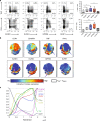
Progressive expression of KLRs and GPR56 is associated with cytokine production during CD4+ memory T-cell development. a Association of surface marker expression with cytokine expression in CD4+ T cells from peripheral blood of healthy individuals shown as exemplary dot plots and summarizing box and whisker plots (% of cytokine expressing cells within total or marker positive subset, whiskers extend to the minimum and maximum) of n = 5. Cells were restimulated with PMA and Ionomycin. b Representative t-SNE plots showing surface marker and cytokine expression pattern of pre-gated CD4+ T cells (excluding CD25highCD127low Treg cells) upon short-term phorbol 12-myristate 13-acetate (PMA)/Ionomycin stimulation. The highlighted area marks CD4+ TEM and TEMRA cells identified by the absence of CCR7 and the expression pattern of CD45RA. c Wanderlust analysis based on the trajectory of CD45RA and CCR7. Relative median surface marker and intracellular cytokine expression within CD4+ T cells from blood of five healthy individuals upon short-term PMA/Ionomycin stimulation are shown as described within Methods. P-values were determined by non-parametric matched-pairs Friedman’s test with post-hoc Dunn’s multiple comparison test. *p < 0.05, **p < 0.01
Fig. 4
Fig. 4
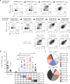
Combinational expression of KLR’s and GPR56 recapitulates human CD4+ memory T-cell development. a Exemplary dot plots of TNF and IFN-γ production upon short-term PMA/Ionomycin stimulation within conventionally gated (CD45RA/CCR7-based path) or KLR/GPR56-based gated CD4+ memory T cells. The plots are arranged according to the anticipated developmental pathway. b Comparative analysis of TNF/IFN-γ co-producing cell frequencies of conventionally gated TEM and TEMRA cells, and KLR/GPR56-based subsets upon short-term PMA/Ionomycin stimulation (n = 7, interleaved scatters with median bars). Functionality was evaluated according to the amount of cytokine production. c KLR/GPR56-based subset composition within classically gated CD4+ TEM and TEMRA cells (n = 10, mean ± SEM). Statistical analysis by one-way non-parametric Friedman’s test with post-hoc Dunn’s multiple comparison test. *p < 0.05, **p < 0.01, ***p < 0.001, ****p < 0.0001
Fig. 5
Fig. 5
Decreased proportions of KLRF1-expressing CD4+ memory T cells in the liver. a Frequency of TN, TCM, TEM, and TEMRA cells within CD4+ T cells of blood from healthy controls (HC-B, n = 8) as well as blood (LD-B, n = 8) and liver (LD-L, n = 11) of patients with inflammatory liver diseases. Whiskers extend to the minimum and maximum. P-values were determined by non-parametric Kruskal–Wallis test with post-hoc Dunn’s multiple comparison test. *p < 0.05, ****p < 0.0001. b Unsupervised hierarchical cluster analysis of candidate gene expression results at single-cell level in five blood and five liver samples of patients. In total, 152 (blood) and 86 (liver) CD4+ TCM, 249 (blood) and 82 (liver) TEM, as well as 183 (blood) and 114 (liver) TEMRA cells were analyzed. Classification as expressing and non-expressing cells based on individual defined limit of detection (LoD) Ct values. Data are provided with the Source Data file. c Proportions of CD4+ TEM and TEMRA cells and subsets according to KLRB1, KLRG1, GPR56, and KLRF1 protein expression within CD4+ T cells from blood (n = 6) and liver (n = 6) of patients. Data are shown as individual scatter plots with median. Statistical analysis by one-way non-parametric Friedman’s test with post-hoc Dunn’s multiple comparison test. *p < 0.05
Fig. 6
Fig. 6
Increase in intrahepatic cytokine producers of TEM and TEMRA cells. a Subset composition gating within classically gated CD4+ TEM and TEMRA cells of the blood and liver from patients with inflammatory liver diseases (n = 7, individual scatter plots with median). b Proportions of TNF/IFN-γ co-expressing cells upon pre-gating of TEM, TEMRA, or KLR/GPR56-based subsets following PMA/Ionomycin stimulation of PBMCs or liver leukocytes (n = 7 each). Statistical analysis was performed by one-way non-parametric Friedman’s test with post-hoc Dunn’s multiple comparison test. *p < 0.05, **p < 0.01, ***p < 0.001, ****p < 0.0001
Fig. 7
Fig. 7
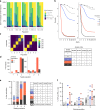
Clonotypic analysis and differentiation capacity of proposed CD4+ T-cell subsets. Clonal space (a), Rényi diversity profile (b, two exemplary), and clonal similarity (c, two exemplary) of TCRβ chain of sorted KLR/GPR56 (population 1), KLRB1+ (population 2), KLRB1+KLRG1+ (population 3), KLRB1+KLRG1+GPR56+ (population 4), and KLRB1+KLRG1+GPR56+KLRF1+ (population 5) CD4+ T cells from healthy controls (n = 5). d Number of TCR clonotypes dominating in source population 4 and 5 identified in target populations 1, 2, and 3. The scaling factor Alpha of the Rényi diversity profile yields the sample diversity with different weighting of the clonotype proportion, see Methods. The clonotypes were verified prior to diversity calculation. The clonal similarity was assessed using the index of Morisita–Horn (1 indicates identity). Data are accessible within the European Nucleotide Archive (ENA) Accession Number PRJEB31283. e KLRB1, KLRG1, GPR56, and KLRF1 protein expression profile upon 48 h anti-CD3/CD28 mAb in vitro stimulation of indicated sorted CD4+ T-cell populations from PBMCs of healthy controls (n = 5). f Proportions of TNF/IFN-γ co-producing cells of in vitro differentiated populations upon 96 h anti-CD3/CD28 mAb in vitro stimulation of indicated sorted CD4+ T-cell populations from PBMCs of healthy controls (n = 5, interleaved scatters with median bars). Due to the low frequency of population 4 and 5 within PBMCs, cytokine analysis was only feasible for starting populations 1, 2, and 3

References

    1. Bedoui S, Heath WR, Mueller SN. CD4( + ) T-cell help amplifies innate signals for primary CD8( + ) T-cell immunity. Immunol. Rev. 2016;272:52–64. doi: 10.1111/imr.12426. - DOI - PubMed
    1. Huber S, Gagliani N, O’Connor W, Jr., Geginat J, Caprioli F. CD4 + T helper cell plasticity in infection, inflammation, and autoimmunity. Mediat. Inflamm. 2017;2017:7083153. doi: 10.1155/2017/7083153. - DOI - PMC - PubMed
    1. Juno JA, et al. Cytotoxic CD4 T cells-friend or foe during viral infection? Front Immunol. 2017;8:19. doi: 10.3389/fimmu.2017.00019. - DOI - PMC - PubMed
    1. Zens KD, Farber DL. Memory CD4 T cells in influenza. Curr. Top. Microbiol Immunol. 2015;386:399–421. - PMC - PubMed
    1. Zanetti M. Tapping CD4 T cells for cancer immunotherapy: the choice of personalized genomics. J. Immunol. 2015;194:2049–2056. doi: 10.4049/jimmunol.1402669. - DOI - PubMed

Publication types

"V体育平台登录" MeSH terms

Substances