Skip to main page content (V体育2025版)
U.S. flag

An official website of the United States government

Dot gov

The VSports app下载. gov means it’s official. Federal government websites often end in . gov or . mil. Before sharing sensitive information, make sure you’re on a federal government site. .

Https

The site is secure. The https:// ensures that you are connecting to the official website and that any information you provide is encrypted and transmitted securely. V体育官网.

. 2018 Jul 26:7:e37503.
doi: 10.7554/eLife.37503.

"V体育安卓版" Tryparedoxin peroxidase-deficiency commits trypanosomes to ferroptosis-type cell death

Affiliations

Tryparedoxin peroxidase-deficiency commits trypanosomes to ferroptosis-type cell death

Marta Bogacz et al. Elife. .

Abstract

Tryparedoxin peroxidases, distant relatives of glutathione peroxidase 4 in higher eukaryotes, are responsible for the detoxification of lipid-derived hydroperoxides in African trypanosomes. The lethal phenotype of procyclic Trypanosoma brucei that lack the enzymes fulfils all criteria defining a form of regulated cell death termed ferroptosis. Viability of the parasites is preserved by α-tocopherol, ferrostatin-1, liproxstatin-1 and deferoxamine. Without protecting agent, the cells display, primarily mitochondrial, lipid peroxidation, loss of the mitochondrial membrane potential and ATP depletion. Sensors for mitochondrial oxidants and chelatable iron as well as overexpression of a mitochondrial iron-superoxide dismutase attenuate the cell death. Electron microscopy revealed mitochondrial matrix condensation and enlarged cristae. The peroxidase-deficient parasites are subject to lethal iron-induced lipid peroxidation that probably originates at the inner mitochondrial membrane. Taken together, ferroptosis is an ancient cell death program that can occur at individual subcellular membranes and is counterbalanced by evolutionary distant thiol peroxidases VSports手机版. .

Keywords: Trypanosoma brucei; cell biology; ferroptosis; trypanothione; tryparedoxin peroxidase V体育安卓版. .


"VSports最新版本" Conflict of interest statement

MB, RK No competing interests declared

Figures
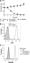
Figure 1.
Figure 1.. Ferroptosis inhibitors protect the Px I-III KO cells.
The mutant cells were either kept in Trolox-supplemented medium or transferred into medium containing 100 µM Dfx, 100 nM ferrostatin-1 or 200 nM liproxstatin-1 or no addition. (A) Every hour viable cells were counted. The data are the mean ± SD of three independent experiments. (B) The cells were kept for 2 hr in medium ± Trolox, incubated for 30 min with 10 µM H2DCFDA in medium + Trolox and subjected to flow cytometry measuring DCF fluorescence. (C) BODIPY was added and after 2 hr incubation the cells were analyzed by flow cytometry. (B and C) show representative histograms from two independent experiments.
Figure 1—figure supplement 1.
Figure 1—figure supplement 1.. Protection of Px I-III KO cells by Trolox, Dfx and α-Toc.
The cells were incubated in the presence of (A) different concentrations of Trolox, (B) various concentrations of Dfx, (C) in medium supplemented with 10 µM Trolox or 20 µM Dfx alone or in combination and (D) different concentrations of α-Toc. (E) The cells were pre-cultured for 18 hr in the presence or absence of 10 µM α-Toc in medium containing Trolox, washed with PBS and transferred into medium ± α-Toc. After different times, viable cells were counted and the percentage relative to the start cell density was calculated. The data are the mean ± SD of three independent experiments.
Figure 2.
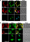
Figure 2.. Px I-III KO cells are affected in mitochondrial matrix – but not outer membrane – immuno-staining.
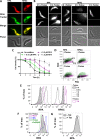
The parasites were incubated for 1 or 2 hr in medium ± Trolox, treated with MitoTracker (red) and subjected to immunofluorescence microscopy using antibodies against (A) mtTXNPx (green) or (B) VDAC (green). Nuclear and kinetoplast DNA were visualized by DAPI staining (blue). Merge, overlay of the respective three signals. Phase, phase contrast image. Scale bare 10 µm.
Figure 2—figure supplement 1.
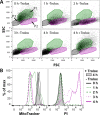
Figure 2—figure supplement 1.. Mitochondrial damage precedes plasma membrane leakage.
The Px I-III KO cells were incubated ± Trolox for up to 4 hr, stained with MitoTracker or propidium iodide and subjected to flow cytometry. (A) Forward- (FSC) and side-scatter (SSC) DotPlots of MitoTracker-treated cells. The cells were gated in three sub-populations (P1-3) representing cells with normal FSC and SSC (P1; green), cells with reduced SSC (P2; magenta), and mainly dead cells (P3; grey). (B) Histograms of MitoTracker and propidium iodide signals of all single cells. The figure shows representative results of two independent experiments.
Figure 3.
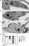
Figure 3.. PC Px I-III KO T. brucei undergo strinking ultrastructural changes at their single mitochondrion.
Parasites kept in the presence or absence of Trolox were fixed, processed and subjected to transmission electron microscopy as described under Materials and methods. Electron micrographs of representative cells incubated for (A) 2 hr in Trolox-containing medium and (B and C) 2 hr in Trolox-free medium. N, nucleus; LP, lipid droplet; G, glycosome; ER, endoplasmic reticulum; K, kinetoplast; GA, Golgi apparatus; M, mitochondrion; A, acidocalcisome; black arrow heads, normal cristae; white arrow heads, enlarged cristae. The inserts in (A) and (C) show higher magnifications of mitochondria to highlight the normal and altered morphology, respectively. The double white arrow heads point to the three membranes that surround a dilated cristae. Scale bars 500 nm. (D) Quantification of different phenotypes observed. In the presence of Trolox or for ≤0.5 hr without the antioxidant, the mutant cells were indistinguishable from wild type parasites (not shown). The cytosol was densely packed and the mitochondrion had a comparable or even lower electron density, defined as normal. In the absence of Trolox, the Px I-III KO parasites displayed a time-dependent darkening of the mitochondrion and lightening of the cytosol. Only parasites that clearly displayed an elongated cell body in the electron micrographs were incorporated in the analysis. The number of cells inspected at the different time points is given above the columns. The percentage of each phenotype in the total number of inspected cells is depicted.
Figure 3—figure supplement 1.
Figure 3—figure supplement 1.. Short-term incubation of Px I-III KO cells in Trolox-free medium results in mitochondrial alterations without changes at other organelles, whereas prolonged incubation can finally lead to plasma membrane blebs.
Electron micrographs of Px I-III KO parasites kept for (A) 1 hr and (B) 2 hr in Trolox-free medium. (A) The intact elongated mitochondrion is darker than the cytosol. Other subcellular structures appear to be unaffected. (B) One of the very few cells that after 2 hr in Trolox-free medium displayed already a plasma membrane damage. The high magnification shows vesicular structures blebbing out of the plasma membrane (double arrow heads). N, nucleus; LP, lipid droplet; G, glycosome; ER, endoplasmic reticulum; M, mitochondrion; A, acidocalcisome. Scale bar 500 nm.
Figure 4.
Figure 4.. MitoSOX Red exerts a dual effect on the Px I-III KO cells, sensing oxidant production and acting as protecting agent.
(A) The cells were pre-loaded with MitoSOX in Trolox-containing medium, transferred into medium ± Trolox, incubated for 2 hr, and stained with MitoTracker Green. Nuclear (large dot) and kinetoplast (small dot) DNA were visualized by Hoechst 33342 staining. Representative fluorescence microscopy images are depicted. Merge, overlay of all three signals. Phase, phase contrast image. Cells kept in the presence of Trolox displayed a very small red MitoSOX signal that co-localized with the kinetoplast (upper panel). After 2 hr in the absence of Trolox, most cells still had normal morphology but many of them displayed a more intense kinetoplast MitoSOX signal (lower two panels). Scale bar 10 µm. (B–D) The cells were incubated ± MitoSOX in Trolox-containing medium, transferred into medium ± Trolox, incubated for up to 5 hr, stained with DAPI, and subjected to flow cytometry. (B) Representative histograms of the 488:585/42 (ex:em) channel (MitoSOX signal of the treated cells and auto-fluorescence of non-treated cells) and DAPI signal of all single cells from samples kept for 5 hr ± Trolox, with or without loading with MitoSOX. (C) MitoSOX fluorescence change between cells kept in the absence and presence of Trolox. The data represent the mean ± error of the mean of two independent experiments. (D) Representative FSC:SSC DotPlots of cells kept for 5 hr in the presence or absence of Trolox, with or without loading with MitoSOX. The cells were gated in three sub-populations (P1-3) representing cells with normal FSC and SSC (P1; green), those with reduced SSC (P2; magenta), and severely damaged or dead cells (P3; grey).
Figure 4—figure supplement 1.
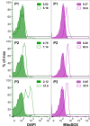
Figure 4—figure supplement 1.. The P2 sub-population displays the strongest increase in MitoSOX fluorescence.
The Px I-III KO cells were loaded with MitoSOX for 10 min in Trolox-supplemented medium, washed with PBS and re-suspended in medium ± Trolox. After 5 hr incubation, the cells were stained with DAPI and subjected to flow cytometry. The histograms represent overlays of the DAPI (green) and MitoSOX (magenta) fluorescence signals of cells kept in the presence (tinted peak) or absence (transparent peak) of Trolox. The upper, central and lower panels show the fluorescence intensities for the P1, P2, and P3 subsets, representing cells with normal FSC:SSC, decreased SSC and dead cells, respectively, as defined in Figure 4. The percentage of cells with increased signals (black range line) are given in each histogram. The figure shows representative results of one of two independent experiments.
Figure 5.
Figure 5.. Cellular ATP levels rapidly decrease when Px I-III KO cells are in medium lacking a protecting agent.
The cells were incubated ± Trolox in (A) SDM-79 medium or (B) MEM-Pros medium. After 0 to 4 hr, aliquots of each sample were removed and (A) cells with normal morphology or only highly motile cells were counted, or (B) the cells were treated with PI and the fluorescence was measured by flow cytometry. The remaining cells were treated with ATPlite one-step solution and the luminescence was measured in the plate reader. The data are given as percentage of the respective value at 0 hr that was set as 100%. They represent the mean ± SD of three independent experiments.
Figure 6.
Figure 6.. Lipid peroxidation in the Px I-III KO cells originates at the mitochondrion.
(A) Px I-III KO parasites in Trolox-supplemented medium were incubated for 1.5 hr with MitoPerOx, stained with MitoTracker Green and subjected to life cell fluorescence microscopy. Representative images are depicted. Merge, overlay of both signals. Phase, phase contrast image. Scale bar 10 µm. (B) The cells were transferred into medium ± Trolox. BODIPY or MitoPerOx was added and after different times cells were analyzed by flow cytometry. Histograms of BODIPY (upper panel) and MitoPerOx (lower panel) fluorescence of all single cells at 520 nm from a representative experiment are depicted. (C) Quantitative analysis of the percentage of cells with increased BODIPY and MitoPerOx signals (gating as depicted in B). The data represent the mean ± SD of three independent experiments.
Figure 7.
Figure 7.. Px I-III KO cells that overexpress mitochondrial SODA require less Dfx for survival.
(A) Total lysates of 5 × 10⁶ Px I-III KO/SODA-myc cells cultured for 18 hr ± tetracycline (Tet) were subjected to Western blot analysis using antibodies against myc as well as aldolase for loading control. (B) Px I-III KO/SODA-myc cells cultured for 18 hr in the presence of Tet were treated with MitoTracker (red, upper panel) followed by antibodies against Myc (green) or simultaneously with antibodies against Myc and cytosolic 2-Cys-peroxiredoxin (cTXNPx) (red; lower panel). Nuclear and kinetoplast DNA were stained with DAPI (blue) and the cells were subjected to immunofluorescence microscopy. Merge, overlay of the Myc with the MitoTracker or cTXNPx signal. Phase + DAPI, phase contrast image with the nucleus (large dot) and the kinetoplast (small dot) visualized by DAPI staining. Scale bar 10 µm. (C) Px I-III KO and Px I-III KO/SODA-myc cells cultured for 18 hr ± Tet in Trolox-supplemented medium were transferred into Trolox-free medium and incubated ± Tet and ± 25 µM Dfx. Every hour viable cells were counted. The data represent the mean ± SD of three independent experiments. (D) Px I-III KO/SODA-myc cells cultured for 18 hr ± Tet in Trolox-supplemented medium were transferred into Trolox-free medium and incubated ± Tet and ± 25 µM Dfx for 1–4 hr starting with the longest time point. Cells were stained with PI and subjected to flow cytometry. Data show the mean + SEM of the PI fluorescence of all single cells from two experiments. Data were analyzed by two-way ANOVA with Bonferroni post-test, *p<0.05.
Figure 8.
Figure 8.. Mitochondrial iron is involved in the death phenotype of Px I-III-deficient cells.
(A) Cells were pre-loaded with 150 nM RPA or 10 nM RPAC in PBS, re-transferred into Trolox-supplemented medium, stained with MitoTracker Green and subjected to life cell imaging. Merge, overlay of the RPA and RPAC fluorescence, respectively, with the MitoTracker signal. Phase, phase contrast image. (B) Cells pre-loaded with 150 nM RPA or 10 nM RPAC were transferred into medium ± Trolox and incubated for up to 3 hr. The upper panels show the RPA or RPAC fluorescence, the lower ones the respective phase contrast pictures. (A and B) Representative parasites are depicted. Scale bars 10 µm. (C) The cells were pre-loaded with 0.15, 0.5 or 1.5 µM RPA, transferred into medium – Trolox and incubated for 6 hr. Every hour, viable cells were counted. Cells not treated with RPA (no addition) served as control. The data are the mean ± SD of three independent experiments. (D) FSC:SSC dot plots of cells that were treated with 150 nM RPA or RPAC and incubated for 3 hr ± Trolox. P1 (green), P2 (magenta) and P3 (grey) gates determined as defined in Figure 4. A representative histogram of two independent experiments is shown. (E) RPA and RPAC fluorescence of cells stained with 50 nM RPA or 1 nM RPAC and incubated ± Trolox for 0–3 hr. (F) RPA fluorescence of cells incubated for 2 hr in medium supplemented with 100 µM Trolox ± 100 µM Fe(III)/HQ complex, 100 nM ferrostatin-1 or 100 µM Dfx and stained with 50 nM RPA. (G) RPA fluorescence of Px I-III KO/SODA-myc cells grown for 20 hr in the presence or absence of Tet and stained with 50 nM RPA. (E–G) Representative histograms from one of at least three independent experiments are depicted.

VSports在线直播 - References

    1. Alberto ME, Russo N, Grand A, Galano A. A physicochemical examination of the free radical scavenging activity of Trolox: mechanism, kinetics and influence of the environment. Physical Chemistry Chemical Physics. 2013;15:4642–4650. doi: 10.1039/c3cp43319f. - DOI - PubMed
    1. Beig M, Oellien F, Garoff L, Noack S, Krauth-Siegel RL, Selzer PM. Trypanothione reductase: a target protein for a combined in vitro and in silico screening approach. PLoS Neglected Tropical Diseases. 2015;9:e0003773. doi: 10.1371/journal.pntd.0003773. - DOI - PMC - PubMed
    1. Biebinger S, Wirtz LE, Lorenz P, Clayton C. Vectors for inducible expression of toxic gene products in bloodstream and procyclic Trypanosoma brucei. Molecular and Biochemical Parasitology. 1997;85:99–112. doi: 10.1016/S0166-6851(96)02815-0. - DOI - PubMed
    1. Brigelius-Flohé R, Maiorino M. Glutathione peroxidases. Biochimica et Biophysica Acta (BBA) - General Subjects. 2013;1830:3289–3303. doi: 10.1016/j.bbagen.2012.11.020. - DOI - PubMed
    1. Budde H, Flohé L, Hecht HJ, Hofmann B, Stehr M, Wissing J, Lünsdorf H. Kinetics and redox-sensitive oligomerisation reveal negative subunit cooperativity in tryparedoxin peroxidase of Trypanosoma brucei brucei. Biological Chemistry. 2003;384:619–633. doi: 10.1515/BC.2003.069. - DOI (V体育平台登录) - PubMed

"VSports" Publication types

V体育官网 - MeSH terms