Skip to main page content (VSports注册入口)
U.S. flag

An official website of the United States government

Dot gov

The . gov means it’s official. Federal government websites often end in VSports app下载. gov or . mil. Before sharing sensitive information, make sure you’re on a federal government site. .

Https

The site is secure. The https:// ensures that you are connecting to the official website and that any information you provide is encrypted and transmitted securely V体育官网. .

. 2018 Oct;37(40):5435-5450.
doi: 10.1038/s41388-018-0315-z. Epub 2018 Jun 5.

The glutathione redox system is essential to prevent ferroptosis caused by impaired lipid metabolism in clear cell renal cell carcinoma

Affiliations

"V体育官网" The glutathione redox system is essential to prevent ferroptosis caused by impaired lipid metabolism in clear cell renal cell carcinoma

Heike Miess (V体育2025版) et al. Oncogene. 2018 Oct.

Abstract

Metabolic reprogramming is a prominent feature of clear cell renal cell carcinoma (ccRCC). Here we investigated metabolic dependencies in a panel of ccRCC cell lines using nutrient depletion, functional RNAi screening and inhibitor treatment. We found that ccRCC cells are highly sensitive to the depletion of glutamine or cystine, two amino acids required for glutathione (GSH) synthesis. Moreover, silencing of enzymes of the GSH biosynthesis pathway or glutathione peroxidases, which depend on GSH for the removal of cellular hydroperoxides, selectively reduced viability of ccRCC cells but did not affect the growth of non-malignant renal epithelial cells. Inhibition of GSH synthesis triggered ferroptosis, an iron-dependent form of cell death associated with enhanced lipid peroxidation. VHL is a major tumour suppressor in ccRCC and loss of VHL leads to stabilisation of hypoxia inducible factors HIF-1α and HIF-2α. Restoration of functional VHL via exogenous expression of pVHL reverted ccRCC cells to an oxidative metabolism and rendered them insensitive to the induction of ferroptosis. VHL reconstituted cells also exhibited reduced lipid storage and higher expression of genes associated with oxidiative phosphorylation and fatty acid metabolism VSports手机版. Importantly, inhibition of β-oxidation or mitochondrial ATP-synthesis restored ferroptosis sensitivity in VHL reconstituted cells. We also found that inhibition of GSH synthesis blocked tumour growth in a MYC-dependent mouse model of renal cancer. Together, our data suggest that reduced fatty acid metabolism due to inhibition of β-oxidation renders renal cancer cells highly dependent on the GSH/GPX pathway to prevent lipid peroxidation and ferroptotic cell death. .


Conflict of interest statement

Author Information

The authors declare no other competing financial interests.

Figures
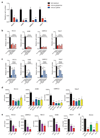
Figure 1
Figure 1. Renal cancer cells are sensitive to glutamine and cystine depletion
A) RCC4, A498, UMRC2 and 769-P cells were seeded in full medium. After 24h, medium was replaced to normal medium or medium deprived of glutamine or cystine for 72h and cell numbers were determined. Values represent mean cell number ±SEM relative to full medium (n=3). B) RCC4, A498, UMRC2 and 769-P cells were seeded in full medium. After 24h, cells were transferred to glutamine-free medium containing the different supplements and antioxidants. Cells were fixed after 72h and cell numbers were determined. Values represent mean cell number ±SEM relative to full medium (n=3). C) RCC4, A498, UMRC2 and 769-P cells were seeded in full medium. After 24h, cells were transferred to cystine-free medium containing the different supplements and antioxidants. Cells were fixed after 72h and cell numbers were determined. Values represent mean cell number ±SEM relative to full medium (n=3). D) RCC4, A498, UMRC2 and 769-P cells were transfected with siRNA oligonucleotides targeting SLC7A11, SLC1A5, GSR or GLS or a non-targeting control (CTRL). Cell numbers were determined 96h post transfection. Values represent mean cell number ±SEM (n=3). E) RCC4, A498, UMRC2, 786-O and 769-P cells were transfected with siRNA oligonucleotides targeting GCLC, GSS or a non-targeting control (CTRL). Cell number was determined 96h post transfection. Values represent mean cell number ±SEM (n=3). F) RCC4 cells were transfected with siRNA oligonucleotides targeting SLC7A11, GSR, GCLC, GLS or a non-targeting control (CTRL). After 96 hours, cells were lysed and levels of reduced glutathione (GSH) were determined by mass spectrometry. Values represent mean peak intensity normalised to protein ±SEM (n=4). *p≤0.05; **p≤0.01; ***p≤0.005; ****p≤0.001 unpaired two-tailed Student’s t-test.
Figure 2
Figure 2. Functional siRNA screen identifies glutathione peroxidases as essential enzymes in renal cancer cells
A) RCC4, A498, UMRC2, 786-O and 769-P cells were transfected with siRNA oligonucleotides targeting 230 different metabolic enzymes, nutrient transporters and metabolic regulators. 96h post transfection, number of remaining cells was determined and used to calculate z-scores. Graph displays mean z-scores across all cell lines and selected genes are highlighted. B) Table showing z-scores of selected genes in each of the five cell lines. C) RCC4, A498, 786-O and 769-P cells were transfected with siRNA oligonucleotides targeting MYC, G6PD, DHCR24, GPX3 or GPX4 or a non-targeting control (RF). Cell numbers were determined 96h post transfection. Values represent mean cell number ±SEM (n=3). *p≤0.05; **p≤0.01; ***p≤0.005; ****p≤0.001 unpaired two-tailed Student’s t-test.
Figure 3
Figure 3. Inhibition of GSH synthesis induces ferroptosis in ccRCC cells
A) Diagram illustrating the mechanisms of GSH synthesis and induction of ferroptosis. B) RCC4 and 786-O cells were treated with the indicated concentrations of Erastin or solvent (DMSO) either in the presence or absence of ferrostatin (fer-1) for 72h. Cell viability was determined by crystal violet staining. Values represent mean ±SEM relative to solvent-treated controls (n=3). C) RCC4 and 786-O cells were treated with the indicated concentrations of BSO or solvent (DMSO) either in the presence or absence of ferrostatin (fer-1) for 72h. Cell viability was determined by crystal violet staining. Values represent mean ±SEM relative to solvent-treated controls (n=3). D) RCC4 cells were transfected with siRNA oligonucleotides targeting SLC7A11, UBB or non-targeting controls (CTRL) and treated with 4 μM ferrostatin (fer-1) or solvent for 96 h. Cell viability was determined by crystal violet staining. Values represent mean ±SEM relative to solvent-treated controls (n=3). E) RCC4 cells were treated with the indicated concentrations of BSO or solvent (DMSO) for 24h. GSH levels were determined by mass spectrometry. Values represent mean ±SEM (n=2). F) RCC4 and 786-O cells were treated with the indicated concentrations of BSO or solvent (DMSO) with or without different concentrations of hydrogen peroxide (H2O2) for 72 hours. Cell viability was determined by crystal violet staining. Values represent mean ±SEM relative to solvent-treated controls (n=3). *p≤0.05; **p≤0.01; ***p≤0.005; ****p≤0.001 unpaired two-tailed Student’s t-test.
Figure 4
Figure 4. Restoration of pVHL function leads to ferroptosis resistance in ccRCC cells
A) Isogenic RCC4 cells were generated by expression of functional VHL (RCC4-VHL) or empty vector (RCC4-EV) . Analysis of protein expression shows reduced expression of HIF-1α and HIF-2α in VHL-restored cells. Expression of HA-tagged pVHL was also confirmed. β-Actin was used as loading control. B) mRNA levels of VEGFA and PDHK1 in isogenic RCC4 cell lines. Values represent mean ±SD relative to RCC4-EV and are normalised to B2M (n=2). C) Phosphorylation of AKT (serine 473) and pS6 (serines 235 and 236) was determined by immunoblotting using phospho-specific antibodies. D) Oxygen consumption rates (OCR) were determined in RCC4-EV and RCC4-VHL cells using a Seahorse Bioanalyzer and used to calculate basal respiration, maximal respiration in the presence of FCCP, spare respiratory capacity and ATP-dependent respiration. Values represent mean ±SEM of 6 replicates. E) Genes showing differential essentiality in RCC4-EV and RCC4-VHL cells (Δz-score≥1). F) RCC4-EV and RCC4-VHL cells were treated with 1μM Erastin or solvent (DMSO) in the presence or absence of 4μM ferrostatin (fer-1) for 72h. Cell viability was determined by crystal violet staining. Values represent mean ±SEM relative to solvent-treated controls (n=3). G) RCC4-EV and RCC4-VHL cells were treated with the indicated concentrations of BSO or solvent (DMSO) in the presence or absence of ferrostatin (fer-1) for 72h. Cell viability was determined by crystal violet staining. Values represent mean ±SEM relative to solvent treated controls (n=3). H) RCC4-EV and RCC4-VHL cells were treated with the indicated concentrations of BSO or solvent (DMSO) together with increasing concentrations of H2O2 for 72 hours. Cell viability was determined by crystal violet staining. Values represent mean ±SEM relative to solvent-treated controls (n=3). *p≤0.05; **p≤0.01; ***p≤0.005; ****p≤0.001 unpaired two-tailed Student’s t-test.
Figure 5
Figure 5. Induction of ferroptosis in ccRCC cells is mediated by lipid peroxidation
A) RCC4-EV cells were treated with the indicated concentrations of Erastin for 24h. Lipid peroxidation was determined using the BODIPY 581/591 C11 reagent. Values represent mean ±SEM relative to solvent-treated controls (n=3). B) Relative amounts of saturated (SFA), mono-unsaturated (MUFA) and poly-unsaturated (PUFA) fatty acids in RCC4-EV and RCC4-VHL cells. Values represent mean percentages ±SEM of the total fatty acid content (n=3). C) RCC4-EV and RCC4-VHL cells were cultured for 48 hours in full medium before fixation and staining with Nile Red to detect lipid droplets (LD). Cells were imaged using a Cellomics high content microscope. D) Average numbers of LDs per cell, average LD size (pixel per LD) and average LD staining intensity (fluorescence intensity units, FIU). Values represent mean ± SEM (n=3). E) GSEA results of expression data from RCC4-EV and RCC4-VHL cells from . Enrichment plots for two gene sets from the hallmark database associated with oxidative phosphorylation and fatty acid metabolism are shown. F) Expression of CPT1A in RCC4-EV and RCC4-VHL cells. Values represent mean ±SEM relative to RCC4-EV and are normalised to ACTB (n=3). G) RCC4-EV and RCC4-VHL cells were treated with the indicated concentrations of BSO or solvent (DMSO) together with 100 μM of Etomoxir or 2 μM of Oligomycin for 72h. Cell viability was determined by crystal violet staining. Values represent mean ±SEM relative to solvent-treated controls (n=7). H) Diagram illustrating the link between induction of β-oxidation and oxidative phosphorylation (OxPhos) and protection from ferroptosis by pVHL. *p≤0.05; **p≤0.01; ***p≤0.005; ****p≤0.001 unpaired two-tailed Student’s t-test.
Figure 6
Figure 6. Inhibition of GSH synthesis blocks renal tumour growth
A) Detection of GSH in normal kidney tissue. Corresponding sections of normal renal cortex were stained for in situ protein-bound GSH. H&E staining is shown for comparison. G=glomerulus, PT=proximal tubulus, DT=distal tubulus. B) Detection of in situ protein-bound GSH in tissue from ccRCC patients. Representative sections of grade 1 and grade 3 tumours are shown with corresponding H&E staining for comparison. Insets indicate nuclear morphology of grade 1 and grade 3 tumours. C) Quantification of GSH staining in 12 low- and 25 high-grade ccRCC tumours. Relative staining intensities were scored in a blinded fashion. Statistical comparison was performed using a two-tailed Chi-square test with 95% confidence intervals. D) Transgenic mice with the γ-glutamyl transferase (GGT) promoter driving the tetracycline transactivator protein (tTA) and MYC under the control of the tetracycline-responsive element (MYC-GGT-tTA) were treated with 100 μg/ml doxycycline in the drinking water. To induce the formation of renal cell carcinomas (RCC), mice were shifted to normal drinking water to induce expression of MYC. After 2 weeks of MYC activation, one cohort of mice was treated with 20 mM BSO in the drinking water. Kidney volumes were monitored by magnetic resonance imaging (MRI) over a period of 3 weeks (BSO-treated) and compared to untreated MYC-expressing mice (untreated RCC) and kidneys from normal controls (normal kidney). Values show mean kidney volume ±SEM (n=3). E) Representative MRI images indicating differences in kidney volume in BSO treated animals. F) Kidney weight at endpoint of D in the 3 cohorts. G) Representative histological sections of kidneys at endpoint of D illustrating the restoration of normal tissue morphology by BSO treatment. ****p≤0.001 unpaired two-tailed Student’s t-test.

VSports在线直播 - References

    1. Avissar N, Ornt DB, Yagil Y, Horowitz S, Watkins RH, Kerl EA, et al. Human kidney proximal tubules are the main source of plasma glutathione peroxidase. Am J Physiol. 1994;266:C367–375. - PubMed
    1. Bensaad K, Favaro E, Lewis CA, Peck B, Lord S, Collins JM, et al. Fatty acid uptake and lipid storage induced by HIF-1alpha contribute to cell growth and survival after hypoxia-reoxygenation. Cell reports. 2014;9:349–365. - PubMed
    1. Beroukhim R, Brunet JP, Di Napoli A, Mertz KD, Seeley A, Pires MM, et al. Patterns of gene expression and copy-number alterations in von-hippel lindau disease-associated and sporadic clear cell carcinoma of the kidney. Cancer research. 2009;69:4674–4681. - PMC - PubMed
    1. Bierl C, Voetsch B, Jin RC, Handy DE, Loscalzo J. Determinants of human plasma glutathione peroxidase (GPx-3) expression. The Journal of biological chemistry. 2004;279:26839–26845. - PubMed
    1. Bligh EG, Dyer WJ. A rapid method of total lipid extraction and purification. Can J Biochem Physiol. 1959;37:911–917. - "VSports在线直播" PubMed

V体育安卓版 - Publication types

Substances

"V体育平台登录" LinkOut - more resources