Skip to main page content
U.S. flag

An official website of the United States government

Dot gov

The . gov means it’s official VSports app下载. Federal government websites often end in . gov or . mil. Before sharing sensitive information, make sure you’re on a federal government site. .

Https

The site is secure. The https:// ensures that you are connecting to the official website and that any information you provide is encrypted and transmitted securely V体育官网. .

. 2017 Aug;62(8):2021-2034.
doi: 10.1007/s10620-017-4576-0. Epub 2017 Apr 19.

Intestinal Alkaline Phosphatase Attenuates Alcohol-Induced Hepatosteatosis in Mice

Affiliations

Intestinal Alkaline Phosphatase Attenuates Alcohol-Induced Hepatosteatosis in Mice

Sulaiman R Hamarneh et al. Dig Dis Sci. 2017 Aug.

Abstract

Background and aims: Bacterially derived factors from the gut play a major role in the activation of inflammatory pathways in the liver and in the pathogenesis of alcoholic liver disease VSports手机版. The intestinal brush-border enzyme intestinal alkaline phosphatase (IAP) detoxifies a variety of bacterial pro-inflammatory factors and also functions to preserve gut barrier function. The aim of this study was to investigate whether oral IAP supplementation could protect against alcohol-induced liver disease. .

Methods: Mice underwent acute binge or chronic ethanol exposure to induce alcoholic liver injury and steatosis ± IAP supplementation. Liver tissue was assessed for biochemical, inflammatory, and histopathological changes. An ex vivo co-culture system was used to examine the effects of alcohol and IAP treatment in regard to the activation of hepatic stellate cells and their role in the development of alcoholic liver disease V体育安卓版. .

Results: Pretreatment with IAP resulted in significantly lower serum alanine aminotransferase compared to the ethanol alone group in the acute binge model V体育ios版. IAP treatment attenuated the development of alcohol-induced fatty liver, lowered hepatic pro-inflammatory cytokine and serum LPS levels, and prevented alcohol-induced gut barrier dysfunction. Finally, IAP ameliorated the activation of hepatic stellate cells and prevented their lipogenic effect on hepatocytes. .

Conclusions: IAP treatment protected mice from alcohol-induced hepatotoxicity and steatosis. Oral IAP supplementation could represent a novel therapy to prevent alcoholic-related liver disease in humans VSports最新版本. .

Keywords: Alcoholic liver disease; Fatty liver; Intestinal alkaline phosphatase; Stellate cells V体育平台登录. .


V体育平台登录 - Conflict of interest statement

Conflict of interests: There are none to declare.

VSports app下载 - Figures

Figure 1
Figure 1
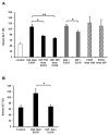
Oral IAP supplementation inhibits alcohol induced liver damage. Serum ALT Levels: (A) single alcohol dose, (B) 3-dose alcohol binge model. Values are expressed as mean ± SEM. Statistical significance between the groups was tested using one-way analysis of variance with Tukey’s multiple comparison posttests. *p < 0.05, **p < 0.01.
Figure 2
Figure 2
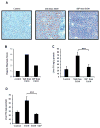
Oral IAP supplementation ameliorates alcohol-induced liver steatosis. In the 3-dose alcohol binge model, frozen liver tissues were obtained to determine (A) Oil Red O stain, (B) Hepatic steatosis score (0–4+). Liver triglyceride content was measured in (C) 3-dose alcohol binge model, (D) chronic alcohol model. Results were compared to mice untreated with alcohol and alcohol-alone treated mice. Values are expressed as mean ± SEM. Statistical significance between the groups was tested using one-way analysis of variance with Tukey’s multiple comparison posttests. ***p < 0.001.
Figure 3
Figure 3
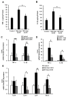
IAP treatment prevents alcohol-induced liver inflammation and the subsequent sensitivity to pro-inflammatory stimuli. In the 3-dose alcohol binge model, liver inflammatory cytokine levels: (A) TNF-α and (B) IL-1β were measured using ELISA. Liver inflammatory cytokine mRNA levels were determined by qPCR: (C) 3-dose alcohol binge model and (D) chronic alcohol model. (E) Liver Tlr4 and co-receptor mRNA expression levels from mice using the 3-dose alcohol binge model. Values are expressed as mean ± SEM. Statistical significance between the groups was tested using one-way analysis of variance with Tukey’s multiple comparison posttests; *p < 0.01, **p < 0.01, ***p < 0.001.
Figure 4
Figure 4
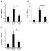
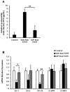
Treatment with oral IAP prevents alcohol-induced endotoxemia. Serum LPS in (A) 3-dose alcohol binge model and (B) chronic alcohol model. (C) Portal serum LPS in 3-dose alcohol binge model. Values are expressed as mean ± SEM. Statistical significance between the groups was tested using one-way analysis of variance with Tukey’s multiple comparison posttests; *p < 0.05, **p < 0.01.
Figure 5
Figure 5
Oral supplementation with IAP prevents alcohol-induced gut barrier dysfunction and junctional protein losses. In the 3-dose alcohol binge model (A) 4-kda Dextran-FITC permeation in in vivo intestinal loop model and (B) Intestinal mRNA levels of junctional proteins measured by qPCR. Values are expressed as mean ± SEM. Statistical significance between the groups was tested using one-way analysis of variance with Tukey’s multiple comparison posttests; *p < 0.05, **p < 0.01.
Figure 6
Figure 6
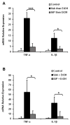
Oral supplementation with IAP prevents alcohol-induced intestinal inflammation. Inflammatory cytokine mRNA levels: TNF-α and IL-1β in both the (A) 3-dose binge and (B) chronic alcohol models. Values are expressed as mean ± SEM. Statistical significance between the groups was tested using one-way analysis of variance with Tukey’s multiple comparison posttests; *p < 0.05, ***p < 0.001.
Figure 7
Figure 7
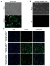
IAP treatment prevents alcohol-induced stellate cell activation and the lipogenic effect on hepatocytes ex vivo. Primary hepatocytes and stellate cells were isolated from mouse liver (A and B). Fat accumulation in hepatocytes were assessed with BODIPY staining (C and D) after being co-cultered with Hepatic stellate cells from mice exposed to 3 doses of binge EtOH gavages +/− pretreatment with IAP. qRT-PCR was performed to determine inflammatory cytokine and chemokine mRNA levels in co-cultured hepatocytes (E). Values are expressed as mean ± SEM. Statistical significance between two groups was tested using the two-tailed Student’s t test.; *p < 0.05.
Figure 7
Figure 7
IAP treatment prevents alcohol-induced stellate cell activation and the lipogenic effect on hepatocytes ex vivo. Primary hepatocytes and stellate cells were isolated from mouse liver (A and B). Fat accumulation in hepatocytes were assessed with BODIPY staining (C and D) after being co-cultered with Hepatic stellate cells from mice exposed to 3 doses of binge EtOH gavages +/− pretreatment with IAP. qRT-PCR was performed to determine inflammatory cytokine and chemokine mRNA levels in co-cultured hepatocytes (E). Values are expressed as mean ± SEM. Statistical significance between two groups was tested using the two-tailed Student’s t test.; *p < 0.05.

"V体育官网入口" References

    1. Miniño AM, Murphy SL, Xu J, et al. Deaths: final data for 2008. Natl Vital Stat Rep. 2011 Dec 7;59:1–126. - "V体育平台登录" PubMed
    1. Varma V, Webb K, Mirza DF. Liver transplantation for alcoholic liver disease. World J Gastroenterol. 2010 Sep 21;16:4377–93. - PMC - PubMed
    1. O’Shea RS, Dasarathy S, McCullough AJ. Alcoholic liver disease. HEPATOLOGY. 2010;51:307–328. - PubMed
    1. Celli R, Zhang X. Pathology of Alcoholic Liver Disease. J Clin Transl Hepatol. 2014 Jun 15;2:103–109. - PMC - PubMed
    1. Thurman RG. Alcoholic liver injury involves activation of Kupffer cells by endotoxin. Am J Physiol Gastrointest Liver Physiol. 1998;275:G605–G611. - PubMed

Publication types

"V体育ios版" MeSH terms