<u date-time="4PUAmNL"></u><sub date-time="CgnrYX"></sub><sub date-time="WPWGWN"><small dropzone="2jGQ0r"></small></sub> VSports注册入口 - Skip to main page content
U.S. flag

An official website of the United States government

Dot gov

The . gov means it’s official. Federal government websites often end in . gov or . mil. Before sharing sensitive information, make sure you’re on a federal government site VSports app下载. .

Https

The site is secure. The https:// ensures that you are connecting to the official website and that any information you provide is encrypted and transmitted securely. V体育官网.

. 2017 Feb 1;198(3):1320-1333.
doi: 10.4049/jimmunol.1601486. Epub 2016 Dec 28.

IL-15 Enables Septic Shock by Maintaining NK Cell Integrity and Function

Affiliations

IL-15 Enables Septic Shock by Maintaining NK Cell Integrity and Function

Yin Guo et al. J Immunol. .

Abstract

Interleukin 15 is essential for the development and differentiation of NK and memory CD8+ (mCD8+) T cells. Our laboratory previously showed that NK and CD8+ T lymphocytes facilitate the pathobiology of septic shock. However, factors that regulate NK and CD8+ T lymphocyte functions during sepsis are not well characterized. We hypothesized that IL-15 promotes the pathogenesis of sepsis by maintaining NK and mCD8+ T cell integrity. To test our hypothesis, the pathogenesis of sepsis was assessed in IL-15-deficient (IL-15 knockout, KO) mice. IL-15 KO mice showed improved survival, attenuated hypothermia, and less proinflammatory cytokine production during septic shock caused by cecal ligation and puncture or endotoxin-induced shock VSports手机版. Treatment with IL-15 superagonist (IL-15 SA, IL-15/IL-15Rα complex) regenerated NK and mCD8+ T cells and re-established mortality of IL-15 KO mice during septic shock. Preventing NK cell regeneration attenuated the restoration of mortality caused by IL-15 SA. If given immediately prior to septic challenge, IL-15-neutralizing IgG M96 failed to protect against septic shock. However, M96 caused NK cell depletion if given 4 d prior to septic challenge and conferred protection. IL-15 SA treatment amplified endotoxin shock, which was prevented by NK cell or IFN-γ depletion. IL-15 SA treatment also exacerbated septic shock caused by cecal ligation and puncture when given after the onset of sepsis. In conclusion, endogenous IL-15 does not directly augment the pathogenesis of sepsis but enables the development of septic shock by maintaining NK cell numbers and integrity. Exogenous IL-15 exacerbates the severity of sepsis by activating NK cells and facilitating IFN-γ production. .


Figures

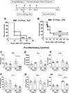
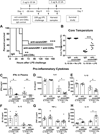
Figure 1
Figure 1. IL-15 KO are resistant to CLP-induced septic shock
Wild type and IL-15 KO mice were subjected to CLP and were monitored for 7-day survival (A). Body temperature (B) was measured at 6 and 18 hours after CLP. The median value is designated in Figure 1B. * p < 0.05, *** p < 0.001, compared to wild type (WT) mice. n=11–14 mice per group. Data are representative of two to three separate experiments.
Figure 2
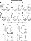
Figure 2. IL-15 KO mice exhibit attenuated proinflammatory cytokine production during CLP-induced septic shock
Blood was harvested at 6 and 18 hours after CLP challenge for measurement of pro-inflammatory cytokines (A–F). Neutrophil numbers in peritoneal cavity (G) and bacterial colony forming units in blood and peritoneal fluid (H–J) were also measured at designated time points in wild type and IL-15 KO mice. * p < 0.05, ** p < 0.01, *** p < 0.001 compared to wild type mice at designated time points. n=6–8 mice per group. Data are representative of two separate experiments.
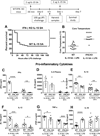
Figure 3
Figure 3. IL-15 KO are resistant to LPS-induced septic shock
In a dose escalation study, IL-15 KO and wild type mice received intraperitoneal injection with 100 or 150 µg LPS and were observed for 7-day survival (A and B). Body temperature was measured at 24 hours after 100 or 150 µg LPS injection (A and B). ** p < 0.01, *** p < 0.001, **** p < 0.0001 when compared to wild type mice; n=8–23 mice per group. Data are representative of two to four separate experiments.
Figure 4
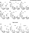
Figure 4. IL-15 KO have attenuated proinflammatory cytokine production and organ injury after LPS-induced septic shock
Blood was harvested at 6 and 24 hours after 150 µg LPS challenge for measurement of proinflammatory cytokines in plasma (A–F). AST (aspartate transaminase), BUN (blood urea nitrogen) and creatinine (G–I) concentrations in plasma were also measured at 24 hours post 150 µg LPS. * p < 0.05, ** p < 0.01, *** p < 0.001, **** p < 0.0001 when compared to wild type mice. n=8 mice per group. Data are representative of two separate experiments.
Figure 5
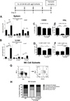
Figure 5. Low dose IL-15 SA treatment restores NK and memory CD8+ T cells in IL-15 KO mice
IL-15 KO mice were treated with 0.125 µg IL-15 SA for 4 consecutive days. At 24 hours following the last treatment, splenic (A) and liver (B) NK (CD3NK1.1+), NKT (CD3+NK1.1+) and memory CD8+ T (CD8+CD44high) lymphocyte numbers were measured using flow cytometry. The activation status (CD69 expression) and IFNγ expression by NK (C and D) and memory CD8+ T cells (E and F) upon IL-15 SA treatment were also analyzed. For IFNγ expression, splenocytes from wild type mice and vehicle- and IL-15 SA-treated IL-15 KO mice were restimulated with PMA/ionomycin for 5 hours to amplify intracellular cytokine signaling (for details see method session). In the representative dot plot, NK cells are divided into four subpopulations based on CD11b and CD27 expression, namely precursor (CD11blowCD27low), immature (I, CD11blowCD27high), mature pro-inflammatory (II, CD11bhighCD27high) and mature cytotoxic (III, CD11bhighCD27low) NK cells (G). The graph in Figure H shows the relative number of splenic NK subsets among intact wild type mice, vehicle- and IL-15 SA-treated IL-15 KO mice. * p < 0.05, ** p < 0.01, *** p < 0.001, **** p < 0.0001 compared to designated groups. n=4–12 mice per group. Data are representative of two to four separate experiments.
Figure 6
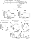
Figure 6. Low dose IL-15 SA treatment restores IL15-mediated lethality to septic shock in IL-15 KO mice
IL-15 SA (0.125 µg) treatment was initiated 4 days prior to septic challenge and continued throughout the experimental period to maintain regenerated NK and mCD8+ T cell populations. Mice were monitored for survival rate over 7 days during CLP- or 150 µg LPS-induced sepsis (A and B). Core temperature was measured at 24 hours after 150 µg LPS challenge (C). Vehicle-treated wild type and IL-15 KO mice served as control. Blood was collected from wild type and IL-15 KO mice at 24 hours after 150 µg LPS challenge for measurement of IL-6 and IL-1β (D and E) as well as ALT, AST, BUN and creatinine (F and G) concentrations in plasma. * p < 0.05, ** p < 0.01, **** p < 0.0001 compared to designated groups. n=7–14 mice per group. Data are representative of two to four separate experiments.
Figure 7
Figure 7. Neutralization of NK cells ablates IL-15 SA treatment-induced restoration of lethality to septic shock in IL-15 KO mice
IL-15 KO mice received anti-asialoGM1 or anti-CD8α at 24 hours prior to the initiation of IL-15 SA (0.125 µg) and NK and memory CD8+ T lymphocyte counts were measured at 24 hours after the lastIL-15 SA treatment. Dotted lines represent the baseline levels of NK and memory CD8+ T cells in wild type controls (A and C). A survival study was also undertaken to assess the effect of anti-asialoGM1 or anti-CD8α on the survival of IL-15 SA-treated IL-15 KO mice upon 150 µg LPS challenge (B and D). Isotype-specific IgG served as control. * p < 0.05, ** p < 0.01, *** p < 0.001, **** p < 0.0001 compared to designated groups. n=5–15 mice per group. Data are representative of two to four separate experiments.
Figure 8
Figure 8. Acute neutralization of IL-15 does not mediate resistance to septic shock
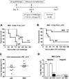
Wild type mice received 20 µg of M96, an IL-15 neutralizing antibody i.p. at 2 hours prior to CLP or 150 µg LPS challenge and a survival study was followed (A and B). Specific IgG serve as control. Concentrations of IFNγ, IL-6, IL-1β, IL-12, TNF-α and IL-10 in the plasma were measured at 6 and 24 hours after 150 µg LPS challenge (C–H). * p < 0.05, ** p < 0.01 compared to IgG control. n=5–10 mice per group Data are representative of two to three separate experiments.
Figure 9
Figure 9. Prolonged neutralization of IL-15 mediates resistance to septic shock
Wild type mice received 20 µg of IL-15 neutralizing antibody M96 or IgG i.p. at 4 days prior to (day -4) and at the time of CLP or 150 µg LPS challenge (day 0). A survival study was undertaken over 7 days (A and B). n=8–11 mice per group. Core temperature was measured at 24 hours after 150 µg LPS challenge (C). n= 9 mice per group. Splenic and hepatic NK cell number was measured at day 0 without LPS challenge (D). n= 6–9 mice/group. ** p < 0.01, **** p < 0.0001, compared to IgG control. n=8–11 mice per group in survival studies. Data are representative of two to three separate experiments.
Figure 10
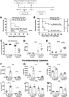
Figure 10. High dose IL-15 SA pre-treatment accentuates lethality to septic shock
Wild type mice received vehicle or IL-15 SA at 2 µg 30 minutes prior to and 24 hours after CLP or LPS (100 µg) and survival studies were followed (A, B). Core temperature, AST and BUN levels, and IFNγ, IL-6, IL-1β, IL-12, TNF-α and IL-10 concentrations in the plasma were measured at 24 hours after 100 µg LPS (C–K)..* p < 0.05, ** p < 0.01, *** p < 0.001, compared to vehicle wild type control. n=5–20 mice per group. Data are representative of two to three separate experiments.
Figure 11
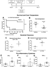
Figure 11. High dose IL-15 SA post-treatment accentuates lethality to septic shock
Wild type mice (16- to 20-week-old) received vehicle or IL-15 SA (2 µg) at 2 and 18 hours after CLP challenge and survival rate was monitored over 7 days (A). Core temperature, bacterial counts in blood and peritoneal fluid as well as IFNγ, IL-6 and TNF-α concentrations in the plasma were measured at 18 hours after LPS (B–G). * p < 0.05, ** p < 0.01, *** p < 0.001, **** p < 0.0001 compared to vehicle wild type control. n= 8–9 mice per group. Data are representative of two separate experiments.
Figure 12
Figure 12. Neutralization of NK cells and CD8+ T cells combined, NK cells, but not CD8 T cells alone, mediates resistance of IL-15 SA treated mice to septic shock
Wild type mice received anti-asialoGM1 and/or anti-CD8α i.p. at 24 hours prior to IL-15 SA treatment and then were challenged with 100 µg LPS and a second dose of IL-15 SA at 24 hours thereafter. Survival rate was monitored over 7 days (A). Isotype-specific IgG serve as control. Core temperature and IFNγ, IL-6, IL-1β, IL-12, TNF-α and IL-10 concentrations in the plasma were measured at 6 and/or 24 hours after 100 µg LPS (B–H). ** p < 0.01, *** p < 0.001, **** p < 0.0001, compared to IgG wild type mice that were treated with 2 µg IL-15 SA. n=5–10 mice per group. Data are representative of two to three separate experiments.
Figure 13
Figure 13. IFNγ KO mice are resistant to IL-15 SA-induced accentuation of septic shock
IFNγ KO mice received intraperitoneal injection with 2 µg of IL-15 SA 30 minutes prior to and 24 hours after LPS (100 µg) challenge. A survival study was performed over 7 days (A). Core temperature and IFNγ, IL-6, IL-1β, IL-12, TNF-α and IL-10 concentrations in the plasma were measured at 6 and/or 24 hours after 100 µg LPS (B–H). * p < 0.05, ** p < 0.01, *** p < 0.001, **** p < 0.0001 compared to wild type control treated with IL-15 SA. n=5–10 mice per group. Data are representative of two separate experiments.

References

    1. Puzanov IJ, Bennett M, Kumar V. IL-15 can substitute for the marrow microenvironment in the differentiation of natural killer cells. Journal of immunology. 1996;157:4282–4285. - PubMed
    1. Huntington ND, Legrand N, Alves NL, Jaron B, Weijer K, Plet A, Corcuff E, Mortier E, Jacques Y, Spits H, Di Santo JP. IL-15 trans-presentation promotes human NK cell development and differentiation in vivo. The Journal of experimental medicine. 2009;206:25–34. - PMC - PubMed
    1. Mortier E, Woo T, Advincula R, Gozalo S, Ma A. IL-15Ralpha chaperones IL-15 to stable dendritic cell membrane complexes that activate NK cells via trans presentation. The Journal of experimental medicine. 2008;205:1213–1225. - PMC (VSports注册入口) - PubMed
    1. Zhang X, Sun S, Hwang I, Tough DF, Sprent J. Potent and selective stimulation of memory-phenotype CD8+ T cells in vivo by IL-15. Immunity. 1998;8:591–599. - "VSports手机版" PubMed
    1. Schluns KS, Williams K, Ma A, Zheng XX, Lefrancois L. Cutting edge: requirement for IL-15 in the generation of primary and memory antigen-specific CD8 T cells. Journal of immunology. 2002;168:4827–4831. - PubMed

Publication types

Substances