"VSports" Skip to main page content
U.S. flag

An official website of the United States government

Dot gov

The . gov means it’s official. Federal government websites often end in VSports app下载. gov or . mil. Before sharing sensitive information, make sure you’re on a federal government site. .

Https

The site is secure. The https:// ensures that you are connecting to the official website and that any information you provide is encrypted and transmitted securely. V体育官网.

. 2016 Sep 27;17(1):206-220.
doi: 10.1016/j.celrep.2016.08.092.

"V体育ios版" Rapid and Efficient Generation of Regulatory T Cells to Commensal Antigens in the Periphery

Affiliations

Rapid and Efficient Generation of Regulatory T Cells to Commensal Antigens in the Periphery

Katherine Nutsch et al. Cell Rep. .

V体育ios版 - Abstract

Commensal bacteria shape the colonic regulatory T (Treg) cell population required for intestinal tolerance. However, little is known about this process. Here, we use the transfer of naive commensal-reactive transgenic T cells expressing colonic Treg T cell receptors (TCRs) to study peripheral Treg (pTreg) cell development in normal hosts. We found that T cells were activated primarily in the distal mesenteric lymph node VSports手机版. Treg cell induction was rapid, generating >40% Foxp3(+) cells 1 week after transfer. Contrary to prior reports, Foxp3(+) cells underwent the most cell divisions, demonstrating that pTreg cell generation can be the dominant outcome from naive T cell activation. Moreover, Notch2-dependent, but not Batf3-dependent, dendritic cells were involved in Treg cell selection. Finally, neither deletion of the conserved nucleotide sequence 1 (CNS1) region in Foxp3 nor blockade of TGF-β (transforming growth factor-β)-receptor signaling completely abrogated Foxp3 induction. Thus, these data show that pTreg cell selection to commensal bacteria is rapid, is robust, and may be specified by TGF-β-independent signals. .

Keywords: CNS1 Foxp3; Notch2-dependent dendritic cells; TGFβ; commensal microbiota; pTreg; peripheral regulatory T cells V体育安卓版. .


VSports注册入口 - Figures

Figure 1
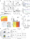
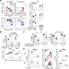
Figure 1. A TCR transgenic model for peripheral Treg cell selection to commensal antigens
(A) Effect of host age on Treg cell selection. Left, sorted naive (CD44loCD62LhiCD4+) CT2/CT6 Tg cells (5×10⁴) were injected into 1 wk old congenically marked Foxp3gfp lymphoreplete mice. The percentage of transferred cells that become Foxp3+ in the MLN was analyzed by flow cytometry at the indicated time point. Right, naïve Tg cells were injected into 1,2, 3 or 14-wk-old hosts and analyzed 1 wk later (expt=2–3, n=2+). (B) Changes in putative tTreg cell markers with age in polyclonal Treg cells. Foxp3gfp mice were analyzed by Nrp-1 or intracellular staining for Helios at different ages (expt=2–3, n=4–6). (C) Effect of host age on bacterial composition in the gut. 16s rDNA was analyzed in terminal fecal pellets from 1.5–14wk old mice. Changes in phyla (left) and selected families (right) are shown. Families shown on right are those that increase from 1.5 to 3 wk old mice by ≥1% (5–15 mice/age). BH adjusted Mann-Whitney U p-values are used. (D) Transplant of fecal material from older mice can induce CT2/CT6 Treg cell development in neonatal mice. One wk old mice were gavaged with fecal material from 1 or 3 wk old mice concurrent with i.p. injection of 2.5–5×10⁴ each of CT2/CT6 Tg cells. TCR Tg cells were analyzed by flow cytometry for CD25 and Foxp3 at 3 days (expt=2, n=3). (E) Expression of Helios and Nrp-1 on Tg cells. Naïve Tg cells were injected into 3–4 wk old mice and analyzed for Helios and Nrp-1 expression after 1 and 5 wks. Representative FACS plots and summary graphs show Tg+ Treg cells versus endogenous polyclonal T cells from the MLN (expt=2–3, n=2–5). See also Figure S1.
Figure 2
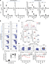
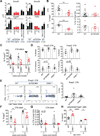
Figure 2. Kinetics and biogeography of T cell activation of CT2 and CT6
(A) Analysis of early time points after transfer of naïve CT6/CT2 T cells. Naïve Tg cells were injected into 3–4 wk old mice and analyzed after 1–2 days by flow cytometry for CD25 or Foxp3IRES-GFP (expt=2, n=2–4). Tissues: SI, small intestine; PP, Peyer’s patch; dMLN, distal MLN; rem. MLN, remaining MLN; pLN, peripheral LN (pooled axial, brachial and inguinal). (B,C) Proliferation of CT2/CT6 cells after transfer. CTV labelled naïve CT2/CT6 cells were injected into 3–4 wk old mice. Foxp3 expression and cell division were assessed by flow cytometry of dMLN at the indicated times. Representative FACS plots of CT2 (expt=2–4, n=2–5) (B), and of CT2/CT6 at day 7 after transfer (expt=4) (C) are shown. Data include points from Figure 1A. In (C), the percentage cells that have undergone any (left) or 3+ divisions (right) by CTV are shown. (D) Increased TCR activation in colonic Treg cells during homeostasis. T cells were analyzed from 6–8 wk old mice Tg for Nur77-GFP, a reporter for recent TCR activation. Nrp-1hi and Helioshi were used as markers for tTreg cells. Numbers in representative FACS plots of Foxp3+ cells indicate frequency in the quadrant. Summary data are the percentage Nur77GFP by Helios or Nrp-1 subset. Asterisk indicates significance in Nrp1lo or Helioslo colon versus spleen (expt=2, n=1–2). See also Figure S2.
Figure 3
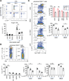
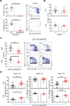
Figure 3. IL-10 reporter is induced in the colon post Foxp3 induction
(A) Induction of IL-10 reporter on CT2 cells. Naïve 10BiT CT2 Tg cells were injected into 3–4 wk old mice and analyzed at the indicated time points. Representative FACS plots and summary of 10BiT in Foxp3+ cells are shown (expt=2, n=2–4). (B,C) Analysis of polyclonal CD25+ Treg cells for IL-10 reporter (B) and correlation with TCR activation (C). 2–3 month old 10BiT Nur77GFP mice were analyzed by flow cytometry (expt=2, n=1–2). Representative FACS plots and summary plots are shown (B,C). The frequency of Helioslo/Nrp-1lo by IL-10Thy1.1 expression is show in (B). The frequency of Nur77GFPhi by IL-10Thy1.1 expression in the CD25+ subset is shown (C). See also Figure S3.
Figure 4
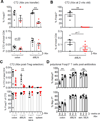
Figure 4. Antibiotics affect pTreg cell induction of CT2 but not stability of Treg cell percentage
(A,B) Effect of antibiotics on CT2 pTreg cell selection at weaning (A) or before (B). In (A), weaned mice were treated with VAMN in drinking water for 5 (squares) or 14 days (triangles) prior to transfer of naïve CT2 Tg cells. One wk later, CT2 cells were analyzed by flow cytometry for Foxp3 expression and the percentage of CT2 cells within the CD4 population. In (B), 2 wk old mice were gavaged with VAMN for 1 wk prior to transfer of CT2. (C) Effect of antibiotics on differentiated CT2 pTreg cells. Naïve CT2 cells were transferred into 3 wk old mice and allowed to develop into Treg cells for 2 wks. Mice were then treated with VAMN in drinking water for 3 wks and then analyzed by flow cytometry. Data shown are the percentage of CT2 cells amongst the entire CD4 population and the frequency of Foxp3+ cells within CT2 cells (expt=2, n=2+). (D) Effect of antibiotics on polyclonal Treg cells. Foxp3gfp mice were treated with VAMN in drinking water for 2–3 wks before assessing the expression of Nrp-1 and Helios in Foxp3+ cells (expt=3, n=2–3). See also Figure S4.
Figure 5
Figure 5. Effects of host age on Foxp3 induction and maintenance
(A–C) Treg cells generated in older mice show lower proliferation, Foxp3, and CD25 expression. Naïve CTV labelled CT2 Tg cells were injected into 3 or 14 wk old mice. CT2 dMLN cells were assessed for Foxp3 expression and cell division at 1 wk (A), with representative FACS plots on the left, and cell divisions summarized on the right (expt=2+, includes data in Figure 1A,2C). CD25 expression is shown in (B). Foxp3 percentage and expression level at 1 and 5 wks after transfer is summarized in (C) (expt=2+, n≥2). (D) Pre-existing cells can inhibit subsequent pTreg selection. 3–4 wk old littermates were injected with PBS or naïve CT2 or CT6 cells. One wk later, congenically marked naïve CTV labelled CT2/CT6 cells was injected into all hosts. 4 days later, cells from the second transfer were analyzed for Foxp3 expression and CTV dilution (expt=2, n=2–4). See also Figure S5.
Figure 6
Figure 6. Partial reduction of Treg cell selection with impaired TGFβ receptor signaling and CNS1-deficiency
(A) Analysis of TGFβ dependent genes. For in vitro experiments, sorted naïve polyclonal T cells were stimulated with anti-CD3/CD28 with the indicated TGFβ condition. After 2 days, TGFβ-responsive genes (Table S1) were analyzed by qPCR. For in vivo experiments, 1.5×10⁵ naïve CT6 cells were injected into WT mice. After 2 days, CD25+Foxp3 cells were sorted and gene expression assessed by qPCR. Data represent fold-change over in vitro αTGFβ condition (in vitro expt=2, n=1; in vivo expt=5, n=1 for naïve and n=1–2 post-transfer of 4–5 pooled mice). (B) Treg cell selection with impaired TGFβ receptor signaling. Naive CT2 (5×10⁴) or CT6 (2×10⁵) cells on a WT or dnTGFβRII background were injected into 3–4 wk old mice, and analyzed 1 wk later. Data show frequency of Foxp3+ cells amongst CT2/CT6 Tg cells in the dMLN (left), and the percentage of Tg cells in the total CD4+ population (right). (C) Impaired Treg cell selection in CNS1-deficient CT6 cells. Naïve CT6 Tg cells (WT or CNS1−/−) were injected into 3–4 wk old mice. Cells from the dMLN were analyzed at the indicated time point (expt=2–3, n=2–4). (D) Analysis of proliferation. The percentage of divided cells with the indicated mutations is shown (expt=2, n=2–4 pools of 2 mice). (E) Assessment of IFNγ expression. Naïve CT6 Tg cells (2×10⁵) (WT, dnTGFβRII or CNS1−/−) were stained with CTV and injected into 3–4 wk old hosts. 1 wk later, cells were stimulated with PMA/Ionomycin for intracellular staining. Representative plots are shown (left) and summarized (right) (expt=2, n=2–4 pools of 2 mice). (F–G) CNS1-independent Treg cell selection of CT6 cells. (F) Naïve WT or CNS1−/− CT6 Tg cells were injected into 3–4 wk old mice, and the colons analyzed 1 or 5 wks later for the frequency of Foxp3+ CT6 cells (F) and of CT6 cells amongst the total CD4+ T cell population (G) (expt=3, n=1–3). (H) Helios expression in polyclonal colonic CNS1−/− Treg cells with age. CNS1−/− mice were cross-fostered to limit variability in microbiota (expt=2–3, n=1–3). See also Figure S6–7.
Figure 7
Figure 7. Role of Notch2-dependent DCs in Foxp3 induction to commensal antigens
(A) Conventional DCs cells are necessary for activation of CT2. Bone marrow chimeras with WT or Zbtb46-DTR donors were generated as described in Supplemental Materials. CTV labelled naïve CT2 cells were injected into DC-depleted mice and analyzed after 1 wk (expt=2–3, n=2–5). (B) Loss of CD103+CD11b DCs does not decrease pTreg cells. Naïve Tg cells were injected into BATF3+/− or −/− littermates and analyzed at 1 wk for Foxp3 (expt=2–3, n=1–3). (C–D) Notch2 dependent DCs are involved in pTreg cell generation. Naïve Tg cells were stained with CTV and injected into Notch2cKO mice. Foxp3 expression and representative FACS plots (C), and the summary of effector cell frequency (CD44hiCD62Llo) and cell divisions (D) are shown for dMLN cells 1 wk after transfer (expt=4, n=2–5).

"VSports" References

    1. Akimova T, Beier UH, Wang L, Levine MH, Hancock WW. Helios expression is a marker of T cell activation and proliferation. PLoS One. 2011;6:e24226. - PMC - PubMed
    1. Arpaia N, Campbell C, Fan X, Dikiy S, van der Veeken J, Deroos P, Liu H, Cross JR, Pfeffer K, Coffer PJ, Rudensky AY. Metabolites produced by commensal bacteria promote peripheral regulatory T-cell generation. Nature. 2013 - PMC - PubMed
    1. Atarashi K, Tanoue T, Shima T, Imaoka A, Kuwahara T, Momose Y, Cheng G, Yamasaki S, Saito T, Ohba Y, et al. Induction of colonic regulatory T cells by indigenous Clostridium species. Science. 2011;331:337–341. - "V体育ios版" PMC - PubMed
    1. Bautista JL, Lio CW, Lathrop SK, Forbush K, Liang Y, Luo J, Rudensky AY, Hsieh CS. Intraclonal competition limits the fate determination of regulatory T cells in the thymus. Nat Immunol. 2009;10:610–617. - "VSports" PMC - PubMed
    1. Belkaid Y, Hand TW. Role of the microbiota in immunity and inflammation. Cell. 2014;157:121–141. - "V体育官网" PMC - PubMed

Publication types

"V体育2025版" MeSH terms

Substances

V体育官网入口 - LinkOut - more resources