Skip to main page content
U.S. flag

An official website of the United States government

Dot gov

The . gov means it’s official. Federal government websites often end in . gov or . mil. Before sharing sensitive information, make sure you’re on a federal government site. VSports app下载.

Https

The site is secure V体育官网. The https:// ensures that you are connecting to the official website and that any information you provide is encrypted and transmitted securely. .

. 2016 Nov 17;128(20):2435-2449.
doi: 10.1182/blood-2016-04-710632. Epub 2016 Aug 29.

Disulfide HMGB1 derived from platelets coordinates venous thrombosis in mice

Affiliations

Disulfide HMGB1 derived from platelets coordinates venous thrombosis in mice

Konstantin Stark et al. Blood. .

"V体育官网" Abstract

Deep venous thrombosis (DVT) is one of the most common cardiovascular diseases, but its pathophysiology remains incompletely understood. Although sterile inflammation has recently been shown to boost coagulation during DVT, the underlying molecular mechanisms are not fully resolved, which could potentially identify new anti-inflammatory approaches to prophylaxis and therapy of DVT VSports手机版. Using a mouse model of venous thrombosis induced by flow reduction in the vena cava inferior, we identified blood-derived high-mobility group box 1 protein (HMGB1), a prototypical mediator of sterile inflammation, to be a master regulator of the prothrombotic cascade involving platelets and myeloid leukocytes fostering occlusive DVT formation. Transfer of platelets into Hmgb1-/- chimeras showed that this cell type is the major source of HMGB1, exposing reduced HMGB1 on their surface upon activation thereby enhancing the recruitment of monocytes. Activated leukocytes in turn support oxidation of HMGB1 unleashing its prothrombotic activity and promoting platelet aggregation. This potentiates the amount of HMGB1 and further nurtures the accumulation and activation of monocytes through receptor for advanced glycation end products (RAGE) and Toll-like receptor 2, leading to local delivery of monocyte-derived tissue factor and cytokines. Moreover, disulfide HMGB1 facilitates formation of prothrombotic neutrophil extracellular traps (NETs) mediated by RAGE, exposing additional HMGB1 on their extracellular DNA strands. Eventually, a vicious circle of coagulation and inflammation is set in motion leading to obstructive DVT formation. Therefore, platelet-derived disulfide HMGB1 is a central mediator of the sterile inflammatory process in venous thrombosis and could be an attractive target for an anti-inflammatory approach for DVT prophylaxis. .


Figures

Figure 1.
Figure 1.
Presence of HMGB1 in the developing venous thrombus. (A) Immunofluorescence staining of cross sections of the IVC for HMGB1 (green) and DAPI (blue) 1, 6, 12, and 48 hours after flow reduction. Arrowheads show HMGB1 deposition, dotted line indicates endothelium; bar, 200 µm. (B) Left, Scanning electron microscopy of the IVC 6 hours after flow reduction, showing the intact endothelium covered by cell aggregates and fibrin; bar, 5 µm. Right, Three-dimensional reconstruction of images from intravital 2-photon microscopy showing the vessel wall (second harmonic generation, red), with adherent platelets (yellow) and neutrophils (green); bar, 50 µm. Box, Platelet-myeloid leukocyte aggregates indicated by arrowheads at higher magnification. (A-B) Images representative of n = 3 experiments.
Figure 2.
Figure 2.
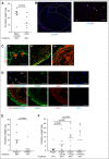
Blood-derived HMGB1 causes venous thrombosis. (A) Thrombus weight in Balb/c mice treated with PBS (n = 8) compared with BoxA (n = 6). (B) Left, Immunofluorescence staining of cross-sections of the IVC for propidium iodide (PI) (red) and DAPI (blue) 6 hours after flow reduction. Dotted line indicates endothelium; bar, 200 µm. Right, Higher magnification of intraluminal propidium iodide–positive cells (arrowheads); bar, 70 µm. (C) Immunofluorescence staining of cross-sections of the IVC 1, 6, and 12 hours after flow reduction for CD41 (red) and HMGB1 (green). Nuclei are counterstained with DAPI (blue). Dotted line indicates endothelium; bar, 100 µm. Images representative of n = 3 experiments. (D) Immunofluorescence staining of cross-sections of the IVC 48 hours after flow reduction. Top, Presence of HMGB1 on NETs (arrowheads) as shown by staining for HMGB1 (green), Ly6G (red), and DAPI (blue); bar, 20 µm. Bottom, HMGB1 (green) colocalizes with platelets (red). Dotted line indicates endothelium; bar, 100 µm. Nuclei are counterstained with DAPI (blue). Images representative of n = 3 experiments. (E) Thrombus weight in Hmgb1+/+ fetal liver cell chimeras (n = 7) compared with Hmgb1−/− chimeras (n = 6). (F) Thrombus weight in Hmgb1−/− bone marrow chimeras (n = 6) compared with Hmgb1−/− chimeras receiving wild-type platelets (n = 10), Hmgb1−/− platelets (n = 8), or wild-type neutrophils (n = 9). (A,E,F) Line indicates mean. The Student t test was used to compare results between 2 groups, 1-way ANOVA followed by LSD–post hoc test for 3 groups.
Figure 3.
Figure 3.
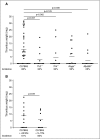
Redundant pattern recognition receptors mediate the prothrombotic effects of HMGB1. (A) Thrombus weight in C57Bl/6 mice (n = 32) compared with Tlr2−/− mice (n = 12), Tlr4−/− mice (n = 12), Rage−/− mice (n = 14), Myd88−/− mice (n = 12). Lines indicate mean. One-way ANOVA followed by LSD–post hoc test was used to compare results between groups. (B) Thrombus weight in C57Bl/6 mice receiving vehicle (n = 13) compared with anakinra s.c. (n = 8). Lines indicate mean. The Student t test was used to compare results between groups.
Figure 4.
Figure 4.
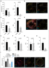
HMGB1 induces NET formation trough RAGE in DVT. (A-I) Quantification of neutrophils and NETs. Results are mean ± SEM. (A) BoxA (n = 5) compared with control (n = 5). (B) Immunofluorescence staining for Ly6G (red) and MPO (green) from cross-sections of the IVC 48 hours after flow reduction of BoxA-treated (bottom) or control mice (top). Nuclei are counterstained with DAPI (blue); bar, 200 µm. (C) Hmgb1−/− (n = 3) fetal liver cell chimeras compared with Hmgb1+/+ chimeras (n = 5). (D) Immunofluorescence staining for Ly6G (red) and MPO (green) from cross-sections of the IVC 48 hours after flow reduction of Hmgb1+/+ (top) or Hmgb1−/− fetal liver cell chimeras (bottom). Nuclei are counterstained with DAPI (blue); bar, 200 µm. (E) Quantification of NETs in Rage−/− mice compared with control (n = 5 each). (F) Quantification of NETs in Tlr2−/− mice compared with control (n = 3 each). (G) Quantification of NETs in Tlr4−/− mice compared with control (n = 3 each). (H) Quantification of NETs in Myd88−/− mice compared with control (n = 3 each). (I) Left, NET formation capacity shown as NETs/neutrophil (n = 5 each) in Hmgb1−/− chimeras compared with Hmgb1−/− chimeras receiving wild-type platelets or wild-type neutrophils. Dotted line indicates mean in Hmgb1+/+ bone marrow chimeras. Right, Immunofluorescence staining for Ly6G (red) and MPO (green) from cross-sections of the IVC 48 hours after flow reduction in Hmgb1−/− chimeras compared with Hmgb1−/− chimeras receiving wild-type platelets, Hmgb1−/− platelets or wild-type neutrophils. (n = 3 each). Nuclei are counterstained with DAPI (blue); bar, 200 µm. The Student t test was used to compare results between 2 groups, 1-way ANOVA followed by LSD–post hoc test for 3 groups.
Figure 5.
Figure 5.
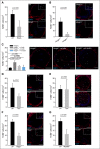
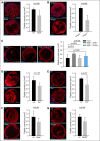
Platelet derived HMGB1 promotes monocyte recruitment. (A-G) Left, Quantification of monocytes. Results are mean ± SEM. Right, Immunofluorescence staining for F4/80 (red) from cross-sections of the IVC. Nuclei are counterstained with DAPI (blue); arrowheads indicate individual monocytes at higher magnification; bar, 200 µm. Images representative of n = 3 experiments. (A) BoxA compared with control (n = 5 each). (B) Hmgb1−/− (n = 3) fetal liver cell chimeras compared with Hmgb1+/+ chimeras (n = 5). (C) Hmgb1−/− chimeras compared with Hmgb1−/− chimeras receiving wild-type platelets, Hmgb1−/− platelets, or wild-type neutrophils. Dotted line indicates mean in Hmgb1+/+ bone marrow chimeras (n = 3 each). (D) Tlr2−/− compared with control (n = 4 each). (E) Tlr4−/− compared with control (n = 4 each). (F) Rage−/− compared with control (n = 5 each). (G) Myd88−/− compared with control (n = 4 each). The Student t test was used to compare results between 2 groups, 1-way ANOVA followed by LSD–post hoc test for 3 groups.
Figure 6.
Figure 6.
HMGB1 contributes to platelet accumulation in venous thrombosis. (A-G) Left, Immunofluorescence staining for CD41 (red) from cross-sections of the IVC. Nuclei are counterstained with DAPI (blue); bar, 200 µm. Images representative of n = 3 experiments. Right, Quantification of platelet-covered area. Results are mean ± SEMT. (A) BoxA compared with control (n = 5 each). (B) Hmgb1−/− fetal liver cell chimeras (n = 3) compared with Hmgb1+/+ chimeras (n = 5). (C) Left, Immunofluorescence for CD41 in Hmgb1−/− chimeras compared with Hmgb1−/− chimeras receiving wild-type platelets, Hmgb1−/− platelets, or wild-type neutrophils (n = 3 each). Right, Quantification of platelet-covered area. Dotted line indicates mean in Hmgb1+/+ bone marrow chimeras (n = 3 each). (D) Tlr2−/− compared with control (n = 3 each). (E) Tlr4−/− compared with control (n = 3 each). (F) Rage−/− compared with control (n = 5 each). (G) Myd88−/− compared with control (n = 3 each). The Student t test was used to compare results between 2 groups, 1-way ANOVA followed by LSD–post hoc test for 3 groups.
Figure 7.
Figure 7.
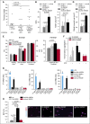
The prothrombotic effects of HMGB1 depend on the redox state. (A) Thrombus weight in Hmgb1−/− chimeras (n = 10) treated with buffer compared with Hmgb1−/− chimeras (n = 5 each) receiving 3S- or disulfide HMGB1. Lines indicate mean. (B) Quantification of NETs, (C), monocytes, (D), and platelet covered area from immunofluorescence stainings of Hmgb1−/− chimeras receiving buffer, 3S-, or disulfide HMGB1 (n = 3 each). (E) Extem (left) and Fibtem (right) 6 hours after IV injection of 3S- (n = 3), disulfide HMGB1 (n = 3), or sulfonyl (n = 3) compared with control (n = 5). (F) Fold increase in whole-blood aggregation after incubation with 3S-, disulfide, or sulfonyl HMGB1 compared with control stimulated with buffer. This was followed by stimulation by ADP in the groups indicated (n = 4-6). (G) Results from RT-PCR for TF (left), IL-1β (middle), and IL-6 (right) of peripheral blood human monocytes incubated with different redox forms of HMGB1 and LPS for 3 hours shown as fold increase compared with control stimulated with the respective buffer (n = 3 each). (H) Left, Quantification of NET formation in vitro after stimulation of isolated human neutrophils with buffer, 3S-, disulfide, or sulfonyl HMGB1 (n = 3 each). Right, Representative images of immunofluorescence stainings for MPO (red) and Hoechst (blue); bar, 50 µm. Results are mean ± SEM. *P < .05. The Student t test was used to compare results between 2 groups, 1-way ANOVA followed by LSD–post hoc test for 3 groups.

Comment in

References

    1. Heit JA. Epidemiology of venous thromboembolism. Nat Rev Cardiol. 2015;12(8):464-474. - PMC - PubMed
    1. Brill A, Fuchs TA, Savchenko AS, et al. . Neutrophil extracellular traps promote deep vein thrombosis in mice. J Thromb Haemost. 2012;10(1):136-144. - "VSports最新版本" PMC - PubMed
    1. Engelmann B, Massberg S. Thrombosis as an intravascular effector of innate immunity. Nat Rev Immunol. 2013;13(1):34-45. - PubMed
    1. Mackman N. New insights into the mechanisms of venous thrombosis. J Clin Invest. 2012;122(7):2331-2336. - VSports手机版 - PMC - PubMed
    1. Martinod K, Wagner DD. Thrombosis: tangled up in NETs. Blood. 2014;123(18):2768-2776. - PMC - PubMed

Publication types

MeSH terms

Substances (VSports注册入口)