V体育ios版 - Skip to main page content
U.S. flag

An official website of the United States government

Dot gov

The . gov means it’s official VSports app下载. Federal government websites often end in . gov or . mil. Before sharing sensitive information, make sure you’re on a federal government site. .

Https

The site is secure V体育官网. The https:// ensures that you are connecting to the official website and that any information you provide is encrypted and transmitted securely. .

. 2015 Nov 17;43(5):923-32.
doi: 10.1016/j.immuni.2015.10.009. Epub 2015 Nov 10.

Caspase-11 Requires the Pannexin-1 Channel and the Purinergic P2X7 Pore to Mediate Pyroptosis and Endotoxic Shock

Affiliations

Caspase-11 Requires the Pannexin-1 Channel and the Purinergic P2X7 Pore to Mediate Pyroptosis and Endotoxic Shock

Dahai Yang et al. Immunity. .

Abstract

The noncanonical inflammasome induced by intracellular lipopolysaccharide (LPS) leads to caspase-11-dependent pyroptosis, which is critical for induction of endotoxic shock in mice. However, the signaling pathway downstream of caspase-11 is unknown. We found that cytosolic LPS stimulation induced caspase-11-dependent cleavage of the pannexin-1 channel followed up by ATP release, which in turn activated the purinergic P2X7 receptor to mediate cytotoxicity. In the absence of P2X7 or pannexin-1, pyroptosis induced by cytosolic LPS was abrogated. Cleavage of pannexin-1 required the catalytic activity of caspase-11 and was essential for ATP release and P2X7-mediated pyroptosis VSports手机版. Priming the caspase-11 pathway in vivo with LPS or Toll-like receptor-3 (TLR3) agonist resulted in high mortality in wild-type mice after secondary LPS challenge, but not in Casp11(-/-), Panx1(-/-), or P2x7(-/-) mice. These results reveal a critical role for pannexin-1 and P2X7 downstream of caspase-11 for pyroptosis and susceptibility to sepsis induced by the noncanonical inflammasome. .


Figures

Figure 1
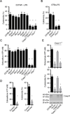
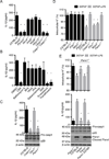
Figure 1. Activation of caspase-11 by intracellular LPS induces pyroptosis via ATP-mediated P2X7 signaling
(A–E) BMMs were primed overnight with LPS and then transfected with ultrapure S. minnesota RE595 LPS packaged with DOTAP or simulated with CTP plus LPS. (A) Cytotoxicity in C57BL/6, Nlrp3−/−, Pycard−/−, Nlrc4−/−, Aim2−/−, Nlrp6−/−, Nlrp12−/−, Casp11−/−, Casp1−/−/Casp11−/− and P2X7−/− BMMs was examined 2 hrs after transfection. (B) Cytotoxicity in C57BL/6, Nlrp3−/−, Casp11−/− and P2X7−/− BMMs was examined 8 hrs after CTB plus LPS stimulation. (C) Extracellular ATP in BMMs culture medium was measured 15 min after LPS transfection. (D) Wild-type BMMs were treated with apyrase (25 U/mL) or left untreated (control) and the amounts of extracellular ATP and cytotoxicity were measured after LPS transfection. (E) Extracellular ATP and cytotoxicity after LPS transfection in Casp11−/− BMMs reconstituted with lentiviral vectors expressing wild-type caspase-11, caspase-11 C254S or vector alone. Immunoblotting for caspase-11 in wild-type BMMs is shown as a control. (A–E) Data are representative of at least 3 experiments. Graphs show mean ± SD of triplicate cultures, * p<0.05, ** p<0.01. See also Figure S1.
Figure 2
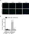
Figure 2. Yo-Pro-1 uptake is induced after transfection with DOTAP or DOTAP plus LPS in LPS-primed BMMs
(A–B) BMMs were primed overnight with LPS and then transfected with ultrapure S. minnesota RE595 LPS packaged with DOTAP. (A) Yo-Pro-1 uptake was measured 2 hrs after treatment with DOTAP or DOTAP plus LPS in BMMs from indicated wild-type and mutant mice by fluorescence microscopy. Representative microscopic images are shown. (B) Percentage of Yo-Pro-1 positive cells was calculated from different fields with 25–30 cells per field at each condition. (A–B) Graph show mean ± SD of six fields. Data are representative of at least 3 experiments. * p<0.05. See also Figure S2 and S3.
Figure 3
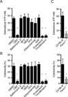
Figure 3. Pannexin-1 is critical for ATP-induced pyroptosis induced by cytosolic LPS
(A–C) BMMs were primed overnight with LPS and then transfected with ultrapure S. minnesota RE595 LPS packaged with DOTAP. (A) Wild-type BMMs were treated with DMSO, CBX (50 µM), bafilomycin A (50 nM), BFA (1 µM), 18GA (50 µM), FFA (50 µM), glibenclamide (100 µM), Gd3 (100 µM), probenecid (100 µM) and trovafloxacin (100 µM), and extracellular ATP was measured 15 min after LPS transfection. (B) Cytotoxicity was measured 2 hrs after LPS transfection. Treatment with inhibitors was as in (A). (C) ATP release was measured 15 min after LPS transfection, and cytotoxicity was determined 2 hrs after transfection in wild-type and Panx1−/− BMMs. (A–C) Data are representative of at least 3 experiments. Graphs show mean ± SD of triplicate cultures, * p<0.05, ** p<0.01. See also Figure S4 and S5.
Figure 4
Figure 4. Activation of caspase-1 leads to pannexin-1 cleavage and ATP release
(A–C) BMMs were primed overnight with LPS and then transfected with ultrapure S. minnesota RE595 LPS packaged with DOTAP. (A) Immunoblotting for pannexin-1 before and after LPS transfection in wild-type (C57BL/6), Casp11−/− and Panx1−/− BMMs. (B) Pannexin-1 cleavage after LPS transfection in Casp11−/− BMMs reconstituted with lentiviral vectors expressing wild-type caspase-11, caspase-11 C254S and vector alone. Immunoblotting for caspase-11 and β-actin are also shown. (C) ATP release (upper panel), cytotoxicity (middle panel) and pannexin-1 cleavage (lower panel) were measured after LPS transfection in Panx1−/− BMMs reconstituted with wild-type pannexin-1, pannexin-1 D378A lentiviruses or control vector. Immunoblotting for pannexin-1 and caspase-11 in wild-type BMMs is also shown as a control. (A–C) Data are representative of at least 3 experiments. Graphs show mean ± SD of triplicate cultures, * p <0.05. See also Figure S6.
Figure 5
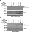
Figure 5. Caspase-11 cleaves pannexin-1
(A) HEK293T cells were co-transfected with indicated pannexin-1 and caspase-11 expression vectors, cultured for 18–24 hrs, and pannexin-1 cleavage was assessed by immunoblotting. (B) In vitro cleavage of purified pannexin-1 proteins after incubation with indicated caspase-11 proteins. Immunoblotting for pannexin-1 and caspase-11 is shown. (A–B) Data are representative of at least 3 experiments.
Figure 6
Figure 6. Noncanonical inflammasome pathway engages the pannexin-1 channel to induce K+ efflux and NLRP3 activation
(A–E) BMMs were primed with LPS and then transfected with ultrapure S. minnesota RE595 LPS packaged with DOTAP. (A) IL-1β release from wild-type (C57BL/6), Nlrp3−/−, Pycard−/−, Nlrc4−/−, Aim2−/−, Nlrp6−/− Nlrp12−/−, Casp11−/− and Casp1−/−/Casp11−/− BMMs was measured by ELISA. (B) Wild-type BMMs were pre-treated with various inhibitors as in Fig 3 (A), IL-1β release was measured 2 hrs after LPS transfection. (C) IL-1β release and caspase-1 processing in C57BL/6, Casp11−/−, P2X7−/− and Panx1−/− BMMs were measured by ELISA and immunoblotting. (D) Intracellular K+ was measured 30 min after treatment of BMMs with DOTAP or DOTAP plus LPS. (E) Intracellular K+, IL-1β release, caspase-1 and pannexin-1 processing were assessed after LPS transfection of Panx1−/− BMMs reconstituted with wild-type, pannexin-1 D378A or control vector. DOTAP-treated Panx1−/− BMMs and LPS-transfected wild-type (C57BL/6) BMMs are shown as controls. (A–E) Data are representative of at least 3 experiments. Graphs show mean ± SD of triplicate cultures, * p<0.05. See also Figure S7.
Figure 7
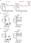
Figure 7. Critical role of pannexin-1 and P2X7 in endotoxic shock induced by activation of the noncanonical inflammasome
(A) Survival of mice primed with 400 µg/kg E. coli O111:B4 LPS (left) or 10 mg/kg poly(I:C) (right), and then challenged with 10 mg/kg E. coli O111:B4 LPS 7 hrs later. N=15 mice in C57BL/6 or Nlrp3−/− groups; N=17 mice in P2X7−/− or Casp11−/− groups; N=12 mice in Panx1−/− group. Data shown are from at least 2 representative experiments, * p<0.05, ** p<0.01, NS=not significant. (B–C) IL-1β and TNF-α levels in mouse sera were measured 2 hrs after re-challenge with LPS. N=8 mice in C57BL/6 or Nlrp3−/− groups; N=7 mice in P2X7−/− or Casp11−/− groups; N=6 mice in Panx1−/− group. (A–C) Data are representative of at least 3 experiments. Error bars indicate SD of technical replicates, * p<0.05, ** p<0.01, NS=not significant.

Comment in

References

    1. Allam R, et al. Mitochondrial apoptosis is dispensable for NLRP3 inflammasome activation but non-apoptotic caspase-8 is required for inflammasome priming. EMBO Rep. 2014;15(9):982–990. - PMC - PubMed
    1. Bartllet R, Stokes L, Sluyter R. The P2X7 receptor channel: recent developments and the use of P2X7 antagonists in models of disease. Pharmacol. Rev. 2014;66(3):638–675. - PubMed
    1. Bauernfeind FG, et al. Cutting edge: NF-kappaB activating pattern recognition and cytokine receptors license NLRP3 inflammasome activation by regulating NLRP3 expression. J. Immunol. 2009;183(2):787–791. - PMC - PubMed
    1. Boyd-Tressler A, et al. Chemotherapeutic drugs induce ATP release via caspase-gated pannexin-1 channels and a caspase/pannexin-1-independent mechanism. J. Biol. Chem. 2014;289:27246–27263. - PMC - PubMed
    1. Broz P, et al. Caspase-11 increases susceptibility to Salmonella infection in the absence of caspase-1. Nature. 2012;490:288–291. - PMC (V体育ios版) - PubMed

"V体育安卓版" Publication types

MeSH terms