Skip to main page content
U.S. flag

An official website of the United States government

Dot gov

The . gov means it’s official. Federal government websites often end in . gov or VSports app下载. mil. Before sharing sensitive information, make sure you’re on a federal government site. .

Https

The site is secure V体育官网. The https:// ensures that you are connecting to the official website and that any information you provide is encrypted and transmitted securely. .

. 2015 Sep 22;112(38):11771-6.
doi: 10.1073/pnas.1505056112. Epub 2015 Sep 8.

Loop recognition and copper-mediated disulfide reduction underpin metal site assembly of CuA in human cytochrome oxidase

Affiliations

Loop recognition and copper-mediated disulfide reduction underpin metal site assembly of CuA in human cytochrome oxidase

Marcos N Morgada et al. Proc Natl Acad Sci U S A. .

VSports app下载 - Abstract

Maturation of cytochrome oxidases is a complex process requiring assembly of several subunits and adequate uptake of the metal cofactors VSports手机版. Two orthologous Sco proteins (Sco1 and Sco2) are essential for the correct assembly of the dicopper CuA site in the human oxidase, but their function is not fully understood. Here, we report an in vitro biochemical study that shows that Sco1 is a metallochaperone that selectively transfers Cu(I) ions based on loop recognition, whereas Sco2 is a copper-dependent thiol reductase of the cysteine ligands in the oxidase. Copper binding to Sco2 is essential to elicit its redox function and as a guardian of the reduced state of its own cysteine residues in the oxidizing environment of the mitochondrial intermembrane space (IMS). These results provide a detailed molecular mechanism for CuA assembly, suggesting that copper and redox homeostasis are intimately linked in the mitochondrion. .

Keywords: CuA site; Sco proteins; cytochrome oxidase; metal site assembly; metallochaperones V体育安卓版. .


Conflict of interest statement

The authors declare no conflict of interest.

Figures

Fig. 1.
Fig. 1.
Design of the chimeric COX II* protein. (A) Comparison of the structures of the soluble COX II domains from Thermus thermophilus ba3 oxidase [Protein Data Bank (PDB) ID code 2CUA; green] and human cytochrome c oxidase (red). The latter was modeled based on the structure of the bovine oxidase (PDB ID code 1V54) (46). (B) Model of the chimeric COX II* colored in green in the region coming from the Thermus oxidase and in red in the loops taken from the human protein. (C) Sequence alignment of the loops corresponding to the COX II subunits from Thermus thermophilus (Tt), bovine (Bt), human (Hs), and the chimeric COX II*.
Fig. S1.
Fig. S1.
Arrangement of the COX II subunit within the structure of different oxidases. Eukaryotic COX II (Left; PDB ID code 1OCC for bovine COX) (9) displays several interactions with other subunits of the complex, whereas prokaryotic COX II (Right; PDB ID code 1EHK for Tt COX) (47) displays a small region of interaction with the rest of the complex. Gray lines define approximate limits of the lipid membrane surrounding the complexes.
Fig. S2.
Fig. S2.
Cu(I) uptake of Apo-COX II* followed by NMR. 1H-15N HSQC spectra of apo-COX II*2HS (green) and upon addition of two equivalents of Cu(I) (purple). The spectrum of the metallated form has been assigned, showing that the main difference is the appearance of cross peaks belonging to the ligands loops (Right) or to the two other loops surrounding the metal site (Left).
Fig. S3.
Fig. S3.
Reduction of Apo-COX II*S-S to Apo-COX II*2SH followed by NMR. 1H-15N HSQC spectra of Apo-COX II*S-S (cyan) and Apo-COX II*2SH (green).
Fig. 2.
Fig. 2.
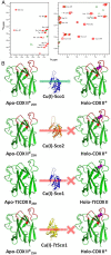
Human Sco1 specifically recognizes the loops of human COX II to assemble its CuA site. (A) 15N, 1H HSQC spectrum of Apo-COX II*2SH (green) and upon addition of two equivalents of Cu(I)-Sco1 (red), showing complete copper transfer. (B) Summary of the copper transfer experiments (spectra shown in Supporting Information). The green arrow indicates the stoichiometric transfer of two Cu(I) ions from Sco1 to COX II*, and the red lines indicate unsuccessful Cu(I) transfer assays.
Fig. S4.
Fig. S4.
Cu(I) transfer from Cu(I)-Sco1 to Apo-TtCOX II2SH followed by NMR. 1H-15N HSQC spectra of TtCOX II2SH (green) and upon addition of two equivalents of Cu(I)-Sco1 (red). Arrows indicate cross peaks that do not correspond neither to apo nor to the metallated form of the protein.
Fig. S5.
Fig. S5.
Cu(I) transfer from Cu(I)-TtSco1 to Apo-COX II*2SH followed by NMR. 1H-15N HSQC spectra of COX II*2SH (green) and upon addition of two equivalents of Cu(I)-TtSco1 (pink), showing that TtSco1 is not able to transfer Cu(I) ions to the oxidase. The position of the expected cross peaks corresponding to the holo form protein absent in the final spectrum are indicated in blue.
Fig. S6.
Fig. S6.
Cu(I) transfer from Cu(I)-Sco2 to Apo-COX II*2SH followed by NMR. 1H-15N HSQC spectra of COX II*2SH (green) and upon addition of two equivalents of Cu(I)-Sco2 (orange). The position of the expected cross peaks corresponding to the holo form protein shifted in the final spectrum are indicated in blue.
Fig. 3.
Fig. 3.
Electron transfer reaction followed by AMS-reacted, nonreducing SDS/PAGE. (A) Sco12SH and Sco22SH mixed with COX II*S-S and (B) Cu(I)-bound forms of the Sco proteins mixed with COX II*S-S. All mixtures were done in a 1:1 ratio, except when indicated. The proteins in their different states are also reported as reference. Signs + and – indicate if the AMS reactive was added to the mixture. Yellow and red arrows indicate where oxidized and reduced COX II* show up, respectively, upon addition of AMS to the mixture.
Fig. S7.
Fig. S7.
Redox potential of the CX3C motif of Apo-COX II*. (A) Fluorescence emission spectra of oxidized (50 mM phosphate buffer, pH 7.0, 0.01 mM GSSG; black line) and reduced (50 mM phosphate buffer, pH 7.0, 200 mM GSH; red line) Apo-COX II* upon excitation at 280 nm. (B) The redox equilibrium of Apo-COX II* with different [GSH]2/GSSG ratios is shown. Data processing and determination of the equilibrium constant was previously described (3). After nonlinear regression, a value of Keq = 23 ± 2 mM was determined for the Apo-COX II*/glutathione equilibrium, corresponding to a redox potential of −288 ±3 mV for the Apo-COX II* S-S –2SH redox couple.
Fig. S8.
Fig. S8.
Electron transfer followed by AMS-reacted, nonreducing SDS/PAGE. Sco12SH and Cu(I)-Sco1 mixed with COX II*S-S. Mixtures were done in a 1:1 and 2:1 ratio Sco1:COX II*. The proteins in their different states are also reported as reference. Signs + and – indicate if the AMS reactive was added to the mixture.
Fig. S9.
Fig. S9.
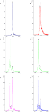
MALDI-TOF-MS spectra of the iodoacetamide (IAM)-derivatized samples of (A) Sco12SH showing the presence a dominant species labeled as 1, corresponding to the Sco1(IAM)2 adduct, indicating that the two reduced Cys at the active site are in the reduced form; (B) Cu(I)-Sco1 showing the presence of three species corresponding to Sco1(IAM)2, Sco1(IAM)1, and Sco1, labeled as 1, 2, and 3, respectively; (C) Sco2S-S with the presence of the presence a dominant species corresponding to the Sco2(IAM) adduct, where Cys115 (far from the active site) is alkylated, and the two Cys at the active site are oxidized, not being alkylated, labeled as 4; minority species 5 and 6 correspond to Sco2(IAM)2 and Sco2(IAM)3; (D) a 1:1 mixture of Sco12SH and Sco2S-S, where the peaks of the reference spectra are preserved, showing that there is no redox reaction; (E) a 1:1 mixture of Cu(I)-Sco12SH and Sco2S-S, where the peaks of the reference spectra are preserved, showing that there is no redox reaction.
Fig. S10.
Fig. S10.
Electron transfer from Cu(I)-Sco2 to Apo-COX II*S-S followed by NMR. 1H-15N HSQC spectra of Apo-COX II*S-S (cyan) and upon addition of one equivalent of Cu(I)-Sco2 (blue). The disappearance of several cross peaks compared with the oxidized form of the protein is typical of spectra of COX II*2SH.

References

    1. Macomber L, Imlay JA. The iron-sulfur clusters of dehydratases are primary intracellular targets of copper toxicity. Proc Natl Acad Sci USA. 2009;106(20):8344–8349. - PMC - PubMed
    1. Rae TD, Schmidt PJ, Pufahl RA, Culotta VC, O’Halloran TV. Undetectable intracellular free copper: The requirement of a copper chaperone for superoxide dismutase. Science. 1999;284(5415):805–808. - PubMed
    1. O’Halloran TV, Culotta VC. Metallochaperones, an intracellular shuttle service for metal ions. J Biol Chem. 2000;275(33):25057–25060. - PubMed
    1. Robinson NJ, Winge DR. Copper metallochaperones. Annu Rev Biochem. 2010;79:537–562. - PMC - PubMed
    1. Banci L, et al. The Atx1-Ccc2 complex is a metal-mediated protein-protein interaction. Nat Chem Biol. 2006;2(7):367–368. - PubMed (VSports)

"VSports手机版" Publication types

MeSH terms

LinkOut - more resources