Skip to main page content (VSports最新版本)
U.S. flag

An official website of the United States government

Dot gov

The . gov means it’s official. Federal government websites often end in . gov or . mil VSports app下载. Before sharing sensitive information, make sure you’re on a federal government site. .

Https

The site is secure V体育官网. The https:// ensures that you are connecting to the official website and that any information you provide is encrypted and transmitted securely. .

. 2015 Apr 9;125(15):2435-44.
doi: 10.1182/blood-2014-07-590232. Epub 2015 Feb 11.

Lung parenchyma-derived IL-6 promotes IL-17A-dependent acute lung injury after allogeneic stem cell transplantation

Affiliations

Lung parenchyma-derived IL-6 promotes IL-17A-dependent acute lung injury after allogeneic stem cell transplantation

Antiopi Varelias et al. Blood. .

"VSports app下载" Abstract

Idiopathic pneumonia syndrome (IPS) is a relatively common, frequently fatal clinical entity, characterized by noninfectious acute lung inflammation following allogeneic stem cell transplantation (SCT), the mechanisms of which are unclear. In this study, we demonstrate that immune suppression with cyclosporin after SCT limits T-helper cell (Th) 1 differentiation and interferon-γ secretion by donor T cells, which is critical for inhibiting interleukin (IL)-6 generation from lung parenchyma during an alloimmune response. Thereafter, local IL-6 secretion induces donor alloantigen-specific Th17 cells to preferentially expand within the lung, and blockade of IL-17A or transplantation of grafts lacking the IL-17 receptor prevents disease. Studies using IL-6(-/-) recipients or IL-6 blockade demonstrate that IL-6 is the critical driver of donor Th17 differentiation within the lung. Importantly, IL-6 is also dysregulated in patients undergoing clinical SCT and is present at very high levels in the plasma of patients with IPS compared with SCT recipients without complications. Furthermore, at the time of diagnosis, plasma IL-6 levels were higher in a subset of IPS patients who were nonresponsive to steroids and anti-tumor necrosis factor therapy. In sum, pulmonary-derived IL-6 promotes IPS via the induction of Th17 differentiation, and strategies that target these cytokines represent logical therapeutic approaches for IPS. VSports手机版.


V体育官网入口 - Figures

Figure 1
Figure 1
IFN-γ secretion is suppressed whereas IL-6 is dysregulated after clinical SCT. Plasma (A) IFN-γ and (B) IL-6 levels were measured prior to SCT conditioning (day −7) and up to day 90 post-SCT while receiving calcineurin-based immune suppression (CsA or tacrolimus and MTX). IFN-γ levels after transplant (beyond day 0) are not significantly different to that of pre transplant (day −7). IL-6: ***P = .0007 vs day +7. (C) Plasma IL-6 levels in HLA-matched (n = 29) vs HLA-mismatched (n = 21) recipients at day +7 post-SCT. (D) Plasma IL-6 levels at day +7 post-SCT in recipients receiving either TBI (n = 12) vs reduced intensity conditioning (fludarabine/melphalan; n = 38). **P = .003.
Figure 2
Figure 2
CsA effects on IFN-γ and IL-6 following experimental BMT. (A) IL-6 levels measured in sera of B6D2F1 recipients who received lethal irradiation doses of either 900 or 1300 cGy on day −1 and transplanted with B6.WT BM and splenic T cells on day 0. Data from 2 combined replicate experiments is shown, n = 10 per group. Day 4 vs 7: ****P < .0001; **P = .003. (B) Sera IL-6 levels over time in B6D2F1 recipients irradiated with 1100 cGy on day −1 and transplanted on day 0 with allo BMT grafts consisting of either WT or IL-6−/− T cells with B6.WT BM. Data from 2 combined replicate experiments is shown, n = 10 per group. (C) IFN-γ, TNF, or (D) IL-6 levels were measured in sera from B6D2F1 recipients treated with saline or CsA and transplanted with B6.WT BM and splenic T cells. CsA was administered at 50 mg/kg per dose daily for 7 days. Data from 3 to 7 combined replicate experiments is shown, n = 29 to 30 per group (day 4), 37 or 38 per group (day 7), and 11 to 16 per group (day 11). Day 4, saline vs CsA: ****P < .0001, IFN-γ; ***P = .005, IL-6; **P = .007, TNF. Day 7, saline vs CsA: ****P < .0001, IFN-γ and TNF; *P = .01, IL-6. Day 11, *P < .05. (E) Th17 B6.IL-17-eYFP fate map reporter cells were enumerated in spleen and lung harvested on day 7 from B6D2F1 recipients treated with saline or CsA, and transplanted with B6.WT BM and splenic T cells (n = 9 to 13 per group). CsA was administered at 5 or 50 mg/kg per dose daily for 7 days. Data from 3 combined replicate experiments is shown; *P = .04, saline vs CsA.
Figure 3
Figure 3
IL-6 is generated by recipient lung epithelium and endothelium. G-CSF mobilized BALB/c.WT grafts were transplanted into lethally irradiated B6.WT and B6.IFN-γR−/− recipients (n = 5 to 12 per group combined from 2 experiments). Sera were obtained on days 1 to 5 and soluble protein extracts were prepared from homogenized lung, liver, and GI tract tissue harvested on day 5 post-SCT. (A) Serum IL-6 levels. (B) IL-6 and TNF levels measured in soluble protein extracts from lung, liver, and GI tract tissue. ****P < .0001; ***P < .001; **P < .01. (C) IL-17A, G-CSF, and GM-CSF levels measured in soluble protein extracts from lung tissue on day 5. ***P = .0008; G-CSF, **P = .002; *P = .011. (D) Lung tissue was harvested from B6.WT and B6.IFN-γR−/− recipients (n = 4 to 10 combined from 2 replicate experiments) of G-CSF mobilized BALB/c CD45.1+ grafts and cells were sort purified based on donor hematopoietic (CD45.1+), recipient hematopoietic (CD45.2+), or recipient nonhematopoietic (CD45.1negCD45.2neg) congenic markers at day 5 post-SCT. Cytokine expression was analyzed by real time quantitative reverse transcription PCR (qRT-PCR) for IL-6, TGFβ, and IL-21 transcripts. mRNA expression was determined relative to the expression of the housekeeping gene, β2M. IL-6: ***P = .0006, host hematopoietic WT vs IFN-γR−/−; ****P < .0001, host nonhematopoietic WT vs IFN-γR−/−; *P = .03, host hematopoietic IFN-γR−/− vs host nonhematopoietic IFN-γR−/−; IL-21: *P = .02, donor hematopoietic WT vs IFN-γR−/−; **P = .001, host nonhematopoietic WT vs IFN-γR−/−; TGFβ: **P = .003, host hematopoietic WT vs IFN-γR−/−; ****P < .0001, host nonhematopoietic WT vs IFN-γR−/−. (E) IL-6 mRNA expression quantified in sort purified pulmonary endothelial cells (CD45negCD31+) and epithelial cells (CD45negCD326+) from lung tissue harvested at day 5 post-SCT (n = 5 to 9 per group combined from 2 replicate experiments). **P = .007, epithelial cells, WT vs IFN-γR−/− recipients.
Figure 4
Figure 4
The lung contains large numbers of preformed IL-17A secreting recipient γδ T cells. (A) G-CSF mobilized BALB/c.WT (CD45.1+) grafts were transplanted into lethally irradiated (CD45.2+) B6.WT and B6.IFN-γR−/− recipients (n = 8 to 10 per group combined from 2 experiments). Lung tissue was harvested and cells were sort purified based on donor hematopoietic (CD45.1+) or recipient hematopoietic (CD45.2+) congenic markers at day 5 post-SCT. IL-17A expression was analyzed by qRT-PCR and normalized to β2M. ****P < .0001; **P < .01. (B) G-CSF mobilized BALB/c.WT (CD45.1+) grafts were transplanted into B6.IL-17-eYFP fate map reporter recipients (n = 3 per group) and flow cytometry undertaken on lung cells before and after alloSCT. Representative plots demonstrating that 1% to 2% of lung cells report for IL-17A even prior to alloSCT (top panel) and that these cells are predominantly (72% to 86%) γδTCR+ T cells (middle panel). The majority of all γδTCR+ T cells (63% to 76%) report for IL-17A, even prior to alloSCT (bottom panel). (C) The recipient IL-17A reporter+ γδTCR+ T cells persist in the lung for at least 5 days after SCT. (D) Representative plots of recipient T cells in the lungs of B6.WT or B6.IFN-γR−/− recipients 3 days after alloSCT, and (E) total numbers (n = 5 per group), *P < .01.
Figure 5
Figure 5
Th17 cells accumulate specifically in the lungs in the absence of IFN-γ signaling. Splenocytes from G-CSF mobilized B6.IL-17-eYFP fate map reporter donors (A-D) or TEa TCR transgenic donors (E-F) were transplanted into lethally irradiated B6D2F1 recipients (n = 3 to 6 per group). Control or anti–IFN-γ mAb (500 μg/dose) was administered every alternate day starting from day 0 and tissues harvested on days 7, 9, or 12. (A) Representative lung pathology in control vs IFN-γ blocked allograft recipients at day 12 post-SCT are shown. (B) Representative dot plots showing the frequency of donor CD4+ IL-17eYFP+ T cells in lung tissue at day 12 post-SCT with anti–IFN-γ or control mAb treatment. (C) Time course data showing accumulation of donor CD4+ IL-17eYFP+ T cells in the lung over time ± IFN-γ blockade. **P = .002, control vs IFN-γ treated groups at day 12. (D) Comparison of donor CD4+ IL-17eYFP+ T-cell frequencies at day 12 post-SCT in different tissues ± IFN-γ blockade. ****P < .0001, control vs IFN-γ treated groups. (E) Alloantigen-specific TEa transgenic T-cell expansion in whole body and individual organs at day 12 post-SCT. Quantified luciferin signals (left) and representative bioluminescence images (right) are shown (n = 7 to 9 per group). Light emission is presented as photons per second per cm2 per steer radiant (ph s−1 cm−2 sr−1). Total flux of organ is presented as photons per second (ph s−1). Lung BLI: ***P = .0003, control vs IFN-γ treated groups. (F) Frequency and absolute number of IL-17A+ TEa T cells in lung at day 12 post-SCT ± IFN-γ blockade (n = 10 per group). ***P = .0007, ****P < .0001, control vs IFN-γ treated groups. All data representative of at least 2 combined replicate experiments. Ing LN, inguinal lymph node; mLN = mesenteric lymph node.
Figure 6
Figure 6
Recipient-derived IL-6 induces Th17-dependent IPS. Splenocytes from G-CSF mobilized BALB/c.WT donors were transplanted into B6.IFN-γR−/− recipients. Control or anti–IL-6R mAb (500 μg/dose) was administered on days −1 and +3. Lung and spleen tissue were harvested between days 7 to 11 after SCT. (A) Absolute number of donor CD4+ IL-17A+ T cells were calculated for both lung and spleen. Data from 2 replicate experiments is shown (n = 9 to 10 per group). Lung: ****P < .0001, Spleen: *P = .04, control vs anti–IL-6R mAb groups. (B) Donor CD4+ populations were isolated from lung and spleen tissues and cultured ex vivo with plate bound CD3/28 mAbs. IL-17A production in supernatants was measured using cytokine bead arrays. Data from 2 replicate experiments is shown ( n = 8 to 11 per group). Lung: *P = .03, Spleen: ****P < .0001, control vs anti–IL-6R mAb groups. (C) Splenocytes from G-CSF mobilized B6.IL-17-eYFP fate map reporter donors were transplanted into B6D2F1 recipients. Representative TNF production in fate reporter eYFP+ Th17 cells and eYFPneg T cells 7 days after SCT, mean ± SEM, n = 10 per group from 2 experiments, P < .0001. (D-F) G-CSF mobilized BALB/c.IFN-γ−/− grafts were transplanted into allogeneic B6.IL-6−/− recipients. (D) Representative plots of lung tissue showing intracellular IL-17A expression in donor CD4+ T cells in B6.WT or B6.IL-6−/− recipients are shown. (E) Absolute number of donor CD4+ IL-17A+ T cells in lung and spleen were calculated and data from 2 combined replicate experiments is shown (n = 9 to 12 per group). Lung: *P = .049, B6.WT vs B6.IL-6−/− recipients. (F) IL-17A and TNF production by ex vivo stimulated donor CD4+ populations isolated from lung and spleen (n = 5 to 7 per group). Lung: IL-17A, **P = .003; TNF, **P = .005, B6.WT vs B6.IL-6−/− recipients. (G) BALB/c.WT grafts (T-cell replete or TCD) were transplanted into B6.IFN-γR−/− recipients. Control or anti–IL-17A mAb (M210, 100 μg/dose) was administered every alternate day posttransplant starting from day 0 with lung tissue harvested on day 9. Data from 3 combined replicate experiments is shown (n = 13 to 16 per group). Lung pathology: **P = .002, control vs anti–IL-17A mAb treated groups. (H) BALB/c.WT or BALB/c.IL-17RA−/− donor grafts (T-cell replete or TCD) were transplanted into allogeneic B6.IFN-γR−/− recipients and lung tissue was harvested on days 9 to 11 post-SCT. Data from 3 combined replicate experiments is shown (n = 15 to 16 per T-cell replete group). Lung pathology: **P = .003, WT vs IL-17RA−/− grafts. (I) Mixing experiment utilizing grafts comprised of either BALB/c.IL-17RA−/− BM or T cells in combination with BALB/c.WT BM or T cells transplanted into allogeneic B6.IFN-γR−/− recipients. Lung tissue was harvested on days 7 and 8, digested, and T cells and macrophages enumerated. Data from 2 combined replicate experiments is shown (n = 8 to 10 per group). T cells: **** P < .0001, WT recipients (white bars) vs IFN-γR−/− recipients (black bars) of WT BM + WT T-cell grafts; *P = .01, WT BM + IL-17RA−/− T cell grafts (blue bars) vs WT BM + WT T-cell grafts (black bars) into IFN-γR−/− recipients; *P = .02, IL-17RA−/− BM + IL-17RA−/− T-cell grafts (gray bars) vs WT BM + WT T-cell grafts (black bars) into IFN-γR−/− recipients. Macrophages: ***P = .0004, WT recipients (white bars) vs IFN-γR−/− recipients (black bars) of WT BM + WT T-cell grafts; * P = .02, IL-17RA−/− BM + WT T-cell grafts (orange bars) vs WT BM + WT T-cell grafts (black bars) into IFN-γR−/− recipients.
Figure 7
Figure 7
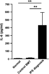
IL-6 is elevated in patients developing IPS. IL-6 levels in plasma from patients at the time of diagnosis of IPS after alloSCT (n = 38), control BMT recipients without evidence of IPS between days 14 and 21 after SCT (control BMT, n = 14) and normal volunteers (n = 13). **P < .01.

V体育平台登录 - Comment in

References

    1. Ferrara JL, Levine JE, Reddy P, Holler E. Graft-versus-host disease. Lancet. 2009;373(9674):1550–1561. - PMC - PubMed
    1. Clark JG, Hansen JA, Hertz MI, Parkman R, Jensen L, Peavy HH. NHLBI workshop summary. Idiopathic pneumonia syndrome after bone marrow transplantation. Am Rev Respir Dis. 1993;147(6 pt 1):1601–1606. - VSports在线直播 - PubMed
    1. Panoskaltsis-Mortari A, Griese M, Madtes DK, et al. American Thoracic Society Committee on Idiopathic Pneumonia Syndrome. An official American Thoracic Society research statement: noninfectious lung injury after hematopoietic stem cell transplantation: idiopathic pneumonia syndrome. Am J Respir Crit Care Med. 2011;183(9):1262–1279. - "V体育官网入口" PMC - PubMed
    1. Yanik G, Cooke KR. The lung as a target organ of graft-versus-host disease. Semin Hematol. 2006;43(1):42–52. - PubMed
    1. Burman AC, Banovic T, Kuns RD, et al. IFNgamma differentially controls the development of idiopathic pneumonia syndrome and GVHD of the gastrointestinal tract. Blood. 2007;110(3):1064–1072. - PubMed

"VSports最新版本" Publication types

MeSH terms