Skip to main page content
U.S. flag

An official website of the United States government

Dot gov

The . gov means it’s official. Federal government websites often end in . gov or . mil. Before sharing sensitive information, make sure you’re on a federal government site VSports app下载. .

Https

The site is secure. The https:// ensures that you are connecting to the official website and that any information you provide is encrypted and transmitted securely V体育官网. .

. 2014 Jun;13(6):1578-88.
doi: 10.1158/1535-7163.MCT-13-0877. Epub 2014 Mar 31.

V体育官网 - Inhibition of mTOR signaling reduces PELP1-mediated tumor growth and therapy resistance

Affiliations

Inhibition of mTOR signaling reduces PELP1-mediated tumor growth and therapy resistance

Vijay K Gonugunta et al. Mol Cancer Ther. 2014 Jun.

Abstract

Proline, Glutamic acid-, and Leucine-rich Protein 1 (PELP1) is a proto-oncogene that modulates estrogen receptor (ER) signaling. PELP1 expression is upregulated in breast cancer, contributes to therapy resistance, and is a prognostic marker of poor survival. In a subset of breast tumors, PELP1 is predominantly localized in the cytoplasm and PELP1 participates in extranuclear signaling by facilitating ER interactions with Src and phosphoinositide 3-kinase (PI3K). However, the mechanism by which PELP1 extranuclear actions contributes to cancer progression and therapy resistance remains unclear. In this study, we discovered that PELP1 cross-talked with the serine/threonine protein kinase mTOR and modulated mTOR signaling VSports手机版. PELP1 knockdown significantly reduced the activation of mTOR downstream signaling components. Conversely, PELP1 overexpression excessively activated mTOR signaling components. We detected the presence of the mTOR signaling complex proteins in PELP1 immunoprecipitates. mTOR-targeting drugs (rapamycin and AZD8055) significantly reduced proliferation of PELP1-overexpressed breast cancer cells in both in vitro and in vivo xenograft tumor models. MCF7 cells that uniquely retain PELP1 in the cytoplasm showed resistance to hormonal therapy and mTOR inhibitors sensitized PELP1cyto cells to hormonal therapy in xenograft assays. Notably, immunohistochemical studies using xenograft tumors derived from PELP1 overexpression model cells showed increased mTOR signaling and inhibition of mTOR rendered PELP1-driven tumors to be highly sensitive to therapeutic inhibition. Collectively, our data identified the PELP1-mTOR axis as a novel component of PELP1 oncogenic functions and suggest that mTOR inhibitor(s) will be effective chemotherapeutic agents for downregulating PELP1 oncogenic functions. .


Conflict of interest statement (VSports手机版)

Disclosure of Potential Conflicts of Interest: Authors have no potential conflict of interests

Figures

Figure 1
Figure 1. PELP1 is needed for optimal activation of the mTOR pathway
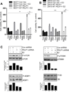
(A, B) MCF7 and ZR75 cells were transiently transfected with nonspecific control siRNA or PELP1 specific siRNA for 72 h, serum starved for 24h and stimulated with 10% serum for 10 min. Status of phosphorylation of mTOR signaling components was analyzed by Western blotting. Quantitation of phosphorylation after normalizing to its respective total protein is shown.(C) Lysates of MCF7 breast cancer cells were subjected to immunoprecipitation with PELP1, Raptor, and Rictor antibodies, and the presence of mTOR and PELP1 in the immunoprecipitates was analyzed by Western blotting.
Figure 2
Figure 2. mTOR inhibitors reduced PELP1-mediated signaling and cell proliferation
(A) Cell viability of MCF7-vec, MCF7-PELP1#20, MCF7-PELP1#13 cells were analyzed after treating the cells with or without 20 nM of rapamycin or AZD8055 in 5% FBS in RPMI medium for 72 h using an MTT assay. Results are the mean value of experiment performed in triplicates. (B) Cell viability of MCF7-vec, and MCF7-PELP1#13 cells were analyzed after treating the cells with or without 40 nM of rapamycin or AZD8055 in 5% DCC in RPMI medium for 72 h using MTT assay. Results are the mean value of experiments performed in triplicates. (C, D) Model cells were plated on cover slips in six well plates, treated with or without 40 nM of Rapamycin or 20 nM AZD8055 in 5% DCC serum in RPMI medium. (C) Ki-67 staining as a marker of proliferation was performed as described in methods section. (D)TUNEL staining was performed as a marker of apoptosis on fixed cells. Quantitation of Ki-67 and TUNEL staining was done as described in the methods section. **, P<0.01; ***, P<0.001; ***, P<0.0001.
Figure 3
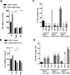
Figure 3. mTOR inhibitors reduced PELP1-mediated estrogen driven growth in breast cancer cells
(A) MCF7 or (B) ZR75 cells (2×10³) stably expressing control vector or PELP1 WT were seeded in 96-well plates and stimulated with E2 (1×10⁻⁸ M) in 5% charcoal-stripped medium for 7 days in the presence or absence of mTOR inhibitors. Cell viability was determined by using an MTT assay. Results are the mean value of experiments performed in triplicates. Student t-test was used for analysis. **, P<0.01; **, P<0.001. (C) ZR75 cells stably expressing control shRNA or PELP1 shRNA were E2 starved for 72h and stimulated with E2 (1×10⁻⁸ M) for 15 min in 5% charcoal stripped media. Status of mTOR signaling was analyzed by Western analysis. Quantitation of phosphorylation after normalizing to its respective total protein is shown.
Figure 4
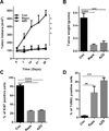
Figure 4. Rapamycin or AZD8055 treatment reduced PELP1-mediated xenograft tumor growth
(A) Six-week-old nude female mice were subcutaneously implanted with MCF7-PELP1 WT cells. After the tumors reached a measurable size, the mice were treated daily with vehicle, Rapamycin or AZD8055 for 4 weeks. Tumor volumes were measured at weekly intervals. (B) Tumor weights are shown in the histogram. (C) Ki-67 expression was analyzed by using IHC and quantitation of Ki-67 staining was done as described in the methods section. (D) TUNEL staining was performed as a marker of apoptosis on tumors that were treated with Rapamycin or AZD8055. Quantitation was done as described in methods. **, P<0.01; ***, P<0.001; ***, P<0.0001.
Figure 5
Figure 5. PELP1 cytoplasmic localization promotes mTOR signaling and tamoxifen resistance
(A) MCF7 cells stably expressing control vector or PELP1-cyto were serum starved for 72 h and stimulated with 10% serum for 10 min. The status of PELP1 expression and S6 phosphorylation was analyzed by Western blotting. Quantitation of S6 phosphorylation after normalizing to its respective total protein is shown. (B) MCF7 control vector, MCF7-PELP1-cyto or MCF7-PELP1-WT cells were treated with vehicle or 2.5µM tamoxifen for 72 h in 5% charcoal-stripped medium and cell viability was assayed by using an MTT assay. (C) MCF7 control vector or MCF7-PELP1-cyto cells were treated with vehicle, 2.5 µM tamoxifen, 20 nM rapamycin or in combination for 7 days in 5% charcoal-stripped medium. After the treatment, cell viability was determined by using an MTT assay. (D) Total lysates from various model cells were analyzed for the expression of PELP1 by Western blotting. (E) MCF7-TamR cells were treated with vehicle, 2.5 µM tamoxifen, 20 nM rapamycin or in combination for 7 days, and cell viability was determined by using an MTT assay. (F) MCF7-HER2 cells were treated with or without 2.5 µM tamoxifen, 20 nM rapamycin or in combination for 7 days, and cell viability was determined by using an MTT assay. Student t-test was used to analyze the data. **P<0.01; ***, P<0.001; ****, P<0.0001.
Figure 6
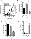
Figure 6. mTOR inhibitors reduce the PELP1–tamoxifen-mediated subcutaneous xenograft tumor growth
(A) Six-week-old nude female mice were subcutaneously implanted with MCF7-PELP1-cyto cells. After the tumors reached a measurable size, the mice were treated daily with vehicle, tamoxifen alone or tamoxifen and AZD8055 together for 4 weeks. Tumor volumes were measured at weekly intervals. (B) Tumor weights are shown in the histogram. (C) Ki-67 expression was analyzed by using IHC, and quantitation was done as described in methods section. (D) TUNEL staining was performed as a marker of apoptosis on tumors. Quantitation was done as described in methods. **, P<0.01; ***, P<0.001; ****, P<0.0001

References

    1. Jemal A, Siegel R, Ward E, Hao Y, Xu J, Thun MJ. Cancer statistics 2009. CA Cancer J Clin. 2009;59:225–249. - PubMed
    1. Osborne CK, Zhao H, Fuqua SA. Selective estrogen receptor modulators: structure, function, and clinical use. J Clin Oncol. 2000;18:3172–3186. - "V体育ios版" PubMed
    1. Mouridsen H, Gershanovich M, Sun Y, Perez-Carrion R, Boni C, Monnier A, et al. Superior efficacy of letrozole versus tamoxifen as first-line therapy for postmenopausal women with advanced breast cancer: results of a phase III study of the International Letrozole Breast Cancer Group. J Clin Oncol. 2001;19:2596–2606. - "V体育官网入口" PubMed
    1. Ali S, Coombes RC. Endocrine-responsive breast cancer and strategies for combating resistance. Nat Rev Cancer. 2002;2:101–112. - PubMed
    1. O'Malley BW, Kumar R. Nuclear receptor coregulators in cancer biology. Cancer Res. 2009;69:8217–8222. - PMC - PubMed

Publication types

MeSH terms