Skip to main page content
U.S. flag

An official website of the United States government

Dot gov

The . gov means it’s official. Federal government websites often end in . gov or . mil. Before sharing sensitive information, make sure you’re on a federal government site. VSports app下载.

Https

The site is secure. The https:// ensures that you are connecting to the official website and that any information you provide is encrypted and transmitted securely V体育官网. .

. 2014 Jan 9;9(1):e84410.
doi: 10.1371/journal.pone.0084410. eCollection 2014.

Effects of dietary glutamine on the homeostasis of CD4+ T cells in mice with dextran sulfate sodium-induced acute colitis (V体育官网)

Affiliations

Effects of dietary glutamine on the homeostasis of CD4+ T cells in mice with dextran sulfate sodium-induced acute colitis

Yuan-Chin Hsiung et al. PLoS One. .

Abstract

This study investigated the effects of dietary glutamine (Gln) on T-helper (Th) and T regulatory (Treg) cell homeostasis and colonic inflammatory mediator expression in mice with dextran sulfate sodium (DSS)-induced colitis. Mice were randomly assigned to 4 groups with 2 normal control (C and G) and 2 DSS-treated groups (DC and DG). The C and DC groups were fed a common semipurified diet, while the G and DG groups received an identical diet except that part of the casein was replaced by Gln, which provided 25% of the total amino acid nitrogen. Mice were fed the diets for 10 days. On day 6, mice in the normal control groups were given distilled water, while those in the DSS groups were given distilled water containing 1. 5% DSS for 5 d. At the end of the experiment, the mice were sacrificed for further examination. Results showed that DC group had higher plasma haptoglobin, colonic weight, immunoglobulin G, inflammatory cytokine and nuclear factor (NF)-κB protein levels VSports手机版. Gln administration lowered inflammatory mediators and NF-κB/IκBα ratio in colitis. Compared with the DC group, the percentages of interleukin-17F and interferon-γ in blood and transcription factors, T-bet and RAR-related orphan receptor-γt, gene expressions in mesenteric lymph nodes were lower, whereas blood Foxp3 was higher in the DG group. Also, DG group had lower colon injury score. These results suggest that Gln administration suppressed Th1/Th17 and Th-associated cytokine expressions and upregulated the expression of Tregs, which may modulate the balance of Th/Treg and reduce inflammatory reactions in DSS-induced colitis. .


Conflict of interest statement

Competing Interests: No conflicts of interest, financial or otherwise, are declared by the authors.

V体育ios版 - Figures

Figure 1
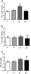
Figure 1. Percentage of Th1/Th2 cell subpopulations in the blood.
Lymphocytes were gated according to their size and granularity using light scatter detectors (FSC/SSC). CD4-positive lymphocytes were considered Th cells and were gated to determine the expression of intracellular cytokines. A, B: Percentages of interferon (IFN)-γ- and interleukin (IL)-4-expressing CD4+ lymphocytes. C: IFN-γ/IL-4 ratio. Data are presented as the mean ± SEM. Differences among groups were analyzed by ANOVA using the Tukey's test. *Significantly differs from the C group. #Significantly differs from the DG group (p<0.05).
Figure 2
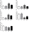
Figure 2. Percentages of Th17/Th22-associated cytokine-producing cell and Treg cell populations in the blood.
CD4-positive lymphocytes were gated to analyze intracellular cytokine expressions by flow cytometry. A: Expression of intracellular interleukin (IL)-17A. B: Expression of intracellular IL-17F. C: Triple expression of intracellular IL-17A, IL-17F, and IL-22. D: IL-22+ and IL-17ATh cells. E: Percentage of Treg cells. Data are presented as the mean ± SEM. Differences among groups were analyzed by an ANOVA using Tukey's test. *Significantly differs from the C group. #Significantly differs from the DG group (p<0.05).
Figure 3
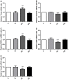
Figure 3. Transcription factor gene expressions of A) T-bet, B) GATA-3, C) ROR-γt, D) the aryl hydrocarbon receptor (AhR), and E) forhead box p3 (Foxp3) in mesenteric lymph nodes (MLNs) were analyzed by a real-time PCR.
Quantitation of mRNA changes was calculated by the comparative CT (2−ΔΔCt) method, and the mRNA expression of C mice was used as a calibrator. Data are shown as the mean ± SEM. *Significantly differs from the C group. # Significantly differs from the DG group (p<0.05).
Figure 4
Figure 4. mRNA expressions of mucosal recovery-related and antiapoptotic genes in colon tissues.
mRNA levels were analyzed by a real-time PCR. A: Expression of Muc2 mRNA. B: Expression of Tff3 mRNA. C: Expression of Hsp72 mRNA. D: Expression of Bcl-xL mRNA. Quantitation of mRNA changes was calculated by the comparative CT (2−ΔΔCt) method, and mRNA expression of C mice was used as a calibrator. Data are shown as the mean ± SEM. *Significantly differs from the C group. #Significantly differs from the DG group (P<0.05).
Figure 5
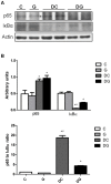
Figure 5. Protein levels of nuclear factor (NF)-κB p65 and inhibitory factor κBα (IκBα) in colon tissues.
a Protein expressions of NF-κB p65 and IκBα. Whole-tissue lysates were analyzed by immunoblotting, and b-actin was used as a loading control. b Densitometric analysis of the blot corrected by the protein loading control. c The ratio of NF-κB p65 to IκBα. Results of the densitometric analysis are shown as the mean ± SEM. *Significantly differs from the C group. #Significantly differs from the DG group (P<0.05).
Figure 6
Figure 6. Protein levels of PARP and cleaved PARP in colon tissues.
A: protein expressions of PARP and cleaved PARP. Whole-tissue lysates were analyzed by immunoblotting, and β-actin was used as a loading control. B: densitometric analysis of the blot corrected by the protein loading control. Results of the densitometric analysis are shown as the mean ± SEM. *Significantly differs from the C group. #Significantly differs from the DG group (P<0.05).
Figure 7
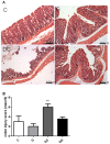
Figure 7. Histopathology of colon tissues.
A: Hematoxylin and eosin staining of colon tissues from mice in the C, G, DC, and DG groups. B: Histological scores of colitis. Data are presented as the mean ± SEM., which were determined as described in “Materials and Methods.” Differences among groups were analyzed by a one-way ANOVA using Tukey's test. *Significantly differs from the C group. #Significantly differs from the DG group (p<0.05).

References

    1. Triantafillidis JK, Nasioulas G, Kosmidis PA (2009) Colorectal cancer and inflammatory bowel disease: epidemiology, risk factors, mechanisms of carcinogenesis and prevention strategies. Anticancer Res 29: 2727–2737. - "VSports手机版" PubMed
    1. Maloy KJ, Powrie F (2011) Intestinal homeostasis and its breakdown in inflammatory bowel disease. Nature 474: 298–306. - PubMed
    1. Kaser A, Zeissig S, Blumberg RS (2010) Inflammatory bowel disease. Annu Rev Immunol 28: 573–621. - PMC - PubMed
    1. Bouma G, Strober W (2003) The immunological and genetic basis of inflammatory bowel disease. Nat Rev Immunol 3: 521–533. - PubMed (VSports最新版本)
    1. Fuss IJ, Neurath M, Boirivant M, Klein JS, de la Motte C, et al. (1996) Disparate CD4+ lamina propria (LP) lymphokine secretion profiles in inflammatory bowel disease. Crohn's disease LP cells manifest increased secretion of IFN-gamma, whereas ulcerative colitis LP cells manifest increased secretion of IL-5. J Immunol 157: 1261–1270. - PubMed

Publication types

"VSports app下载" MeSH terms