Skip to main page content (VSports手机版)
U.S. flag

An official website of the United States government

Dot gov

The . gov means it’s official. Federal government websites often end in . gov or VSports app下载. mil. Before sharing sensitive information, make sure you’re on a federal government site. .

Https

The site is secure. The https:// ensures that you are connecting to the official website and that any information you provide is encrypted and transmitted securely V体育官网. .

. 2006 Aug 15;54(3):223-33.
doi: 10.1002/glia.20364.

Expression and function of calcium-activated potassium channels in human glioma cells

Affiliations

Expression and function of calcium-activated potassium channels in human glioma cells

Amy K Weaver (V体育安卓版) et al. Glia. .

Abstract

Ca(2+)-activated K(+) (K(Ca)) channels are a unique family of ion channels because they are capable of directly communicating calcium signals to changes in cell membrane potential required for cellular processes including but not limited to cellular proliferation and migration. It is now possible to distinguish three families of K(Ca) channels based on differences in their biophysical and pharmacological properties as well as genomic sequence. Using a combination of biochemical, molecular, and biophysical approaches, we show that human tumor cells of astrocytic origin, i. e. glioma cells, express transcripts for all three family members of K(Ca) channels including BK, IK, and all three SK channel types (SK1, SK2, and SK3). The use of selective pharmacological inhibitors shows prominent expression of currents that are inhibited by the BK channel specific inhibitors iberiotoxin and paxilline. However, despite the presence of transcripts for IK and SK, neither clotrimazole, an inhibitor of IK channels, nor apamin, known to block most SK channels inhibited any current VSports手机版. The exclusive expression of functional BK channels was further substantiated by shRNA knockdown experiments, which selectively reduced iberiotoxin sensitive currents. Western blotting of patient biopsies with antibodies specific for all three KCa channel types further substantiated the exclusive expression of BK type KCa channels in vivo. This finding is in sharp contrast to other cancers that express primarily IK channels. .


V体育官网入口 - Figures

Fig. 1
Fig. 1
RT-PCR demonstrates expression of KCa channel mRNA in both glioma cell lines and biopsies. Primers directed against each of the 5 KCa channels were used and are listed in Table 1. mRNA from normal human brain tissue, human glioma tissue, three glioma cells lines, and primary rat cortical astrocytes were probed for expression of each of the five channels. IK channel transcripts were detected in human tumor tissue and D54 and U251 glioma cell lines. mRNA for BK channels was detected in all samples except for rat cortical astrocytes. SK1 and SK3 had identical expression profiles being found in normal human brain as well as all of the malignant cell samples. They were absent in rat cortical astrocytes. SK2 transcripts were detected in all samples except for normal human brain tissue. Negative controls were conducted in the same manner as all other sample; however, they remained on ice during reverse transcription, to ensure that samples were not contaminated with genomic DNA. Results are representative of three independent experiments.
Fig. 2
Fig. 2
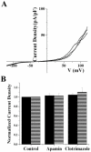
U251 whole cell currents are not sensitive to either apamin or CLT. (A) Representative traces from whole cell recordings of U251 cells in the presence of 750 nM [Ca2+]i while bathing on normal bath (black line), 300 nM apamin (red line) or 10 μM CLT (blue line). Cells were held at -40 mV and ramped from -120 mV to +120 mV during the course of the experiment. (B) Cumulative data (n = 10 cells each) illustrating the difference in normalized current density before and after application of specific inhibitors of SK (apamin) or IK (CLT) channels. Current densities from all treatments were normalized to their respective control (either normal pipette solution, or 750 nM Ca2+.
Fig. 3
Fig. 3
The specific BK channel inhibitor paxilline inhibits ∼90% of the whole cell outward current in glioma cells. (A) Representative traces from two cells before and after application of 2 μM paxilline. Top trace was carried out in the relative absence of intracellular Ca2+, and the bottom traces had 750 nM free Ca2+ in the pipette. (B) Activation curve of BK channels in normal (■) and 750 nM Ca2+ (▲) pipette solution. Application of paxilline completely inhibits activation of BK channels regardless of [Ca2+]i.(C) Current densities before and after treatment with Pax show that BK currents make up almost 90% of the whole cell outward current in glioma cells (n = 10). Current densities under both conditions were normalized to peak current density at 120 mV in normal pipette solution.
Fig. 4
Fig. 4
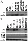
Western blots demonstrate that only protein for BK Channels is expressed in glioma cell lines. (A) Representative blot from four independent experiments. Specific antibodies to hIK1 were used to probe lysates from three glioma cell lines, two primary human glioma cultures, cortical rat astrocytes, and whole rat brain. The K562 leukemia cell line was used as a positive control for expression of IK channel protein. (B) Representative blot from three independent experiments illustrating a lack of expression of SK1, 2, and 3 in the same lysates as listed above. Whole rat brain lysates were used as a positive control. Specific antibodies directed against BK, SK1, SK2, and SK3 channels were used.
Fig. 5
Fig. 5
BK channels are expressed in human glioma tissue. (A) Tissue samples from three human GBM biopsies as well as two normal samples were obtained, and tissue lysates were generated and probed for KCa channel expression as with glioma cell lines. Only BK channels were detected in the tumor samples. (B) Tissue slices from a human GBM biopsy were stained with antibodies against BK channels. The images demonstrate expression of BK channels diffusely in human gliomas. The left panel illustrates immunostaining for BK channels, the middle panel for DAPI, and the right panel represents the merge of the two channels.
Fig. 6
Fig. 6
BK shRNA knocks down the majority of the outward voltage dependent current in glioma cells. (A) Representative traces taken from a whole cell recording of a control cell (left) and a cell three days post shRNA transfection (right). Voltage steps in increments of 20 mV were elicited from -40 mV. Following treatment with shRNA, TEA sensitive and the paxilline sensitive (data not shown) component is lost. (B) Bar graph of whole cell current density illustrates a 4-fold reduction in whole-cell conductance following treatment with BK shRNA. ShRNA constructs directed against AQP4 were used as a negative control to illustrate a lack of off-target effects of the shRNA on BK currents. (C) After 3 days of culture following either no treatment (control), BK shRNA treatment, or treatment with vector plasmid, cell lysates were harvested probed for BK channel expression. Actin levels were used as a loading control. BK channel protein is markedly reduced following treatment with BK shRNA. (D) I/Imax plot from voltage step recordings demonstrates that there is no change in voltage dependence of the remaining BK current following treatment.
Fig. 7
Fig. 7
IbTX and paxilline, two specific inhibitors of BK channels, inhibit glioma cell migration. Representative images from control (A), IbTX treated (B), and paxilline treated (C) cells are shown. Images illustrate cells that have migrated from the top of a transwell filter to the bottom. (D) Cumulative data from three independent experiments. Cells were plated on top of an 8-lm pore transwell filter and allowed to migrate for 4 h in the presence or absence of 100 nM IbTX, 2 μM Pax, or 30 μM NS1619. Cells that had migrated through at the end of 4 h were counted and values were normalized to control. IbTX and Pax inhibited U251 cell migration by 63 and 68%, respectively, while NS1619, a BK channel activator, had no effect on migration.

References

    1. Agresti C, Meomartini ME, Amadio S, Ambrosini E, Serafini B, Franchini L, Volonte C, Aloisi F, Visentin S. Metabotropic P2 receptor activation regulates oligodendrocyte progenitor migration and development. Glia. 2005;50:132–144. - PubMed
    1. Attali B, Wang N, Kolot A, Sobko A, Cherepanov V, Soliven B. Characterization of delayed rectifier Kv channels in oligodendrocytes and progenitor cells. J Neurosci. 1997;17:8234–8245. - PMC - PubMed
    1. Bahia PK, Suzuki R, Benton DC, Jowett AJ, Chen MX, Trezise DJ, Dickenson AH, Moss GW. A functional role for small-conductance calcium-activated potassium channels in sensory pathways including nociceptive processes. J Neurosci. 2005;25:3489–3498. - PMC - PubMed
    1. Baran I. Calcium and cell cycle progression: Possible effects of external perturbations on cell proliferation. Biophys J. 1996;70:1198–1213. - PMC - PubMed
    1. Barfod ET, Moore AL, Lidofsky SD. Cloning and functional expression of a liver isoform of the small conductance Ca2+-activated K+ channel SK3. Am J Physiol Cell Physiol. 2001;280:C836–C842. - "V体育2025版" PubMed

Publication types

MeSH terms

Substances (V体育安卓版)