Skip to main page content
U.S. flag

An official website of the United States government

Dot gov

The VSports app下载. gov means it’s official. Federal government websites often end in . gov or . mil. Before sharing sensitive information, make sure you’re on a federal government site. .

Https

The site is secure. The https:// ensures that you are connecting to the official website and that any information you provide is encrypted and transmitted securely. V体育官网.

. 2004 May;15(5):2156-63.
doi: 10.1091/mbc.e03-12-0894. Epub 2004 Mar 5.

Deoxycholic acid activates beta-catenin signaling pathway and increases colon cell cancer growth and invasiveness

Affiliations

Deoxycholic acid activates beta-catenin signaling pathway and increases colon cell cancer growth and invasiveness

Rama Pai et al. Mol Biol Cell. 2004 May.

Abstract

Colorectal cancer is often lethal when invasion and/or metastasis occur. Tumor progression to the metastatic phenotype is mainly dependent on tumor cell invasiveness. Secondary bile acids, particularly deoxycholic acid (DCA), are implicated in promoting colon cancer growth and progression. Whether DCA modulates beta-catenin and promotes colon cancer cell growth and invasiveness remains unknown. Because beta-catenin and its target genes urokinase-type plasminogen activator receptor (uPAR) and cyclin D1 are overexpressed in colon cancers, and are linked to cancer growth, invasion, and metastasis, we investigated whether DCA activates beta-catenin signaling and promotes colon cancer cell growth and invasiveness. Our results show that low concentrations of DCA (5 and 50 microM) significantly increase tyrosine phosphorylation of beta-catenin, induce urokinase-type plasminogen activator, uPAR, and cyclin D1 expression and enhance colon cancer cell proliferation and invasiveness. These events are associated with a substantial loss of E-cadherin binding to beta-catenin. Inhibition of beta-catenin with small interfering RNA significantly reduced DCA-induced uPAR and cyclin D1 expression. Blocking uPAR with a neutralizing antibody significantly suppressed DCA-induced colon cancer cell proliferation and invasiveness. These findings provide evidence for a novel mechanism underlying the oncogenic effects of secondary bile acids. VSports手机版.


"VSports app下载" Figures

Figure 1.
Figure 1.
Dose- and time-response studies of DCA on tyrosine phosphorylation of β-catenin in colon cancer cells. Serum-starved colon cancer (SW480 and LoVo) cells were treated with either medium only (control) (Ct) or DCA in various concentrations (5–100 μM) to varying time intervals (30 min–2 h). Tyrosine phosphorylation of β-catenin (P-β-cat) was determined by immunoprecipitation (β-catenin monoclonal antibody) and immunoblotting with anti-phosphotyrosine antibody. Total β-catenin in immunoprecipitates was determined by reprobing the same blot with anti-β-catenin antibody. Top, representative blots from three separate experiments performed in triplicate. Bottom, quantitative analysis of tyrosine phosphorylation of β-catenin by determining the ratio between β-catenin protein and tyrosine phosphorylation levels from three separate experiments (mean values ± SE).
Figure 2.
Figure 2.
DCA increases nuclear β-catenin levels. Colon cancer cells (SW480 and LoVo) were treated with either medium only (control) or DCA (5 and 50 μM for 30 min). Nuclear extracts were subjected to immunoblotting using anti-β-catenin antibody. Top, representative blots from three separate experiments performed in triplicate. Bottom, quantitative analysis of nuclear β-catenin levels showing relative densities of bands from three separate experiments (mean values ± SE).
Figure 3.
Figure 3.
DCA causes loss of E-cadherin association with β-catenin. Blots that were used to determine tyrosine phosphorylation of β-catenin in response to DCA were stripped and reprobed by using anti-E-cadherin antibody and subsequently reprobed with anti-β-catenin antibody. Top, representative blots from three separate experiments performed in triplicate. Bottom, quantitative analysis of E-cadherin bound to β-catenin after various treatments by determining the ratio between E-cadherin and total β-catenin levels from three separate experiments (mean values ± SE).
Figure 4.
Figure 4.
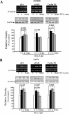
DCA induces uPA, uPAR, and cyclin D1 mRNA and protein expression in colon cancer cells. Top, serum-starved SW480 (A) and LoVo (B) cells were treated either with medium only (control) (Ct) or DCA (5 and 50 μM) for 2 h. Total RNA was isolated and subjected to RT-PCR by using specific primers for uPA, uPAR, cyclin D1, and β-actin. DNA was visualized by ethidium bromide staining. Middle, serum-starved SW480 and LoVo cells were treated either with medium only (control) (Ct) or DCA (5 μM) for 3–24 h. Equal amounts of protein (0.1 mg) were subjected to 10% SDS-PAGE and Western blot analysis performed using uPA-, uPAR-, and cyclin D1-specific antibodies. Representative blots from two separate experiments performed in triplicate. Bottom, quantitative analysis of relative densities of bands of uPA, uPAR, and cyclin D1 proteins normalized to β-actin levels from three separate experiments (mean values ± SE).
Figure 5.
Figure 5.
β-Catenin siRNA treatment significantly reduces DCA-stimulated uPAR protein expression. Colon cancer (SW480) cells were transfected with either siRNA directed against β-catenin or nonspecific RNA by using Oligofectamine. Twenty-four hours after transfection, cells were serum starved for the next 36 h and then treated with or without DCA (5 μM) for 3 h. Cells were lysed in lysis buffer and β-catenin, uPAR, cyclin D1, and β-actin protein expressions were assessed by Western blot analysis by using specific antibodies. This figure is a representative blot from two separate experiments performed in duplicate.
Figure 6.
Figure 6.
Effect of DCA on colon cancer cell proliferation. Serum-starved colon cancer cells were treated with either 1) medium only, 2) varying concentrations of DCA (5–100 μM) for 24 h, 3) uPAR neutralizing antibody, or 4) uPAR neutralizing antibody (4 h) followed by DCA (5 μM, 24 h). Two hours before harvesting, the individual wells were pulsed with [3H]thymidine (0.5 μCi), washed with phosphate-buffered saline, and collected on glass wool fibers filters by using a multiple sample cell harvester, and the radioactivity was measured. Values are presented as mean ± SE of three individual experiments performed in quadruplicate.
Figure 7.
Figure 7.
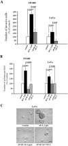
DCA enhances colon cancer cell invasiveness. (A) Serum-starved SW480 and LoVo cells were pretreated with or without uPAR neutralizing antibody, and 4 × 10⁴ cells were seeded in the upper chamber in the presence or absence of uPAR antibody. Medium alone or medium containing DCA was added to the lower chamber. After 24 h, cells on the upper surface of the filter were removed. Filters were fixed and stained. The cells on the lower surface were counted under a microscope (magnification 100×). Five fields were counted per each filter, and four wells were used for each treatment. (B) Serum-starved SW480 and LoVo cells (1 × 10⁴) were suspended in 0.5 ml of 1:2 diluted Matrigel. The cell/Matrigel mixture was plated into 24-well plates and incubated at 37°C. The medium containing uPAR neutralizing antibody (5 μg/ml) with or without DCA (5 μM) was then added in fresh serum-free medium every 2 d. Four wells were used for each treatment. After 15 d, images were captured using a camera attached to an inverted microscope. Quantitative analysis of the number of colonies formed in Matrigel after various treatments. Values are presented as mean ± SE number of colonies from five fields of each of four wells (total 20 fields) after each treatment. (C) Representative image of cancer cell colonies from one of the five fields captured from each well. Compared with nonstimulated control, DCA (5 μM) treatment significantly increased the number and size of the cancer cell colonies formation in Matrigel. Whereas pretreatment of colon cancer cells with uPAR neutralizing antibody (5 μg/ml, 4 h) moderately suppressed the number and size of colonies formed at basal levels, it significantly reduced the number and size of colonies formed in response to DCA (5 μM) treatment. Bar, 50 μM.

"V体育ios版" References

    1. Alexander, N., Wong, C., and Pignatelli, M. (2002). β-Catenin – a linchpin in colorectal carcinogenesis? Am. J. Pathol. 160, 389–401. - PMC - PubMed
    1. Andreasen, P.A., Kjoller, L., Christensen, L., and Duffy, M.J. (1997). The urokinase-type plasminogen activator system in cancer metastasis: a review. Int. J. Cancer 72, 1–22. - PubMed
    1. Andrews, N.C., and Faller, D.V. (1991). A rapid micropreparation technique for extraction of DNA-binding proteins from limiting numbers of mammalian cells. Nucleic Acid Res. 19, 2499. - "V体育ios版" PMC - PubMed
    1. Arber, N., Doki, Y., Han, E.K.-H., Sgambato, A., Zhou, P., Kim, N.-H., Delohery, T., Klein, M.G., Holt, P.R., and Weinstein, I.B. (1997). Antisense to cyclin D1 inhibits the growth and tumorigenicity of human colon cancer cells. Cancer Res. 57, 1569–1574. - PubMed
    1. Arber, N., Hibshoosh, H., Moss, S.F., Sutter, T., Zhang, Y., Begg, M., Wang, S., Weinstein, I.B., and Holt, P.R. (1996). Increased expression of cyclin D1 is an early event in multistage colorectal carcinogenesis. Gastroenterology 110, 669–674. - PubMed (VSports在线直播)

Publication types

MeSH terms

LinkOut - more resources