
Hypoxia Inducible Factor 1 (HIF-1) Recruits Macrophage to Activate Pancreatic Stellate Cells in Pancreatic Ductal Adenocarcinoma

 ,
, "> Figure 1
The correlation between hypoxia inducible factor (HIF)-1α and CD68 expression in specimens of pancreatic ductal adenocarcinoma (PDAC). (A) Immunohistochemical analysis of HIF-1α and CD68 correlative expression in consecutive sections from human PDAC surgical samples (tumor tissue and peritumoral tissue). Left, low expression (intensity grade − and +); Right, high expression (intensity grade ++ and +++) (magnification, 100 or 200); (B) Statistical analysis of CD68 and HIF-1α in tumor tissue and peritumoral tissue; (C) Statistical analysis of CD68 and HIF-1α in the PDAC surgical samples; (D) Analyzed messenger RNA (mRNA) expression profiles of HIF-1α and CD68 in 177 PDAC patients from The Cancer Genome Atlas (TCGA).
"> Figure 2The correlation between HIF-1α and chemical chemokines 2 (CCL2) expression in specimens of PDAC. (A) Immunohistochemical analysis of HIF-1α and CCL2 correlative expression in consecutive sections from human PDAC surgical samples (tumor tissue and peritumoral tissue). Left, low expression (intensity grade − and +). Right, high expression (intensity grade ++ and +++) (magnification, 100 or 200); (B) Association between CCL2 expression levels and the overall survival of patients with PDAC. Patients with PDAC (131) were stratified into two groups according to CCL2 IHC staining intensity. Patients with high CCL2 expression (intensity grade ++ and +++) had much worse overall survival when compared with patients with low CCL2 expression (intensity grade − and +). p = 0.014 was determined with a log-rank test; (C) Statistical analysis of immunohistochemical results of CCL2 expression in tumor tissue and peritumoral tissue. p Value was calculated by the Spearman rank correlation test; (D) Statistical analysis of immunohistochemical results of HIF-1α and CCL2 expression in 131 human PDAC surgical samples. p Value was calculated by the Spearman rank correlation test; (E) Analyzed mRNA expression profiles of HIF-1α and CCL2 in 177 PDAC patients from TCGA.
"> Figure 2 Cont.The correlation between HIF-1α and chemical chemokines 2 (CCL2) expression in specimens of PDAC. (A) Immunohistochemical analysis of HIF-1α and CCL2 correlative expression in consecutive sections from human PDAC surgical samples (tumor tissue and peritumoral tissue). Left, low expression (intensity grade − and +). Right, high expression (intensity grade ++ and +++) (magnification, 100 or 200); (B) Association between CCL2 expression levels and the overall survival of patients with PDAC. Patients with PDAC (131) were stratified into two groups according to CCL2 IHC staining intensity. Patients with high CCL2 expression (intensity grade ++ and +++) had much worse overall survival when compared with patients with low CCL2 expression (intensity grade − and +). p = 0.014 was determined with a log-rank test; (C) Statistical analysis of immunohistochemical results of CCL2 expression in tumor tissue and peritumoral tissue. p Value was calculated by the Spearman rank correlation test; (D) Statistical analysis of immunohistochemical results of HIF-1α and CCL2 expression in 131 human PDAC surgical samples. p Value was calculated by the Spearman rank correlation test; (E) Analyzed mRNA expression profiles of HIF-1α and CCL2 in 177 PDAC patients from TCGA.
"> Figure 3HIF-1α regulated the expression of CCL2 in PDAC cells, PC, N and H indicate positive control, normoxia, and hypoxia, respectively. (A) HPDE6-C7,BxPC3 and MiaPaca-2 cells were transfected with siHIF-1α (50 nmol/L) and pcDNA3.1-HIF-1α plasmids (4 µg) for 48 h, respectively; the mRNA expression levels of HIF-1α and CCL2 were assessed by qRT-PCR. The experiments were performed thrice independently. * p < 0.05 vs. control. Cells without treatment and normal pancreatic cell with transfection treatment were adopted as two negative controls. Vascular endothelial growth factor (VEGF) was used as positive control; (B) Western blot analysis confirmed the protein level expression correlation between HIF-1α and CCL2 in PDAC cell lines. The experiments were performed thrice independently. VEGF was used as positive control; (C) The DNA sequence of the CCL2 promoter (left) and the DNA sequence of the VEGF promoter (right); (D) Chromatin immunoprecipitation analysis in MiaPaCa-2 cells. The PCR products of VEGF promoter were used as positive control. PC, N and H indicate positive control, normoxia, and hypoxia, respectively.
"> Figure 4HIF-1α promoted the recruitment of macrophages. (A) A comparison of the ability of recruiting macrophages by HPDE6-C7, BxPC3, and MiaPaCa-2 cells transfected with pcDNA3.1 and pcDNA3.1-HIF-1α plasmids (4 µg) for 48 h using Boyden chambers. The experiments were performed thrice independently. * p < 0.05 vs. control (magnification, 100×); (B) A comparison of the ability of recruiting macrophages by HPDE6-C7, BxPC3, and MiaPaCa-2 cells transfected with negative control siRNA and HIF-1α siRNA (50 nmol/L) for 48 h using Boyden chambers. The experiments were performed thrice independently. * p < 0.05 vs. control (magnification, 100×).
"> Figure 4 Cont.HIF-1α promoted the recruitment of macrophages. (A) A comparison of the ability of recruiting macrophages by HPDE6-C7, BxPC3, and MiaPaCa-2 cells transfected with pcDNA3.1 and pcDNA3.1-HIF-1α plasmids (4 µg) for 48 h using Boyden chambers. The experiments were performed thrice independently. * p < 0.05 vs. control (magnification, 100×); (B) A comparison of the ability of recruiting macrophages by HPDE6-C7, BxPC3, and MiaPaCa-2 cells transfected with negative control siRNA and HIF-1α siRNA (50 nmol/L) for 48 h using Boyden chambers. The experiments were performed thrice independently. * p < 0.05 vs. control (magnification, 100×).
"> Figure 5Macrophages promoted the activation of PSCs. (A) PSCs in co-cultured group were more active than those in the control group; (B) Western-blot analysis verified that the expression of α-smooth muscle actin (α-SMA) in PSCs was significantly higher than that in the control group; in addition, glial fibrillary acidic protein (GFAP) did not change significantly, and β-actin was used as a reference; (C) An immunofluorescence assay confirmed that the main changes in PSCs were ascribed to the disappearance of lipid droplets and the increased expression of α-SMA.
"> Figure 5 Cont.Macrophages promoted the activation of PSCs. (A) PSCs in co-cultured group were more active than those in the control group; (B) Western-blot analysis verified that the expression of α-smooth muscle actin (α-SMA) in PSCs was significantly higher than that in the control group; in addition, glial fibrillary acidic protein (GFAP) did not change significantly, and β-actin was used as a reference; (C) An immunofluorescence assay confirmed that the main changes in PSCs were ascribed to the disappearance of lipid droplets and the increased expression of α-SMA.
">
Abstract
: Hypoxia inducible factor 1 (HIF-1) is a transcription factor composed of two subunits, namely, HIF-1α and HIF-1β, in which HIF-1β is constitutively expressed. HIF-1 upregulates several hypoxia-responsive proteins, including angiogenesis factors, glycolysis solution enzymes, and cell survival proteins. HIF-1 is also associated with the degree of inflammation in the tumor region, but the exact mechanism remains unclear. This study aims to identify the molecular mechanism of recruiting monocytes/macrophages by HIF-1α in pancreatic ductal adenocarcinoma (PDAC) and the effects of macrophages on pancreatic stellate cells (PSCs). Immunohistochemistry (IHC) was performed for cluster of differentiation 68 (CD68), HIF-1α, and chemical chemokines 2 (CCL2). Western blot, real-time quantitative reverse transcription polymerase chain reaction (qRT-PCR), chromatin immunoprecipitation assay, and The Cancer Genome Atlas (TCGA) were used to verify the correlation between HIF-1α and CCL2 at protein and nucleic acid levels. Monocytes/macrophages were co-cultured with PSCs to observe their interaction. Samples showed significant correlation between CD68 and HIF-1α (t-test, p < 0. 05). HIF-1α recruited monocytes/macrophages by promoting CCL2 secretion. Moreover, macrophages could accelerate the activation of PSCs. HIF-1α might promote inflammation and fibrosis of PDAC through CCL2 secretion, which may provide a novel target to treat PDAC patients. Keywords: PDAC; monocytes/macrophages; PSCs; HIF-1; CCL2 V体育官网入口.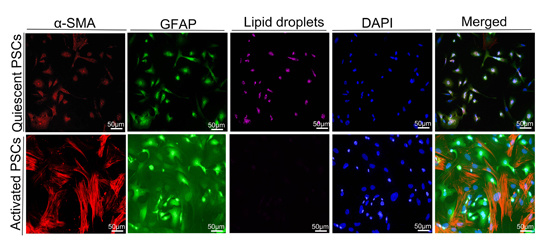
VSports - 1. Introduction
2. Results
2.1. Cluster of Differentiation 68 (CD68) Expression Was Related to Hypoxia Inducible Factor (HIF)-1α in the Samples of Pancreatic Ductal Adenocarcinoma (PDAC) Through Immunohistochemistry (IHC) Staining
2.2. HIF-1α Expression Was Significantly Correlated with Chemical Chemokines 2 (CCL2) at the Tissue Level, and CCL2 Expression Was Related to Patient Prognosis
2.3. HIF-1α Regulated CCL2
2.4. HIF-1α Significantly Promoted the Recruitment of Macrophages
2.5. Macrophages Accelerated the Activation of Pancreatic Stellate Cells (PSCs)
3. Discussion
4. Materials and Methods
4.1. Immunohistochemistry
4.2. Cell Culture and Hypoxic Treatment
4.3. SiRNA Duplexes, Plasmid Constructs, and Transient Transfection
4.4. Western-Blot Analysis
4.5. Real-Time Quantitative Reverse Transcription Polymerase Chain Reaction (qRT-PCR)
4.6. Chromatin Immunoprecipitation Assay
4.7. Isolation and Culture of Monocytes/Macrophages
4.8. Transwell Assay
4.9. Isolation of PSCs
4.10. Immunofluorescence
4.11. Statistical Analysis
5. Conclusions
VSports - Supplementary Materials
Acknowledgments (VSports最新版本)
Author Contributions
Conflicts of Interest (V体育官网)
Abbreviations
| PDAC | Pancreatic ductal adenocarcinoma |
| HIF-1 | Hypoxia inducible factor 1 |
| CCL2 | Chemical chemokines 2 |
| IHC | immunohistochemistry |
| TCGA | The Cancer Genome Atlas |
| PSCs | pancreatic stellate cells |
V体育平台登录 - References
- Semenza, G.L. Hypoxia-inducible factors: Mediators of cancer progression and targets for cancer therapy. Trends Pharmacol. Sci. 2012, 33, 207–214. [Google Scholar (VSports)] [CrossRef] [PubMed]
- Murdoch, C.; Giannoudis, A.; Lewis, C.E. Mechanisms regulating the recruitment of macrophages into hypoxic areas of tumors and other ischemic tissues. Blood 2004, 104, 2224–2234. [Google Scholar] [CrossRef] [PubMed]
- Negus, R.P.; Stamp, G.W.; Relf, M.G.; Burke, F.; Malik, S.T.; Bernasconi, S.; Allavena, P.; Sozzani, S.; Mantovani, A.; Balkwill, F.R. The detection and localization of monocyte chemoattractant protein-1 (MCP-1) in human ovarian cancer. J. Clin. Investig. 1995, 95, 2391–2396. ["VSports注册入口" Google Scholar] [CrossRef] [PubMed]
- Riethdorf, L.; Riethdorf, S.; Gutzlaff, K.; Prall, F.; Loning, T. Differential expression of the monocyte chemoattractant protein-1 gene in human papillomavirus-16-infected squamous intraepithelial lesions and squamous cell carcinomas of the cervix uteri. Am. J. Pathol. 1996, 149, 1469–1476. [Google Scholar (V体育2025版)] [PubMed]
- Graves, D.T.; Barnhill, R.; Galanopoulos, T.; Antoniades, H.N. Expression of monocyte chemotactic protein-1 in human melanoma in vivo. Am. J. Pathol. 1992, 140, 9–14. [Google Scholar] [PubMed]
- Semenza, G.L.; Jiang, B.H.; Leung, S.W.; Passantino, R.; Concordet, J.P.; Maire, P.; Giallongo, A. Hypoxia response elements in the aldolase A, enolase 1, and lactate dehydrogenase A gene promoters contain essential binding sites for hypoxia-inducible factor 1. J. Biol. Chem. 1996, 271, 32529–32537. [Google Scholar] [CrossRef] [PubMed]
- Forsythe, J.A.; Jiang, B.H.; Iyer, N.V.; Agani, F.; Leung, S.W.; Koos, R.D.; Semenza, G.L. Activation of vascular endothelial growth factor gene transcription by hypoxia-inducible factor 1. Mol. Cell. Biol. 1996, 16, 4604–4613. [Google Scholar] [CrossRef] [PubMed]
- Erkan, M.; Reiser-Erkan, C.; Michalski, C.W.; Kong, B.; Esposito, I.; Friess, H.; Kleeff, J. The impact of the activated stroma on pancreatic ductal adenocarcinoma biology and therapy resistance. Curr. Mol. Med. 2012, 12, 288–303. [Google Scholar] [CrossRef] [PubMed]
- Hoffmann, A.C.; Mori, R.; Vallbohmer, D.; Brabender, J.; Klein, E.; Drebber, U.; Baldus, S.E.; Cooc, J.; Azuma, M.; Metzger, R.; et al. High expression of HIF1a is a predictor of clinical outcome in patients with pancreatic ductal adenocarcinomas and correlated to PDGFA, VEGF, and bFGF. Neoplasia 2008, 10, 674–679. [VSports最新版本 - Google Scholar] [CrossRef] [PubMed]
- Zhao, T.; Gao, S.; Wang, X.; Liu, J.; Duan, Y.; Yuan, Z.; Sheng, J.; Li, S.; Wang, F.; Yu, M.; et al. Hypoxia-inducible factor-1α regulates chemotactic migration of pancreatic ductal adenocarcinoma cells through directly transactivating the CX3CR1 gene. PLoS ONE 2012, 7, e43399. [Google Scholar] [CrossRef] [PubMed]
- Gordon, S.; Taylor, P.R. Monocyte and macrophage heterogeneity. Nat. Rev. Immunol. 2005, 5, 953–964. [V体育2025版 - Google Scholar] [CrossRef] [PubMed]
- Lewis, C.E.; Pollard, J.W. Distinct role of macrophages in different tumor microenvironments. Cancer Res. 2006, 66, 605–612. [Google Scholar] [CrossRef] [PubMed]
- Siveen, K.S.; Kuttan, G. Role of macrophages in tumour progression. Immunol. Lett. 2009, 123, 97–102. ["VSports" Google Scholar] [CrossRef] [PubMed]
- Qian, B.Z.; Pollard, J.W. Macrophage diversity enhances tumor progression and metastasis. Cell 2010, 141, 39–51. [VSports app下载 - Google Scholar] [CrossRef] [PubMed]
- Jaiswal, S.; Jamieson, C.H.; Pang, W.W.; Park, C.Y.; Chao, M.P.; Majeti, R.; Traver, D.; van Rooijen, N.; Weissman, I.L. CD47 is upregulated on circulating hematopoietic stem cells and leukemia cells to avoid phagocytosis. Cell 2009, 138, 271–285. [Google Scholar (VSports)] [CrossRef] [PubMed]
- Masamune, A.; Kikuta, K.; Watanabe, T.; Satoh, K.; Hirota, M.; Shimosegawa, T. Hypoxia stimulates pancreatic stellate cells to induce fibrosis and angiogenesis in pancreatic cancer. Am. J. Physiol. Gastrointest. Liver Physiol. 2008, 295, G709–G717. [Google Scholar] [CrossRef] [PubMed]
- Oosterling, S.J.; van der Bij, G.J.; Meijer, G.A.; Tuk, C.W.; van Garderen, E.; van Rooijen, N.; Meijer, S.; van der Sijp, J.R.; Beelen, R.H.; van Egmond, M. Macrophages direct tumour histology and clinical outcome in a colon cancer model. J. Pathol. 2005, 207, 147–155. ["VSports注册入口" Google Scholar] [CrossRef] [PubMed]
- Deshmane, S.L.; Kremlev, S.; Amini, S.; Sawaya, B.E. Monocyte chemoattractant protein-1 (MCP-1): An overview. J. Interferon Cytokine Res. 2009, 29, 313–326. [Google Scholar] [CrossRef] [PubMed]
- Bosco, M.C.; Puppo, M.; Pastorino, S.; Mi, Z.; Melillo, G.; Massazza, S.; Rapisarda, A.; Varesio, L. Hypoxia selectively inhibits monocyte chemoattractant protein-1 production by macrophages. J. Immunol. 2004, 172, 1681–1690. [Google Scholar (V体育安卓版)] [CrossRef] [PubMed]
- Tao, L.L.; Shi, S.J.; Chen, L.B.; Huang, G.C. Expression of monocyte chemotactic protein-1/CCL2 in gastric cancer and its relationship with tumor hypoxia. World J. Gastroenterol. 2014, 20, 4421–4427. [V体育2025版 - Google Scholar] [CrossRef] [PubMed]
- Craig, M.J.; Loberg, R.D. CCL2 (Monocyte Chemoattractant Protein-1) in cancer bone metastases. Cancer Metastasis Rev. 2006, 25, 611–619. [Google Scholar] [CrossRef] [PubMed]


© 2016 by the authors; licensee MDPI, Basel, Switzerland. This article is an open access article distributed under the terms and conditions of the Creative Commons Attribution (CC-BY) license (http://creativecommons.org/licenses/by/4.0/).
Share and Cite
Li, N.; Li, Y.; Li, Z.; Huang, C.; Yang, Y.; Lang, M.; Cao, J.; Jiang, W.; Xu, Y.; Dong, J.; et al. Hypoxia Inducible Factor 1 (HIF-1) Recruits Macrophage to Activate Pancreatic Stellate Cells in Pancreatic Ductal Adenocarcinoma. Int. J. Mol. Sci. 2016, 17, 799. https://doi.org/10.3390/ijms17060799
Li N, Li Y, Li Z, Huang C, Yang Y, Lang M, Cao J, Jiang W, Xu Y, Dong J, et al. Hypoxia Inducible Factor 1 (HIF-1) Recruits Macrophage to Activate Pancreatic Stellate Cells in Pancreatic Ductal Adenocarcinoma. International Journal of Molecular Sciences. 2016; 17(6):799. https://doi.org/10.3390/ijms17060799
Chicago/Turabian StyleLi, Na, Yang Li, Zengxun Li, Chongbiao Huang, Yanhui Yang, Mingxiao Lang, Junli Cao, Wenna Jiang, Yu Xu, Jie Dong, and et al. 2016. "Hypoxia Inducible Factor 1 (HIF-1) Recruits Macrophage to Activate Pancreatic Stellate Cells in Pancreatic Ductal Adenocarcinoma" International Journal of Molecular Sciences 17, no. 6: 799. https://doi.org/10.3390/ijms17060799
APA StyleLi, N., Li, Y., Li, Z., Huang, C., Yang, Y., Lang, M., Cao, J., Jiang, W., Xu, Y., Dong, J., & Ren, H. (2016). Hypoxia Inducible Factor 1 (HIF-1) Recruits Macrophage to Activate Pancreatic Stellate Cells in Pancreatic Ductal Adenocarcinoma. International Journal of Molecular Sciences, 17(6), 799. https://doi.org/10.3390/ijms17060799