Skip to main page content
U.S. flag

An official website of the United States government

Dot gov

The . gov means it’s official. Federal government websites often end in VSports app下载. gov or . mil. Before sharing sensitive information, make sure you’re on a federal government site. .

Https

The site is secure. The https:// ensures that you are connecting to the official website and that any information you provide is encrypted and transmitted securely V体育官网. .

. 1998 Nov 30;143(5):1341-52.
doi: 10.1083/jcb.143.5.1341.

Integration of endothelial cells in multicellular spheroids prevents apoptosis and induces differentiation (V体育平台登录)

Affiliations

V体育官网入口 - Integration of endothelial cells in multicellular spheroids prevents apoptosis and induces differentiation

T Korff et al. J Cell Biol. .

"V体育2025版" Abstract

Single endothelial cells (EC) seeded in suspension culture rapidly undergo apoptosis. Addition of survival factors, such as VEGF and FGF-2, does not prevent apoptosis of suspended EC. However, when cells are allowed to establish cell-cell contacts, they become responsive to the activities of survival factors. These observations have led to the development of a three-dimensional spheroid model of EC differentiation. EC spheroids remodel over time to establish a differentiated surface layer of EC and a center of unorganized EC that subsequently undergo apoptosis. Surface EC become quiescent, establish firm cell-cell contacts, and can be induced to express differentiation antigens (e. g. , induction of CD34 expression by VEGF). In contrast, the unorganized center spheroid cells undergo apoptosis if they are not rescued by survival factors. The responsiveness to the survival factor activities of VEGF and FGF-2 was not dependent on cell shape changes since it was retained after cytochalasin D treatment VSports手机版. Taken together, these findings characterize survival factor requirements of unorganized EC and indicate that polarized surface EC differentiate to become independent of exogenous survival factors. Furthermore, they demonstrate that spheroid cell culture systems are useful not just for the study of tumor cells and embryonic stem cells but also for the analysis of differentiated functions of nontransformed cells. .


VSports在线直播 - Figures

Figure 1
Figure 1
Formation of a standard spheroid of BAE cells. A defined number of cells (3,000) was seeded in nonadhesive 96-well round-bottom plates. As cells sediment over time, they aggregate within 2–4 h after which they remodel to form a compact rounded spheroid within 18 h. Essentially all suspended cells contribute to the formation of a single spheroid. Similar results are obtained with HUVEC. Bar, 200 μm.
Figure 2
Figure 2
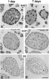
Differentiation of endothelial cell spheroids (A and B) compared with spheroids generated from esophageal epithelial cells (C and D) and C6 glioma cells (E and F). (A) 1-d BAEC spheroid with surface layer of elongated cells and a core of unorganized cells and apoptotic bodies. (B) 7-d BAEC spheroids with surface monolayer and an almost acellular core. (C) 1-d KOP cell spheroid. (D) 7-d KOP cell spheroids with multilayered elongated surface cells and a core of unorganized cells with numerous apoptotic bodies. (E) 1-d C6 glioma spheroid. (F) 7-d C6 glioma spheroids with a necrotic center and a peripheral ring of viable, but undifferentiated surface cells. Bar, 50 μm.
Figure 3
Figure 3
Ultrastructural analysis of a 5-d BAEC spheroid. (A) BAEC spheroids consist of a fully differentiated and polarized surface layer of cells and a core of unorganized cells. The surface cells form a continuous monolayer that may develop electron dense strands indicative of tight junctional cell–cell contacts (magnification in B). The center cells undergo apoptosis as evidenced by the presence of numerous apoptotic bodies (magnification in C). Likewise, cells on the spheroid surface that did not integrate into the monolayer undergo apoptosis (A, arrows). Bars: (A and C) 10 μm; (B) 0.5 μm.
Figure 4
Figure 4
Expression of endothelial cell surface adhesion molecules by HUVEC spheroids. (A) Most spheroid EC express the panendothelial cell marker CD31. (B) Expression of ICAM-1 in HUVEC spheroids after stimulation with TNF-α for 24 h. (C) Expression of VCAM-1 in HUVEC spheroids after stimulation with TNF-α for 24 h. (D) Untreated HUVEC spheroids express barely detectable levels of CD34. (E) Expression of CD34 in HUVEC spheroids after stimulation with VEGF for 24 h. (F and G) Expression of CD34 in cross sections of harvested HUVEC monolayer embedded in paraffin as single cells. CD34 expression is downregulated in two-dimensional culture (F). Stimulation of monolayer cultures with VEGF only minimally increases the number of CD34-positive cells (G). Bar, 50 μm.
Figure 5
Figure 5
Summary of expression pattern of endothelial cell adhesion molecules in monolayer culture and spheroids. EC constitutively express CD31. Intensity of expression does not change after cytokine stimulation. Stimulation of EC with IL-1 or TNF-α induces expression of ICAM-1 and VCAM-1, which is in the spheroids limited to the luminal aspect of the surface monolayer. CD34 is only expressed after VEGF stimulation. The induction of CD34 by VEGF is dependent on the differentiation status of the cells and their microenvironment as it is only inducible in spheroid EC and only minimally in monolayer cells.
Figure 6
Figure 6
Analysis of endothelial cell apoptosis in 1-d BAEC spheroids. Apoptosis of EC was detected either by TUNEL staining (A, random spheroids) or by ethidium bromide staining (B, standard spheroid; calcein AM used as surface stain). Both techniques led to similar results. Apoptosis of BAE cells is limited to the cells in the center of the spheroids (A and B) as well as to surface BAE cells that have not integrated into the surface monolayer (A and B, arrows). (C) Treatment of BAEC spheroids with RAD peptides (30 μM) does not affect endothelial cell apoptosis ([E] hematoxylin-stained sectioned RAD-treated spheroid). (D) Treatment of BAEC spheroids with RGD peptides (30 μM) for 24 h leads to a disruption of the integrity of the surface monolayer and induces surface cell apoptosis ([F] hematoxylin-stained sectioned RGD-treated disintegrating BAEC spheroid). Bar, 50 μm.
Figure 7
Figure 7
Quantitative (A and B) and qualitative (C–F) analysis of endothelial cell apoptosis (A, C, and D, HUVE cells; B, E, and F, BAE cells) after treatment with different cytokines. (A and B) A defined number of spheroids was stimulated with different cytokines for 24 h and the presence of nucleosomal fragmentation products was quantitated by ELISA (see Materials and Methods). Data are expressed as percentage deviation of the level of apoptosis from the untreated control spheroid population (shown in C [HUVEC] and E [BAEC]). The figure shows the mean ± SEM of three different experiments performed in duplicate (*P < 0.05; **P < 0.01). (C) control HUVEC spheroid; (D) VEGF + FGF-2 stimulated HUVEC spheroid. (E) control BAEC spheroid treated with isotype matched nonneutralizing monoclonal antibody to FGF-2 (4 μg/ml). (F) BAEC spheroid treated with neutralizing monoclonal antibody to FGF-2 [4 μg/ml]). Bar, 50 μm.
Figure 7
Figure 7
Quantitative (A and B) and qualitative (C–F) analysis of endothelial cell apoptosis (A, C, and D, HUVE cells; B, E, and F, BAE cells) after treatment with different cytokines. (A and B) A defined number of spheroids was stimulated with different cytokines for 24 h and the presence of nucleosomal fragmentation products was quantitated by ELISA (see Materials and Methods). Data are expressed as percentage deviation of the level of apoptosis from the untreated control spheroid population (shown in C [HUVEC] and E [BAEC]). The figure shows the mean ± SEM of three different experiments performed in duplicate (*P < 0.05; **P < 0.01). (C) control HUVEC spheroid; (D) VEGF + FGF-2 stimulated HUVEC spheroid. (E) control BAEC spheroid treated with isotype matched nonneutralizing monoclonal antibody to FGF-2 (4 μg/ml). (F) BAEC spheroid treated with neutralizing monoclonal antibody to FGF-2 [4 μg/ml]). Bar, 50 μm.
Figure 8
Figure 8
Effect of long-term treatment (4 d) of EC spheroids with the survival factors VEGF and FGF-2 and a neutralizing monoclonal antibody to FGF-2. Cytokines and antibodies were added at the beginning of the experiment and again after 2 d. Spheroids were analyzed after 4 d. (A) control HUVEC spheroid. (B) Control BAEC spheroid. (C) HUVEC spheroid treated with VEGF (50 ng/ml) and FGF-2 (30 ng/ml). (D) BAEC spheroid treated with a neutralizing monoclonal antibody to FGF-2 (4 μg/ml). Bars: (A and C) 30 μm; (B and D) 50 μm.
Figure 9
Figure 9
Analysis of the fate of single nonadherent EC and of their responsiveness to exogenously administered survival factors. The experiments shown were performed with HUVEC, similar results were obtained with BAE cells. (A) HUVEC were grown at low seeding density in nonadhesive tissue culture dishes in the presence of high concentrations of methocel to prevent cellular aggregation (see Materials and Methods). The cells were transferred to adhesive tissue culture dishes after the indicated periods of time and the number of adherent cells per microscopic field of view (MFV) was quantitated after 2 h. Adherence was quantitated by counting the cells that had adhered as single cells (gray bars) and the cells that had adhered in clustered groups that was indicative of cellular aggregation in the methocel medium (black bar). The data shown represent the mean ± SD of three independent experiments performed in triplicate. (B) The reduction of single cell readhesiveness as shown in A is paralleled by a pronounced increase of single cell apoptosis as evidenced by intense DNA laddering. (C) Quantitation of nucleosomal fragmentation by ELISA (see Materials and Methods) to analyze the responsiveness of single suspended HUVEC to exogenously administered survival factors.
Figure 10
Figure 10
Analysis of the effect of cytochalasin D treatment on VEGF and FGF-2 mediated survival factor activities in HUVEC spheroids (quantitation by ELISA; see Materials and Methods; results [mean ± SD of three independent experiments performed in duplicate] are expressed in% of the maximum). (A) Cytochalasin D treatment (5 μg/ml) does not affect apoptosis of aggregated EC. (B) Control HUVEC spheroid. (C) HUVEC aggregation after cytochalasin D treatment. Cytochalasin D–treated HUVEC are responsive to the survival factor activities of VEGF and FGF-2. Treatment of cells with EGTA (5 mM, 24 h) disrupts calcium-dependent cell–cell contacts resulting in highest levels of apoptosis. In contrast to cytochalasin–treated aggregated cells with cell– cell contacts, EGTA-treated single cells are not responsive to VEGF and FGF-2.
Figure 11
Figure 11
Model of endothelial cell organization and differentiation in three-dimensional spheroids. Single suspended EC are neither capable to survive nor responsive to the activities of survival factors and die by apoptosis (anoikis; apoptosis as a consequence of a loss of matrix anchorage). EC that aggregate to form three-dimensional spheroids establish cell–cell contacts that renders the cell survival factor responsive. Subsequently, nontransformed EC in spheroids organize to establish a two compartment system consisting of a peripheral surface monolayer of differentiated, polarized, survival factor–independent cells, and a core of survival factor–dependent unorganized cells. If not continuously exposed to survival factors, the center EC will die by apoptosis to produce a three-dimensional spheroid that consists of an acellular core and a differentiated surface monolayer of EC.

References

    1. Alon T, Hemo I, Itin A, Pe'er J, Stone J, Keshet E. Vascular endothelial growth factor acts as a survival factor for newly formed retinal vessels and has implications for retinopathy of prematurity. Nat Med. 1995;1:1024–1028. - PubMed
    1. Arras M, Mollnau H, Strasser R, Wenz R, Ito WD, Schaper J, Schaper W. The delivery of angiogenic factors to the heart by microsphere therapy. Nat Biotechnol. 1998;16:159–162. - VSports - PubMed
    1. Asahara T, Bauters C, Zheng LP, Takeshita S, Bunting S, Ferrara N, Symes JF, Isner JM. Synergistic effect of vascular endothelial growth factor and basic fibroblast growth factor on angiogenesis in vivo. Circulation. 1995;92:II365–II371. - "V体育ios版" PubMed
    1. Asahara T, Murohara T, Sullivan A, Silver M, van der Zee R, Li T, Witzenbichler B, Schatteman G, Isner JM. Isolation of putative progenitor endothelial cells for angiogenesis. Science. 1997;275:965–967. - PubMed (VSports手机版)
    1. Augustin HG, Voss AK, Pauli BU. Senescence of aortic endothelial cells in culture: effects of basic fibroblast growth factor expression on cell phenotype, migration, and proliferation. J Cell Physiol. 1993;157:279–288. - PubMed

Publication types

MeSH terms

Substances