Skip to main page content
U.S. flag

An official website of the United States government

Dot gov

The . gov means it’s official. Federal government websites often end in . gov or . mil. Before sharing sensitive information, make sure you’re on a federal government site. VSports app下载.

Https

The site is secure. The https:// ensures that you are connecting to the official website and that any information you provide is encrypted and transmitted securely. V体育官网.

Clinical Trial
. 2025 Sep 26;13(9):e012446.
doi: 10.1136/jitc-2025-012446.

Addition of oncolytic virotherapy to clinical isolated limb perfusion for melanoma and sarcoma activates antitumor immunity

Affiliations
Clinical Trial

Addition of oncolytic virotherapy to clinical isolated limb perfusion for melanoma and sarcoma activates antitumor immunity

Andrew J Hayes et al. J Immunother Cancer. .

Abstract

Background: We previously showed that oncolytic virotherapy delivered by isolated limb perfusion (ILP), combined with immune checkpoint inhibition, prevents both local tumor progression and systemic metastases in an animal sarcoma model VSports手机版. .

Methods: We describe a first-in-human phase I/II trial combining oncolytic herpes simplex virus, talimogene laherparepvec (T-VEC), with melphalan and tumor necrosis factor-alpha delivered by ILP, in patients with locally advanced sarcoma or melanoma. V体育安卓版.

Results: T-VEC/ILP is well tolerated, with an overall response rate of 53% in all patients and 44% in sarcoma. Importantly, we report durable complete responses in sarcoma subtypes usually unresponsive to ILP. Translational analysis of longitudinal tumor and blood samples showed that T-VEC induced an inflammatory gene expression profile within injected tumors, which was more sustained in sarcoma than in melanoma. In relation to clinical outcome, responding patients with sarcoma showed a greater increase in gene expression for interferon response after virus treatment than non-responding patients V体育ios版. Analysis of the T-cell repertoire (TCR) in tumor and blood showed that clonality was higher in the tumor, but lower in the blood, in responders following virotherapy, suggesting that virus treatment may expand intratumoral T-cell clones that recognize tumor and/or viral antigens. Increased TCR diversity in the blood was suggestive of a systemic immune response. .

Conclusions: These clinical and translational findings support the further development of oncolytic virotherapy/ILP combinations to activate both systemic and local antitumor immunity, including in tumor types such as sarcoma, which are largely refractory to current treatment with immunotherapy VSports最新版本. .

Keywords: Abscopal; Oncolytic virus; Solid tumor; Surgery. V体育平台登录.


Conflict of interest statement

Competing interests: No, there are no competing interests.

Figures

Figure 1
Figure 1. Clinical trial design. The seroconversion dose of T-VEC was given at 4–6 weeks before ILP, the first treatment T-VEC dose was given 2–3 weeks before ILP and the final treatment dose of T-VEC was given on the day of surgery after induction of anesthesia but before the ILP. Tumor biopsies and blood samples for translational analyses were taken before the seroconversion T-VEC dose (baseline), on the day of surgery after the second T-VEC dose had been administered but before the ILP (pre-ILP) and at 2 weeks after the ILP (post-ILP). A final blood sample only was taken 12 weeks after ILP. ILP, isolated limb perfusion; IT. Intra-tumoural; TNF, tumor necrosis factor; T-VEC, talimogene laherparepvec.
Figure 2
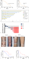
Figure 2. Clinical outcomes of patients undergoing talimogene laherparepvec isolated limb perfusion treatment. (A) Kaplan-Meier curve of overall survival (B) Kaplan-Meier curve of progression-free survival. (C) Swimmers plot displaying individual patient trajectories over time. (D) Waterfall plot showing maximal tumor size fold change from baseline. The dotted lines represent iRECIST criteria for disease progression (>20% increase in size) and partial response (>30% decrease in size); *indicates patients who developed metastatic disease. CR denotes complete response, PR denotes partial response, SD denotes stable disease, PD denotes progressive disease. (E) Clinical photographs of two patients with complete response to treatment at 3 years compared with baseline, arrows indicate lesions before treatment and absence of lesions at 3 years. (F) Kaplan-Meier curves categorized by response to treatment showing overall survival (median survival in non-responder 30 months, not reached in responders). (G) Progression-free survival (median PFS survival in non-responder 7 months, 26 months in responders). iRECIST, immune Response Evaluation Criteria in Solid Tumours.
Figure 3
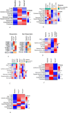
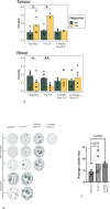
Figure 3. Oncolytic virotherapy and ILP induce transcriptional changes in the tumor and systemic circulation. Tumor biopsy and blood specimens were taken as illustrated in figure 1. (A) Tumor immune cell populations estimated from RNA sequencing transcriptional profiling of tumor biopsies in all patients using human microenvironment cell population counter. (B) Data from (A), subdivided into responders and non-responders. (C) Changes in expression levels of exemplar immune genes over time in responders and non-responders. (D) Transcriptomic deconvolution to demonstrate immune cell composition in peripheral blood in all patients, and (E) comparing responders and non-responders. Tumor immune cell composition over time in melanoma (F) and sarcoma (G) patients. ILP, isolated limb perfusion; NK, natural killer; T-VEC, talimogene laherparepvec.
Figure 4
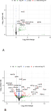
Figure 4. Differentially expressed genes between sarcoma responders and non-responders. (A) Volcano plot of differentially expressed genes between responders and non-responders in patients with sarcoma at baseline (A) and pre-isolated limb perfusion (B). Positive log2 fold change indicates overexpression in responding patients. Genes marked in red pass significance thresholds and the most significant are labeled. FC, fold change; NS, non significant.
Figure 5
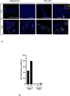
Figure 5. CD8+ T cells higher in selected sarcoma responder versus selected sarcoma non-responder. Patient T007 (responder) and T004 (non-responder) were selected for analysis by mIF. (A) Multiplex immunofluorescence staining of formalin fixed paraffin embedded whole slides for baseline (TP1) and pre-ILP directly following talimogene laherparepvec treatment (TP2). Full slide (left panel) with a selected region (white box) for magnified view (right panel). CD8 (yellow) and DAPI (blue). (B) Quantification of CD8+ T-cell mIF staining. CD8+ T-cell infiltration is shown as normalized cell density (CD8 T-cell count per mm2). ILP, isolated limb perfusion.
Figure 6
Figure 6. TCR sequencing showing changes in tumor microenvironment and systemic antitumor immunity in responders compared with non-responders. (A) TCR sequencing—clonality (1-normalized Shannon Index) of the peripheral and systemic TCR repertoire across time points in responders and non-responders. (B) Triplet CDR3 amino acid kernel clustering of the intratumoral TCR repertoire. (C) Quantification of cluster sizes from (B); green responders, pink non-responders. ILP, isolated limb perfusion; TCR, T-cell repertoire.
Figure 7
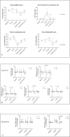
Figure 7. Immune cell activation. (A) Full blood count parameters over time in all patients. Peripheral blood mononuclear cells were surface-stained with anti-CD45, CD3, CD19, CD4, CD8, CD56, CD25, CD127, CD69 and PD-1 antibodies for 30 min at 4°C and analyzed by flow cytometry. Graphs show fold-change versus baseline in percent CD69 (B) and PD-1 (C)-positive cells in the indicated population. P values for responders versus non-responders patients shown for the indicated time point by two-way analysis of variance followed by Šídák multiple comparison test. ILP, isolated limb perfusion; PD-1, programmed cell death protein-1; R, Responder; NR, Non-responder ; WBC, white blood cell.

References

    1. Eggermont AM, Schraffordt Koops H, Klausner JM, et al. Isolated limb perfusion with tumor necrosis factor and melphalan for limb salvage in 186 patients with locally advanced soft tissue extremity sarcomas. The cumulative multicenter European experience. Ann Surg. 1996;224:756–64. doi: 10.1097/00000658-199612000-00011. - V体育2025版 - DOI - PMC - PubMed
    1. Eggermont AMM, de Wilt JHW, ten Hagen TLM. Current uses of isolated limb perfusion in the clinic and a model system for new strategies. Lancet Oncol. 2003;4:429–37. doi: 10.1016/s1470-2045(03)01141-0. - DOI - PubMed
    1. Hayes AJ, Coker DJ, Been L, et al. Technical considerations for isolated limb perfusion: A consensus paper. Eur J Surg Oncol. 2024;50:108050. doi: 10.1016/j.ejso.2024.108050. - DOI - PubMed
    1. Smith HG, Hayes AJ. The role of regional chemotherapy in the management of extremity soft tissue malignancies. Eur J Surg Oncol. 2016;42:7–17. doi: 10.1016/j.ejso.2015.08.165. - "V体育ios版" DOI - PubMed
    1. Deroose JP, Eggermont AMM, van Geel AN, et al. Long-term results of tumor necrosis factor alpha- and melphalan-based isolated limb perfusion in locally advanced extremity soft tissue sarcomas. J Clin Oncol. 2011;29:4036–44. doi: 10.1200/JCO.2011.35.6618. - V体育官网入口 - DOI - PubMed

"VSports注册入口" Publication types

MeSH terms

LinkOut - more resources