Skip to main page content
U.S. flag

An official website of the United States government

Dot gov

The . gov means it’s official. Federal government websites often end in . gov or . mil. Before sharing sensitive information, make sure you’re on a federal government site. VSports app下载.

Https

The site is secure V体育官网. The https:// ensures that you are connecting to the official website and that any information you provide is encrypted and transmitted securely. .

. 2025 Jan 22;482(2):73-86.
doi: 10.1042/BCJ20240569.

Inhibition of RIPK1 or RIPK3 kinase activity post ischemia-reperfusion reduces the development of chronic kidney injury

Affiliations

Inhibition of RIPK1 or RIPK3 kinase activity post ischemia-reperfusion reduces the development of chronic kidney injury

V体育ios版 - Aspasia Pefanis et al. Biochem J. .

Abstract

Ischemia-reperfusion injury (IRI) occurs when the blood supply to an organ is temporarily reduced and then restored. Kidney IRI is a form of acute kidney injury (AKI), which often progresses to kidney fibrosis VSports手机版. Necroptosis is a regulated necrosis pathway that has been implicated in kidney IRI. Necroptotic cell death involves the recruitment of the RIPK1 and RIPK3 kinases and the activation of the terminal effector, the mixed lineage kinase domain-like (MLKL) pseudokinase. Phosphorylated MLKL causes cell death by plasma membrane rupture, driving 'necroinflammation'. Owing to their apical role in the pathway, RIPK1 and RIPK3 have been implicated in the development of kidney fibrosis. Here, we used a mouse model of unilateral kidney IRI to assess whether the inhibition of RIPK1 or RIPK3 kinase activity reduces AKI and the progression to kidney fibrosis. Mice treated with the RIPK1 inhibitor Nec-1s, either before or after IR, showed reduced kidney injury at 24 hr compared with controls, whereas no protection was offered by the RIPK3 inhibitor GSK´872. In contrast, treatment with either inhibitor from days 3 to 9 post-IR reduced the degree of kidney fibrosis at day 28. These findings further support the role of necroptosis in IRI and provide important validation for the contribution of both RIPK1 and RIPK3 catalytic activities in the progression of kidney fibrosis. Targeting the necroptosis pathway could be a promising therapeutic strategy to mitigate kidney disease following IR. .

Keywords: acute kidney injury; ischemia-reperfusion; kidney fibrosis; kinase inhibitor injury; programmed necrosis. V体育安卓版.


Conflict of interest statement

All authors contribute, or have contributed, to a project developing necroptosis pathway inhibitors in collaboration with Anaxis Pharma Pty Ltd. JMM and PJC have received research funding from Anaxis Pharma Pty Ltd V体育ios版. FI is Chair of the SVHM Animal Ethics Committee, declared a conflict of interest and removed himself from any decision processes for the protocols 026/17 and 022/20 used in the present study.

Figures

Figure 1
Figure 1. Mouse models of unilateral kidney ischemia-reperfusion injury.
(a) Model of acute kidney injury (AKI) following ischemia-reperfusion. Nec-1s, GSK´872 or a vehicle control were injected 30 min prior to ischemia, with sample collection at 24 hr. (b) Model of AKI in which Nec-1s was administered 15 min after reperfusion. (c) Model of chronic kidney injury following IR. Mice received Nec-1s, GSK´872 or a vehicle control daily on days 3–9 after IR, with sample collection at day 28. R, right; L, left.
Figure 2
Figure 2. Prophylactic treatment with Nec-1s, but not GSK´872, protects against kidney ischemia reperfusion injury at 24 hr.
(a) Serum creatinine; (b) tubular injury score; (c) representative PAS-stained kidney sections, with arrows indicating necrotic tubules (image obtained using Olympus UPlanApo 40× objective lens; 0,85 N.A. Scale bar 50 μm). **P<0.01, ***P<0.001 compared with sham procedure; ##P<0.01 compared with vehicle treatment. Data presented as mean ± SEM. N=4–8 per group. PAS, periodic acid-Schiff.
Figure 3
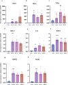
Figure 3. Up-regulation of gene expression of kidney injury marker, pro-inflammatory and necroptosis pathway components at 24 hr after kidney IR is unaffected by prophylactic treatment with Nec-1s or GSK´872.
(a) Kim1; (b) Ngal; (c) Tnfa; (d) Mip2; (e) Il6; (f) Ripk1; (g) Ripk3; (h) Mlkl. **P<0.01, ***P<0.001, ****P<0.0001 compared with sham procedure; #P<0.05, ##P<0.01 compared with vehicle treatment. Data presented as mean ± SEM . n=6–8 per group.
Figure 4
Figure 4. Therapeutic treatment with Nec-1s after ischemia-reperfusion reduces kidney injury at 24 hr.
(a) Serum creatinine; (b) tubular injury score; (c) representative PAS-stained kidney sections following vehicle or Nec-1s treatment with arrows indicating necrotic tubules (image obtained using Olympus UPlanApo 40× objective lens; 0,85 N.A. Scale bar 50 μm). *P 0.05, **P<0.01 compared with sham procedure; #P<0.05 compared with vehicle treatment. Data presented as mean ± SEM. n=4–6 per group. PAS, periodic acid-Schiff.
Figure 5
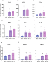
Figure 5. Therapeutic treatment with Nec-1s does not affect gene expression of kidney injury, pro-inflammatory or necroptosis pathway genes at 24 hr following reperfusion.
(a) Kim1; (b) Ngal; (c) Tnfa; (d) Mip2; (e) Il6; (f) IL33; (g) Ripk1; (h) Ripk3; (i) Mlkl. *P<0.05, **P<0.01 compared with sham procedure. Data presented as mean ± SEM. n=4–6 per group.
Figure 6
Figure 6. Inhibition of RIPK1 or RIPK3 after IRI is established reduces kidney fibrosis at day 28.
(a) Kidney fibrosis score at 28 days with representative images of Mason Trichrome staining in each group (image obtained using Olympus UPlanApo 20× objective lens. Scale bar 20 μm). **P<0.01 compared with sham procedure; ##P<0.01 compared with vehicle treatment. Data presented as mean ± SEM. n=5–8 per group.
Figure 7
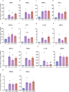
Figure 7. The expression of kidney injury, fibrosis, pro inflammatory and necroptosis pathway genes is up-regulated at day28 post-reperfusion.
(a) Kim1; (b) Ngal; (c) Tgfb; (d) Col1; (e) HIF1a; (f) Et1; (g) Il1b; (h) Mip2; (i) Mcp1; (j) Tnfa; (k) Il10; (l) Ripk1; (m) Ripk3; (n) Mlkl. **P<0.01 compared with sham procedure; #P<0.05, ##P<0.01 compared with vehicle treatment. Data presented as mean ± SEM. n=5–8 per group.

"V体育ios版" References

    1. Bonventre J.V., Yang L. Cellular pathophysiology of ischemic acute kidney injury. J. Clin. Invest. 2011;121:4210–4221. doi: 10.1172/JCI45161. - DOI - PMC - PubMed
    1. Kalogeris T., Baines C.P., Krenz M., Korthuis R.J. Cell biology of ischemia/reperfusion injury. Int. Rev. Cell Mol. Biol. 2012;298:229–317. doi: 10.1016/B978-0-12-394309-5.00006-7. - DOI - PMC - PubMed
    1. Murphy E., Steenbergen C. Ion transport and energetics during cell death and protection. Physiol. (Bethesda). 2008;23:115–123. doi: 10.1152/physiol.00044.2007. - DOI - PMC - PubMed
    1. Kako K., Kato M., Matsuoka T., Mustapha A. Depression of membrane-bound Na+-K+-ATPase activity induced by free radicals and by ischemia of kidney. Am. J. Physiol. Cell Physiol. 1988;254:C330–C337. doi: 10.1152/ajpcell.1988.254.2.C330. - DOI (V体育平台登录) - PubMed
    1. Perico N., Cattaneo D., Sayegh M.H., Remuzzi G. Delayed graft function in kidney transplantation. The Lancet. 2004;364:1814–1827. doi: 10.1016/S0140-6736(04)17406-0. - DOI - PubMed

Publication types (V体育官网)

MeSH terms

Substances

LinkOut - more resources