Skip to main page content
U.S. flag

An official website of the United States government

Dot gov

The . gov means it’s official. Federal government websites often end in VSports app下载. gov or . mil. Before sharing sensitive information, make sure you’re on a federal government site. .

Https

The site is secure. The https:// ensures that you are connecting to the official website and that any information you provide is encrypted and transmitted securely. V体育官网.

. 2023 May 9;120(19):e2300706120.
doi: 10.1073/pnas.2300706120. Epub 2023 May 1.

Angiotensin II receptor inhibition ameliorates liver fibrosis and enhances hepatocellular carcinoma infiltration by effector T cells

Affiliations

Angiotensin II receptor inhibition ameliorates liver fibrosis and enhances hepatocellular carcinoma infiltration by effector T cells

Li Gu et al. Proc Natl Acad Sci U S A. .

V体育2025版 - Abstract

Although viral hepatocellular carcinoma (HCC) is declining, nonviral HCC, which often is the end stage of nonalcoholic or alcoholic steatohepatitis (NASH, ASH), is on an upward trajectory. Immune checkpoint inhibitors (ICIs) that block the T cell inhibitory receptor PD-1 were approved for treatment of all HCC types. However, only a minority of HCC patients show a robust and sustained response to PD-1 blockade, calling for improved understanding of factors that negatively impact response rate and duration and the discovery of new adjuvant treatments that enhance ICI responsiveness. Using a mouse model of NASH-driven HCC, we identified peritumoral fibrosis as a potential obstacle to T cell-mediated tumor regression and postulated that antifibrotic medications may increase ICI responsiveness. We now show that the angiotensin II receptor inhibitor losartan, a commonly prescribed and safe antihypertensive drug, reduced liver and peritumoral fibrosis and substantially enhanced anti-PD-1-induced tumor regression VSports手机版. Although losartan did not potentiate T cell reinvigoration, it substantially enhanced HCC infiltration by effector CD8+ T cells compared to PD-1 blockade alone. The beneficial effects of losartan correlated with blunted TGF-β receptor signaling, reduced collagen deposition, and depletion of immunosuppressive fibroblasts. .

Keywords: NASH-driven HCC; anti-PD-1; liver fibrosis; losartan V体育安卓版. .


Conflict of interest statement

M. K. is the founder and stockholder in Elgia Pharmaceuticals V体育ios版. All other authors declare no competing interest.

VSports在线直播 - Figures

Fig. 1.
Fig. 1.
Losartan potentiates anti-PD-1-induced regression of NASH-driven HCC. (A) Gross liver morphology in HFD-fed MUP-uPA mice that were treated with ctrl IgG, anti-PD-1, losartan+ctrl IgG, and losartan+anti-PD-1 (n = 13 to 15). (B) Liver/body weight ratio in the above mice at the end of the treatments. (C and D) Tumor multiplicity (C) and volume (D) at the end of treatments. Data are presented as mean ± SEM. *P < 0.05, **P < 0.01, ***P < 0.001, ns, not significant (unpaired two-tailed t test and Mann–Whitney U test were used to determine significance).
Fig. 2.
Fig. 2.
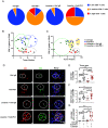
Losartan stimulates intratumoral infiltration by reinvigorated Teff cells. (A) MUP-uPA mice from each group in Fig. 1A were classified into three categories based on the number of CD8+ T cells in their livers (low < 1 million; medium 1 to 4.6 million; high > 4.6 million). The percentages of mice in each category are indicated (n = 13 to 15). (B and C) Correlation between liver CD8+ T cell frequency and treatment outcome determined as either tumor multiplicity (B) or tumor volume (C). (D–G) Frozen liver sections were stained for CD3 and CD8 and counterstained with DAPI. T-tumor. Scale bars, 10 µm (D). Quantification of CD8+CD3+ T cells (E), CD3+CD8- helper “CD4” T cells (F), and CD3CD8+ pDC cells (G) into tumors is based on cell quantitation per high-magnification field (HMF) in tumor and nontumor areas by Image J analysis of 10 fields of per section. Data are presented as mean ± SEM. *P < 0.05, **P < 0.01, ***P < 0.001, ****P < 0.0001, ns, not significant (unpaired two-tailed t test and Mann–Whitney U test).
Fig. 3.
Fig. 3.
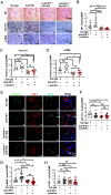
Losartan inhibits NASH-related liver fibrosis. (A) Formalin-fixed paraffin-embedded (FFPE) liver sections were stained with Sirius Red (SR) (Top), collagen I alpha 1 chain (COL1A1) (Middle), and α-SMA (Bottom) antibodies. (Scale bars, 50 µm) T, tumor. (B–D) SR (B), COL1A1 (C), and α-SMA (D) staining intensities per HMF were determined by Image J quantitation of 10 fields per section. (E) Frozen liver sections were stained for COL1A1, FSP1, and α-SMA and examined by fluorescence microscopy. (Scale bars, 10 µm.) Experiments were repeated at least three times. (F–H) Quantification of COL1A1+ (F), α-SMA+ (G), and FSP1+ (H) areas per HMF. Data are presented as mean ± SEM. *P < 0.05, **P < 0.01, ***P < 0.001, ns, not significant (unpaired two-tailed t test and Mann–Whitney U test).
Fig. 4.
Fig. 4.
Losartan promotes stromal remodeling by inhibiting TGF-β expression and signaling. (A) IHC staining of TGF-β1 (Left) and phosphorylated (P)-ERK1/2 (Right) in FFPE liver sections from MUP-uPA mice in the different treatment groups. (Scale bars, 50 µm.) (B) TGF-β1 (Top) and P-ERK1/2 (Bottom) staining intensity per HMF determined by Image J. (C) Immunoblot analysis of the indicated proteins in liver lysates of mice from the indicated treatment groups. (D) Quantification of IL6+α-SMA+ area per HMF by Image J analysis of the data in SI Appendix, Fig. S4E. (E–G) IHC staining of p21 and p16 in FFPE liver sections from the indicated treatment groups (E). Scale bars, 20 µm. p21 (F)- and p16 (G)-positive cells per HMF determined by Image J. Data are presented as mean ± SEM. *P < 0.05, **P < 0.01, ***P < 0.001, ****P < 0.0001, ns, not significant (unpaired two-tailed t test and Mann–Whitney U test).

Update of

References

    1. Llovet J. M., et al. , Hepatocellular carcinoma. Nat. Rev. Dis. Primers 7, 6 (2021). - PubMed
    1. Baglieri J., Brenner D. A., Kisseleva T., The role of fibrosis and liver-associated fibroblasts in the pathogenesis of hepatocellular carcinoma. Int. J. Mol. Sci. 20, 1723 (2019). - PMC - PubMed
    1. Schwabe R. F., Luedde T., Apoptosis and necroptosis in the liver: A matter of life and death. Nat. Rev. Gastroenterol. Hepatol. 15, 738–752 (2018). - PMC - PubMed
    1. Karin M., New insights into the pathogenesis and treatment of non-viral hepatocellular carcinoma: A balancing act between immunosuppression and immunosurveillance. Precis. Clin. Med. 1, 21–28 (2018). - PMC - PubMed
    1. Chidambaranathan-Reghupaty S., Fisher P. B., Sarkar D., Hepatocellular carcinoma (HCC): Epidemiology, etiology and molecular classification. Adv. Cancer Res. 149, 1–61 (2021). - PMC - PubMed

Publication types

"V体育2025版" MeSH terms

Substances (V体育2025版)