Skip to main page content
U.S. flag

An official website of the United States government

Dot gov

The . gov means it’s official. Federal government websites often end in . gov or . mil VSports app下载. Before sharing sensitive information, make sure you’re on a federal government site. .

Https

The site is secure. The https:// ensures that you are connecting to the official website and that any information you provide is encrypted and transmitted securely V体育官网. .

. 2022 Oct 6;11(19):3139.
doi: 10.3390/cells11193139.

"VSports在线直播" Necrostatin-1 Alleviates Lung Ischemia-Reperfusion Injury via Inhibiting Necroptosis and Apoptosis of Lung Epithelial Cells

Affiliations

Necrostatin-1 Alleviates Lung Ischemia-Reperfusion Injury via Inhibiting Necroptosis and Apoptosis of Lung Epithelial Cells

Lingjun Dong et al. Cells. .

"VSports手机版" Abstract

Lung ischemia-reperfusion injury (LIRI) is associated with many diseases, including primary graft dysfunction after lung transplantation, and has no specific and effective therapies. Necroptosis contributes to the pathogenesis of ischemia-reperfusion injury. Necrostatin-1 (Nec-1), the necroptosis inhibitor targeting RIPK1, has been reported to alleviate ischemia-reperfusion injury in various organs VSports手机版. However, the underlying mechanism of Nec-1 in LIRI remains unclear. In this paper, an in vivo LIRI model was built up by left lung hilar clamping in mice, and an in vitro cold ischemia-reperfusion (CI/R) model using BEAS-2B cells was applied to mimic the lung transplantation setting. We found Nec-1 significantly alleviated ischemia-reperfusion-induced lung injury, cytokine releasing, and necroptosis of epithelial cells in mouse lungs. In vitro, Nec-1 also mitigated CI/R-induced cell death and inflammatory responses in BEAS-2B cells, and these protective effects were achieved by simultaneously inhibiting the formation of necrosome and RIPK1-dependent apoptosis. However, Nec-1 decreased the necrosome number but increased the apoptosis level in lung tissues after ischemia reperfusion. We further clarified that Nec-1 could also attenuate lung injury by promoting neutrophil apoptosis from flow cytometry. In conclusion, Nec-1 alleviated lung ischemia-reperfusion injury by inhibiting necroptosis and apoptosis of epithelial cells and promoting the apoptosis of neutrophils. Thus, Nec-1 could be a promising medication against primary graft dysfunction after lung transplantation. .

Keywords: apoptosis; lung epithelial cells; lung ischemia-reperfusion injury; necroptosis; necrostatin-1. V体育安卓版.


Conflict of interest statement

The authors declare no conflict of interest.

"V体育ios版" Figures

Figure 1
Figure 1
Necrostatin-1 alleviated lung injury and inflammatory response caused by ischemia reperfusion (IR) in mice. The lung ischemia-reperfusion injury (LIRI) was induced by clamping the left pulmonary hilum of the mouse for 1 h using a microvascular clip and then removing the clip for reperfusion for 2 h. Nec-1 (1 mg/kg) was administered intraperitoneal injection 1 h before ischemia. (A) Changes in the gross appearance of mouse lungs and the representative HE staining of the lungs. The area between the two yellow cycle was determined as the perivascular cuff area, and the red line indicated the thickness of the epithelium. (B) The ratio of perivascular cuff area to vessel area. (C) The thickness of the epithelial barrier. (D) Lung injury score. (E) Total protein exudation in the bronchoalveolar lavage fluid (BALF). (F,G) The concentration of TNFα and IL-6 in the BALF detected using ELISA. (H,I) The mRNA level of TNFα and IL-6 determined by real-time PCR. (J) The level of IκB and phosphor-IκB in lung tissue homogenates detected by Western blotting. (K,L) Quantification of proteins from Western blots in panel J. n = 6 animals per group. * p < 0.05, ** p < 0.01, *** p < 0.001, compared with Sham; # p < 0.05, ## p < 0.01, ### p < 0.001, compared with LIRI; one-way ANOVA.
Figure 2
Figure 2
Necrostatin-1 inhibited IR-induced necroptosis of lung epithelial cells in mice. The mouse LIRI was induced by clamping the left pulmonary hilum of the mouse for 1 h using a microvascular clip and then removing the clip for reperfusion for 2 h. Nec-1 (1 mg/kg) was administered intraperitoneal injection 1 h before ischemia. (A) Western blotting of necroptosis-related proteins (RIPK1, phosphor-RIPK1, RIPK3, phosphor-RIPK3, MLKL, and phosphor-MLKL) in lung tissue homogenates. (BG) Quantification of proteins from Western blots in panel A. (H) Immunofluorescence staining of phosphor-MLKL and SPC (a marker of lung epithelial cell) in peripheral bronchial epithelium (upper panel) and alveolar epithelium (lower panel). White arrows indicated that pMLKL was localized at airway or alveolar epithelial cells. (I) Percentage of SPC and pMLKL double-positive cells in SPC positive cells. (J) The concentration of HMGB1 in BALF detected using ELISA. (K) Immunohistochemical staining of phosphor-MLKL in different groups. Black arrows indicated the pMLKL-positive areas in the bronchial epithelium. (L) The ratio of the pMLKL-positive area to the whole lung tissue area. n = 6 animals per group. * p < 0.05, ** p < 0.01, *** p < 0.001, compared with Sham; # p < 0.05, ## p < 0.01, ### p < 0.001, compared with LIRI; one-way ANOVA.
Figure 3
Figure 3
Necrostatin-1 mitigated cold ischemia and reperfusion (CI/R)-induced cell death and production of inflammatory cytokines in BEAS-2B cells. BEAS-2B cells were incubated in a 4 °C chamber filled with 50% oxygen for 18 h with ice-cold lung preservation Perfadex® solution to simulate the procedure of clinical donor lung preservation and then subjected to a warm culture medium to mimic reperfusion. Necrostatin-1 (Nec-1, 30 μM) was added to the medium at the beginning of cold-ischemia and sustained during reperfusion. (A) Cell experiment protocol. (B) Morphological changes of BEAS-2B cells in different groups. (C) Cell viability determined using trypan blue exclusive staining. (D) LDH in the cell culture medium. (E) The distribution of HMGB1 in the cells observed by immunofluorescent staining. (F) The counts of cells undergoing HMGB1 cytoplasmic translocation. The percentage of the cells with HMGB1 cytoplasmic translocation to total cells was calculated. (G) The percentage of the optical density of HMGB1 in the cytoplasm to that of the whole cell. (HJ) The level of HMGB1, TNFα and IL-6 in the cell culture supernatant determined using ELISA. (K,L) The mRNA level of TNFα and IL-6 detected using real-time PCR. Three independent experiments were performed. * p < 0.05, ** p < 0.01, *** p < 0.001, compared with Control; # p < 0.05, ## p < 0.01, ### p < 0.001, compared with CI/R. One-way ANOVA.
Figure 4
Figure 4
Necrostatin-1 reduced CI/R-induced cell death through the inhibition of RIPK1-dependent apoptosis and necroptosis in BEAS-2B cells. BEAS-2B cells were incubated in a 4 °C chamber filled with 50% oxygen for 18 h with ice-cold lung preservation Perfadex® solution to simulate the procedure of clinical donor lung preservation and then subjected to a warm culture medium to mimic reperfusion. Necrostatin-1 (Nec-1, 30 μM) was added to the medium at the beginning of cold-ischemia and sustained during reperfusion. (A) Propidium iodide (PI) and Hoechst staining in BEAS-2B cells after CI/R. (B,C) Quantification of necrotic and apoptotic cells in panel A. (D) Western blotting of necroptosis- and apoptosis-related proteins (RIPK1, phosphor-RIPK1, RIPK3, phosphor-RIPK3, MLKL, phosphor-MLKL, cleaved RIPK-1, and Cleaved caspase 3) in BEAS-2B cells. (EL) Quantification of proteins from Western blots in panel D. (M) The colocalization of RIPK1 and RIPK3 determined by immunofluorescence staining. (N) Cell counting of the RIPK1 and RIPK3 double-positive cells. Three independent experiments were performed. * p < 0.05, ** p < 0.01, *** p < 0.001, compared with Control; # p < 0.05, ## p < 0.01, ### p < 0.001, compared with CI/R. One-way ANOVA.
Figure 5
Figure 5
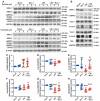
Necrostatin-1 inhibited the formation of necrosome but promoted apoptosis in mouse LIRI. The mouse LIRI was induced by clamping the left pulmonary hilum of the mouse for 1 h using a microvascular clip and then removing the clip for reperfusion for 2 h. Nec-1 (1 mg/kg) was administered intraperitoneal injection 1 h before ischemia. (A) Western blotting of RIPK1, phosphor-RIPK1, RIPK3, and phosphor-RIPK3 in soluble parts (upper panel) and insoluble parts (lower panel) of lung tissue homogenates. n = 3 animals per group. (B) Western blotting of apoptosis-related proteins (cleaved RIPK1, caspase 3, cleaved caspase 3, caspase 8, cleaved caspase 8, and Bax) in lung tissue homogenates. (CH) Quantification of proteins from Western blots in panel B. n = 6 animals per group. * p < 0.05, ** p < 0.01, *** p < 0.001, compared with Sham; # p < 0.05, ## p < 0.01, ### p < 0.001, compared with LIRI; One-way ANOVA.
Figure 6
Figure 6
Necrostatin-1 promoted the apoptosis of neutrophils in mouse LIRI. The mouse LIRI was induced by clamping the left pulmonary hilum of the mouse for 1 h using a microvascular clip and then removing the clip for reperfusion for 2 h. Nec-1 (1 mg/kg) was administered intraperitoneal injection 1 h before ischemia. The left lung tissues were mechanically dissociated and subsequently digested, and then the single-cell suspensions were prepared from lung tissue. (A,B) Gating strategy delineating mouse lung single-cell suspensions. (C) Annexin V and PI staining of single cells prepared from mice lungs. Apoptotic cells were identified by Annexin V positive but PI negative cells. (D) Apoptosis rates of single-cell suspension in lung tissues. (E) Gating strategy delineating lung CD45+ subsets. (F) Percentage of CD45+ cells in total cells isolated from lung tissues. (G) Annexin V and PI staining of CD45+ cells in mice lungs. (H) Apoptosis rates of CD45+ cells. (I) Gating strategy delineating lung neutrophils subsets (CD11b + Ly6G + cells) in lung tissues. (J) Percentage of neutrophils in total cells isolated from lung tissues. (K) Annexin V and PI staining of neutrophils in mice lungs. (L) Apoptosis rates of lung neutrophils. (M) Gating strategy delineating lung macrophage subsets (F4/80 + CD11b + cells). (N) Percentage of macrophages in total cells isolated from lung tissues. (O) Annexin V and PI staining of lung macrophages. (P) Apoptosis rates of lung macrophages. n = 6 animals per group. * p < 0.05, ** p < 0.01, *** p < 0.001, compared with Sham; # p < 0.05, ## p < 0.01, ### p < 0.001, compared with LIRI; one-way ANOVA.

References

    1. Diamond J.M., Arcasoy S., Kennedy C.C., Eberlein M., Singer J.P., Patterson G.M., Edelman J.D., Dhillon G., Pena T., Kawut S.M., et al. Report of the International Society for Heart and Lung Transplantation Working Group on Primary Lung Graft Dysfunction, part II: Epidemiology, risk factors, and outcomes—A 2016 Consensus Group statement of the International Society for Heart and Lung Transplantation. J. Heart Lung Transplant. 2017;36:1104–1113. doi: 10.1016/j.healun.2017.07.020. - DOI - PubMed
    1. Shaver C.M., Ware L.B. Primary graft dysfunction: Pathophysiology to guide new preventive therapies. Expert Rev. Respir. Med. 2017;11:119–128. doi: 10.1080/17476348.2017.1280398. - DOI - PMC - PubMed
    1. Verleden G.M., Glanville A.R., Lease E.D., Fisher A.J., Calabrese F., Corris P.A., Ensor C.R., Gottlieb J., Hachem R.R., Lama V., et al. Chronic lung allograft dysfunction: Definition, diagnostic criteria, and approaches to treatment-A consensus report from the Pulmonary Council of the ISHLT. J. Heart Lung Transplant. 2019;38:493–503. doi: 10.1016/j.healun.2019.03.009. - DOI - PubMed
    1. Chen-Yoshikawa T.F. Ischemia-Reperfusion Injury in Lung Transplantation. Cells. 2021;10:1333. doi: 10.3390/cells10061333. - DOI (VSports) - PMC - PubMed
    1. Capuzzimati M., Hough O., Liu M. Cell death and ischemia-reperfusion injury in lung transplantation. J. Heart Lung Transplant. 2022;41:1003–1013. doi: 10.1016/j.healun.2022.05.013. - "V体育安卓版" DOI - PubMed

Publication types

LinkOut - more resources