<kbd date-time="iugIFmMW"></kbd><del id="15HLw"></del>
Skip to main page content
U.S. flag

An official website of the United States government

Dot gov

The . gov means it’s official. Federal government websites often end in . gov or . mil. Before sharing sensitive information, make sure you’re on a federal government site VSports app下载. .

Https

The site is secure. The https:// ensures that you are connecting to the official website and that any information you provide is encrypted and transmitted securely V体育官网. .

. 2021 Feb 18;184(4):1017-1031.e14.
doi: 10.1016/j.cell.2021.01.016. Epub 2021 Feb 5.

Human gut mycobiota tune immunity via CARD9-dependent induction of anti-fungal IgG antibodies

Affiliations

"VSports最新版本" Human gut mycobiota tune immunity via CARD9-dependent induction of anti-fungal IgG antibodies

"V体育ios版" Itai Doron et al. Cell. .

Abstract

Antibodies mediate natural and vaccine-induced immunity against viral and bacterial pathogens, whereas fungi represent a widespread kingdom of pathogenic species for which neither vaccine nor neutralizing antibody therapies are clinically available VSports手机版. Here, using a multi-kingdom antibody profiling (multiKAP) approach, we explore the human antibody repertoires against gut commensal fungi (mycobiota). We identify species preferentially targeted by systemic antibodies in humans, with Candida albicans being the major inducer of antifungal immunoglobulin G (IgG). Fungal colonization of the gut induces germinal center (GC)-dependent B cell expansion in extraintestinal lymphoid tissues and generates systemic antibodies that confer protection against disseminated C. albicans or C. auris infection. Antifungal IgG production depends on the innate immunity regulator CARD9 and CARD9+CX3CR1+ macrophages. In individuals with invasive candidiasis, loss-of-function mutations in CARD9 are associated with impaired antifungal IgG responses. These results reveal an important role of gut commensal fungi in shaping the human antibody repertoire through CARD9-dependent induction of host-protective antifungal IgG. .

Keywords: B cells; CARD9; CX3CR1 macrophages; Candida albicans; Candida auris; antifungal IgG antibodies; gut fungi; gut-disseminated fungal infections; invasive candidiasis; mycobiome V体育安卓版. .


"VSports最新版本" Conflict of interest statement

Declaration of interests Cornell University has filed a provisional patent application covering inventions described in this manuscript.

Figures

Figure 1.
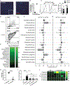
Figure 1.. Systemic IgG antibodies bind gut mycobiota
(A–E) MultiKAP assay. Serum and feces are co-incubated to allow serum IgG to bind to gut microbial epitopes. (A and B) Fecal microbes are distinguished as a Sybrhi population that is absent in similarly stained GF feces (B). (C) Antibody-reactive microbiota (fungi and bacteria) analysis is performed by flow cytometry, and then fungi (SybrhiCFW+) and bacteria (SybrhiCFW+) are enriched in two respective fractions (large and small) through size separation by 900 × g centrifugation. (D) Antibody-bound fungi and bacteria from each sample are sorted by fluorescence-activated cell sorting (FACS), and DNA is isolated and used in downstream applications such as PCR-based quantification and fungal (ITS1) and bacterial (16S) analysis through amplicon-based deep-sequencing. (E) Quantitative PCR-based assessment of the enriched fungal or bacterial population; Mann-Whitney test. CFW+, n = 4; CFW, n = 4. (F and G) Fungi in SPF mouse feces were assessed for IgA, IgG, and IgM binding by multiKAP; no serum incubation (gray), incubation with matched WT SPF mouse serum (black), or B cell-deficient μMT−/− SPF mouse serum (blue). Pooled from two independent experiments; one-way ANOVA followed by Tukey’s multiple comparisons test. −Serum, n = 10; +WT serum, n = 10; +μMT−/− serum, n = 10. (H–K) Fungi in WT SPF mouse feces were assessed for IgG (H and I) and IgM binding (J and K) by multiKAP after incubation with matched WT SPF (black) or WT GF (blue) serum. Mann-Whitney test. SPF, n = 5; GF, n = 5. (L) MultiKAP assay with feces from mice from a dedicated controlled environment room at Weill Cornell Medicine (WCM-CE) and Jackson Laboratory (JAX) or Taconic Bioscience (TAC) facilities with (+serum) or without (−serum) serum co-incubation. Pooled from two independent experiments; one-way ANOVA followed by Tukey’s multiple comparisons test. WCM-CE, n = 7; JAX, n = 9; TAC, n = 7. (M and N) Fungi in SPF WT mouse feces were assessed for IgG1, IgG2b, and IgG3 binding by multiKAP after incubation with matched WT SPF serum. IgG1, n = 5; IgG2b, n = 5; IgG3, n = 5. Error bars indicate SEM. ns, p ≥ 0.05; *p < 0.05; **p < 0.01; ***p < 0.001; ****p < 0.0001. See also Figure S1 and Table S1.
Figure 2.
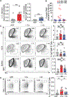
Figure 2.. Enrichment of C. albicans among IgG-coated intestinal microbes identifies the predominant driver of systemic antifungal IgG responses in humans
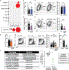
(A and B) Visualization of commensal fungi and bacteria in human feces stained with Sybr Green (DNA), CFW (fungal cells), and the EUB338 FISH probe (bacteria); the scale bar represents 25 μm (A). The percentages of fungal (CFW+Sybr Green+) and bacterial (EUB338+ Sybr Green+) cells in 6 fecal samples from healthy individuals are shown in (B). Each dot represents an individual feces donor. (C) Fungi in feces from healthy human donors were assessed for IgA and IgG binding by multiKAP; no serum incubation (gray), incubation with matched serum (black). Mann-Whitney test, n = 13. (D–G) Combined multiKAP-based sorting and sequencing analysis of antibody-bound species of healthy human mycobiota. (D) Human feces from healthy donors was incubated with matched serum and assessed for IgA and IgG binding by multiKAP. (E) Abundance of the top 15 fungal genera in human fecal samples. (F and G) Relative enrichment of fungal species in antibody-bound fractions, IgG+IgA+ (F) or IgG+IgA (G), expressed relative to the IgGIgA (unbound) population. Confidence interval (CI) values were calculated at the species level. Fungal species represented at 2% or more are depicted. One-sample t test, n = 7. Boxplots represent species’ median and the first to third quartiles, and whiskers represent minimum and maximum values. Starred boxplots indicate fungal species with a mean CI significantly greater or less than 0, indicating preferential enrichment in the antibody-bound or unbound fraction. Non-enriched species are unlabeled. (H) ELISA-based validation of IgG reactivity against fungal lysates from C. albicans (recognized by antifungal antibodies; F and G) or M. restricta (not recognized by antifungal antibodies; F and G) in healthy human serum. Mann-Whitney test. Human sera, n = 10; each dot represents an individual subject. Each dot represents a serum sample from a healthy individual. (I) Serum IgG titers in the serum of germ-free (GF) mice monocolonized with the fungal species identified in Figures 2F and 2G, assessed by ELISA. Serum IgG titers of each group are statistically compared relative to uncolonized controls. One-way ANOVA followed by Tukey’s multiple comparisons test. Data are representative of three independent experiments. GF, n = 6; C. albicans, n = 8; S. cerevisiae, n = 4; M. restricta, n = 6; C. cladosporioides, n = 4. Each dot represents an individual mouse. Error bars indicate SEM. ns, p ≥ 0.05; *p < 0.05; **p < 0.01; ***p < 0.001; ****p < 0.0001. (J) Serum IgG reactivity from untreated (GF) or C. albicans-monocolonized GF mice (Ca-exGF) against lysates from C. albicans, S. cerevisiae, A. amstelodami, M. restricta, and M. globosa pure cultures was measured by ELISA. Squares represent individual mice. Heatmap values represent optical density 405 (OD405) measurements. GF, n = 3; Ca-exGF, n = 5. Error bars indicate SEM. ns, p ≥ 0.05; *p < 0.05; **p < 0.01; ***p < 0.001; ****p < 0.0001. See also Figure S2 and Table S1.
Figure 3.
Figure 3.. Intestinal C. albicans elicits extra-intestinal GC-dependent IgG+ B cell responses
Mycobiota-free altered Schaedler flora (ASF) mice were orally gavaged with PBS (ASF), C. albicans (+Ca), or S. cerevisiae (+Sc). (A) Serum anti-C. albicans IgG antibody production in Ca-colonized ASF mice (A, left) and serum ASCA production in Sc-colonized ASF mice (A, right) compared with ASF controls, as measured by ELISA. (B) qPCR-based 18S rDNA assessment of spleens from ASF mice colonized for 2 weeks with C. albicans or S. cerevisiae or left untreated. (C) Frequency of IgG+IgM among CD19+CD4 B cells in PPs (top), mLNs (center), and spleen (bottom), analyzed by flow cytometry (pooled from two independent experiments). (D) Frequency of GC-B cells among CD19+CD4 B cells in the spleen, analyzed by flow cytometry. One-way ANOVA followed by Tukey’s multiple comparisons test. ASF, n = 8; +Ca, n = 10; +Sc, n = 10. Data are pooled from three independent experiments. Each dot represents an individual mouse. Error bars indicate SEM. ns, p ≥ 0.05; *p < 0.05; **p < 0.01; ***p < 0.001; ****p < 0.0001. See also Figure S3 and Table S1.
Figure 4.
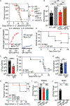
Figure 4.. Antibodies induced through intestinal Candida colonization confer protection against gut-derived and blood-borne systemic candidiasis caused by C. albicans or C. auris
(A and B) WT mice (WT), μMT−/− mice (μMT−/−), and μMT−/− mice adoptively transferred splenic B cells from C. albicans-monocolonized GF mice (mMT−/− +Ca-B) or untreated GF mice (μMT−/− +GF-B) were infected intravenously (i.v.) with C. albicans. A control group of WT mice was left uninfected (WT, NI). Disease morbidity (A) and C. albicans systemic spread to kidney tissue (B) were assessed; log rank Mantel-Cox test (A) and one-way ANOVA followed by Tukey’s multiple comparisons test (B). WT, n = 7; μMT−/−, n = 14; μMT−/− +Ca-B, n = 7; μMT−/− +GF-B, n = 8; WT (NI), n = 5. Data are representative of three independent experiments. (C) Antibody reactivity assessment against C. albicans and S. cerevisiae lysates of purified serum IgG antibodies from C. albicans- (C.alb-IgG, left panel) and S. cerevisiae- (S.cer-IgG, right panel) monocolonized GF mice, respectively measured by ELISA. (D and E) C.alb-IgG protection against cyclophosphamide (Cyc)-induced systemic fungal dissemination in SPF mice intestinally colonized with C. albicans (treatment scheme depicted in Figure S4B). Shown are disease morbidity (D) and systemic C. albicans spread into kidney tissue (H). Statistical analysis was performed using log rank Mantel-Cox test (D) or Mann-Whitney test (E). PBS, n = 7; +C.alb-IgG, n = 9. (F and G) Lack of protection by S.cer-IgG against Cyc-induced systemic fungal dissemination in SPF mice intestinally colonized with C. albicans (treatment scheme depicted in Figure S4B). Shown are survival (F) and systemic C. albicans spread into kidney tissue (G). ns, p > 0.05; log rank Mantel-Cox test (F) and Mann-Whitney test (G). PBS, n = 7; +C.alb-IgG, n = 6. (H–J) Intestinal GF colonization-induced C.aur-IgG protects mice against systemic infection with C. auris. The effect of C.alb-IgG and C.aur-IgG was assessed after i.v. infection with C. auris. Shown are disease morbidity (H) and systemic C. auris spread into kidney (I) and brain tissue (J). Statistical analysis was performed using log rank Mantel-Cox test (H) or one-way ANOVA followed by Tukey’s multiple comparisons test (I and J). PBS, n = 6; +C.alb-IgG, n = 7; +C.aur-IgG, n = 7. Data are representative of two independent experiments. Each dot represents an individual mouse. Error bars indicate SEM. ns, p ≥ 0.05; *p < 0.05; **p < 0.01; ***p < 0.001; ****p < 0.0001. See also Figure S4.
Figure 5.
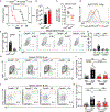
Figure 5.. CARD9 is critical for the GC-dependent generation of antifungal IgG antibodies.
(A–C) Cyc-induced gut-to-system fungal dissemination was induced in Card9+/+ and Card9−/− mice. (A and B) Disease morbidity assessed by survival (A) and C. albicans systemic spread to the kidneys (B). C. albicans detection failed in one mouse, which was excluded from the analysis. (C) Change in anti-C. albicans serum IgG after mice became moribund (14 days after infection) relative to pre-infection titers. (D and E) MultiKAP analysis of anti-fungal antibodies in healthy Card9+/+ and Card9−/− mice. (A–E) Statistical analysis was performed using log rank Mantel-Cox test (A) or Mann-Whitney test (B, C, and E). Card9+/+, n = 6; Card9+/+ +Ca, n = 6; Card9−/− +Ca, n = 7. Data are representative of two independent experiments. (F and G) Systemic IgG and B cell responses of Card9+/+ and Card9−/− mice 2 weeks after intraperitoneal injection with C. albicans. The B cell compartment in blood samples from individual mice was analyzed by flow cytometry. One-way ANOVA followed by Tukey’s multiple comparisons test. Card9+/+ NT, n = 7; Card9+/+ +Ca, n = 6; Card9−/− NT, n = 6; Card9−/− +Ca, n = 6. Data are representative of two independent experiments. (H–K) Systemic IgG and B cell responses of Card9+/+ and Card9−/− mice after intraperitoneal injection with C. albicans. The B cell compartment in spleen samples from individual mice was analyzed by flow cytometry. One-way ANOVA followed by Tukey’s multiple comparisons test. Card9+/+ NT, n = 7; Card9+/+ +Ca, n = 11; Card9−/− NT, n = 6; Card9−/− +Ca, n = 6. Data are representative of three independent experiments. Each dot represents an individual mouse. Error bars indicate SEM. ns, p ≥ 0.05; *p < 0.05; **p < 0.01; ***p < 0.001; ****p < 0.0001. See also Figure S4.
Figure 6.
Figure 6.. Syk-CARD9-dependent signaling in CX3CR1+ MNPs is critical for induction of antifungal IgG antibody responses
(A) Single-cell RNA-seq analysis of CARD9 expression in human biopsies from a single-cell RNA-seq dataset from Smillie et al. (2019). (B and C) Systemic IgG and B cell responses in ΔIRF4 or littermates (Litt) upon intestinal colonization with C. albicans. Data were collected from two independent experiments. Statistical analyses were performed using Mann-Whitney test. Litt, n = 7; ΔIRF4, n = 10. (B) MultiKAP analysis of anti-fungal antibodies in ΔIRF4 and littermate mice. (C) Frequency and absolute numbers of IgG+IgD among splenic CD19+CD4 B cells analyzed by flow cytometry. (D and E) Systemic IgG and B cell responses in ΔCX3CR1 mice and Litt upon intestinal colonization with C. albicans. Data were collected from two independent experiments. Statistical analyses were performed using Mann-Whitney test. Litt, n = 12; ΔCX3CR1, n = 7. (D) MultiKAP analysis of anti-fungal antibodies in ΔCX3CR1 and Litt mice. (E) Frequency and absolute numbers of IgG+IgD among splenic CD19+CD4 B cells analyzed by flow cytometry. (F) Feces of healthy CX3CR1ΔSyk and Litt were assessed by multiKAP for systemic antifungal IgG binding after incubation with matched sera. Data were collected from two independent experiments; Mann-Whitney test. Litt, n = 8; CX3CR1ΔSyk, n = 7. (G) Frequency and absolute numbers of IgG+IgD among splenic CD19+CD4 B cells analyzed by flow cytometry in CX3CR1ΔSyk and Litt mice after oral gavage with C. albicans. Data were collected from three independent experiments; Mann-Whitney test. Litt NT, n = 9; Litt +Ca, n = 12; CX3CR1ΔSyk +Ca, n = 16. (H) MultiKAP analysis of anti-fungal antibodies in healthy TCRβ+/+ and TCRβ−/− mice. Mann-Whitney test TCRβ+/+, n = 9; TCRβ−/−, n = 8. Data are representative of two independent experiments. (I and J) Anti-Candida IgG antibody titers (J) assessed by ELISA in sera from individuals with candidiasis who were non-carriers or carriers of CARD9 loss-of-function mutations (Q289*, R35Q, R70W) (I). Non-carriers: CARD9 sufficient, n = 5; carriers: CARD9 deficient, n = 4. Each dot represents an individual human subject; Mann-Whitney test. Error bars indicate SEM. ns, p ≥ 0.05; *p < 0.05; **p < 0.01; ***p < 0.001; ****p < 0.0001. See also Figures S5.

Comment in

References (V体育官网)

    1. Ansaldo E, Slayden LC, Ching KL, Koch MA, Wolf NK, Plichta DR, Brown EM, Graham DB, Xavier RJ, Moon JJ, and Barton GM (2019). Akkermansia muciniphila induces intestinal adaptive immune responses during homeostasis. Science 364, 1179–1184. - PMC - PubMed
    1. Arumugam M, Raes J, Pelletier E, Le Paslier D, Yamada T, Mende DR, Fernandes GR, Tap J, Bruls T, Batto J-M, et al.; MetaHIT Consortium (2011). Enterotypes of the human gut microbiome. Nature 473, 174–180. - "VSports" PMC - PubMed
    1. Bacher P, Hohnstein T, Beerbaum E, Röcker M, Blango MG, Kaufmann S, Röhmel J, Eschenhagen P, Grehn C, Seidel K, et al. (2019). Human Anti-fungal Th17 Immunity and Pathology Rely on Cross-Reactivity against Candida albicans. Cell 176, 1340–1355.e15. - PubMed
    1. Barbet G, Sander LE, Geswell M, Leonardi I, Cerutti A, Iliev I, and Blander JM (2018). Sensing Microbial Viability through Bacterial RNA Augments T Follicular Helper Cell and Antibody Responses. Immunity 48, 584–598.e5. - VSports - PMC - PubMed
    1. Brown GD, Denning DW, Gow NAR, Levitz SM, Netea MG, and White TC (2012). Hidden killers: human fungal infections. Sci. Transl. Med 4, 165rv13. - PubMed

"V体育平台登录" Publication types

MeSH terms