Skip to main page content
U.S. flag

An official website of the United States government

Dot gov

The . gov means it’s official. Federal government websites often end in . gov or VSports app下载. mil. Before sharing sensitive information, make sure you’re on a federal government site. .

Https

The site is secure V体育官网. The https:// ensures that you are connecting to the official website and that any information you provide is encrypted and transmitted securely. .

Review
. 2020 Dec 23;85(1):e00027-19.
doi: 10.1128/MMBR.00027-19. Print 2021 Feb 17.

Salmonella versus the Microbiome (V体育官网)

Affiliations
Review

Salmonella versus the Microbiome

Andrew W L Rogers et al. Microbiol Mol Biol Rev. .

Abstract

A balanced gut microbiota contributes to health, but the mechanisms maintaining homeostasis remain elusive. Microbiota assembly during infancy is governed by competition between species and by environmental factors, termed habitat filters, that determine the range of successful traits within the microbial community. These habitat filters include the diet, host-derived resources, and microbiota-derived metabolites, such as short-chain fatty acids. Once the microbiota has matured, competition and habitat filtering prevent engraftment of new microbes, thereby providing protection against opportunistic infections. Competition with endogenous Enterobacterales, habitat filtering by short-chain fatty acids, and a host-derived habitat filter, epithelial hypoxia, also contribute to colonization resistance against Salmonella serovars. However, at a high challenge dose, these frank pathogens can overcome colonization resistance by using their virulence factors to trigger intestinal inflammation. In turn, inflammation increases the luminal availability of host-derived resources, such as oxygen, nitrate, tetrathionate, and lactate, thereby creating a state of abnormal habitat filtering that enables the pathogen to overcome growth inhibition by short-chain fatty acids. Thus, studying the process of ecosystem invasion by Salmonella serovars clarifies that colonization resistance can become weakened by disrupting host-mediated habitat filtering. This insight is relevant for understanding how inflammation triggers dysbiosis linked to noncommunicable diseases, conditions in which endogenous Enterobacterales expand in the fecal microbiota using some of the same growth-limiting resources required by Salmonella serovars for ecosystem invasion VSports手机版. In essence, ecosystem invasion by Salmonella serovars suggests that homeostasis and dysbiosis simply represent states where competition and habitat filtering are normal or abnormal, respectively. .

Keywords: Salmonella; colonization resistance; microbiome; microbiota V体育安卓版. .


Figures (V体育安卓版)

FIG 1
FIG 1
Composition of the term microbiome. The microbiome is defined as the microbiota and its environment. The latter is determined by host-derived habitat filters and the diet, which is controlled by host behavior. Host-derived habitat filters shape the size, species composition and biogeography of the microbiota and in turn the microbiota contributes to host nutrition and immune education. Microbiota-nourishing immunity is composed of the microbiota and host-derived habitat filters, which form a host-microbe chimera that functions in conferring colonization resistance.
FIG 2
FIG 2
Habitat filtering in the adult colon. Epithelial hypoxia and dietary fiber filter the habitat in the large intestine to license growth of obligate anaerobic fiber eaters, which drives a dominance of the classes Clostridia and Bacteroidia in the fecal microbiota. Facultative anaerobic bacteria, such as members of the Enterobacterales, remain minority species because epithelial hypoxia limits critical resources they require for overcoming growth inhibition by short-chain fatty acids (acetate, butyrate, and propionate). (Created with BioRender.com.)
FIG 3
FIG 3
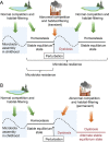
Normal competition and habitat filtering promote homeostasis, microbiota resistance and microbiota resilience. (A) After birth, the microbiota exhibits fluctuations as it assembles to fill nutrient niches created by competition and habitat filtering. Once microbiota assembly is complete, a state of normal competition and habitat filtering maintains homeostasis, characterized by a stable equilibrium state in which the microbiota composition remains invariable over time, a phenomenon termed microbiota resistance. A brief perturbation, such as a disruption of the microbiota with antibiotics, leads to a transient state of abnormal competition and habitat filtering, which causes dysbiotic fluctuation in the microbiota composition. However, once normal competition and habitat filtering resume, the microbiota reassembles to reach an equilibrium state that is functionally similar to that of the community prior to the perturbation. The ability of the microbiota to return to homeostasis after a perturbation is termed microbiota resilience. (B) A lasting perturbation, which can be caused for example by chronic intestinal inflammation, triggers a permanent state of abnormal competition and habitat filtering. As new nutrient niches created by abnormal competition and habitat filtering are filled, the microbiota composition shifts permanently to reach an alternate equilibrium state. Through this process, abnormal competition and habitat filtering maintain a perpetual state of dysbiosis. (Created with BioRender.com.)
FIG 4
FIG 4
Functions of the gut microbiota. Nutrients (e.g., fiber) that evade absorption and degradation by host enzymes in the small intestine enter the colon, where they are converted into fermentation products by the gut microbiota. This metabolic activity of the gut microbiota has been likened to the function of an organ that contributes to host nutrition and immune education. Host-derived habitat filters and the microbiota form a host microbe chimera that performs a third function, termed colonization resistance, which prevents harmful microbes from entering the body. (Created with BioRender.com.)
FIG 5
FIG 5
Intestinal inflammation creates a state of abnormal habitat filtering. (A) During homeostasis, microbiota-derived butyrate maintains high oxygen (O2) consumption in the colonic epithelium through mitochondrial oxidative phosphorylation (246–248). The resulting epithelial hypoxia limits diffusion of oxygen into the gut lumen to preserve anaerobiosis, which maintains a dominance of obligate anaerobic bacteria in the gut microbiota (89). Sulfate-reducing bacteria generate hydrogen sulfide (H2S) (249), which is detoxified by epithelial sulfide oxidases to thiosulfate (S2O32–) (250, 251). (B) During intestinal inflammation, neutrophils and inflammatory monocytes migrate into the intestinal lumen. Inflammatory monocytes are the dominant source of inducible nitric oxide synthase (iNOS), which generates nitric oxide (NO) (252). Nitric oxide can react with superoxide (O2) produced by phagocyte NADPH oxidase (NOX2) to form peroxynitrite (253), which decomposes to nitrate (NO3) in the gut lumen (254). Superoxide is converted by superoxide dismutase (SOD) to hydrogen peroxide (H2O2), which is converted to hypochloric acid (HOCl) by neutrophil myeloperoxidase (MPO). These reactive oxygen species oxidize thiosulfate to tetrathionate (S4O62–) (10). Intestinal inflammation reduces mitochondrial bioenergetics in the colonic epithelium, thereby reducing epithelial oxygen consumption (154). The resulting loss of epithelial hypoxia increases diffusion of oxygen into the intestinal lumen to disrupt anaerobiosis. Catabolism of glucose (Glc) by host cells through aerobic glycolysis increases the luminal concentration of host-derived lactate (59). Through these mechanisms, intestinal inflammation elevates the availability of oxygen, lactate, nitrate and tetrathionate in the colonic lumen to create a state of abnormal habitat filtering that drives an expansion of facultative anaerobic Enterobacterales, which is a microbial signature of dysbiosis in the fecal microbiota (36, 37). (Created with BioRender.com.)
FIG 6
FIG 6
S. Typhimurium uses its virulence factors for ecosystem engineering. During homeostasis, conversion of microbiota-derived butyrate to carbon dioxide (CO2) through mitochondrial oxidative phosphorylation (Ox phos) results in high epithelial oxygen (O2) consumption, which maintains epithelial hypoxia. Epithelial cells detoxify microbiota-derived hydrogen sulfide (H2S) by conversion into thiosulfate (S2O32–). Upon entry, S. Typhimurium uses its virulence factors to invade the intestinal epithelium (T3SS-1) and survive in macrophages in host tissue (T3SS-2). However, prior to the development of host responses, anaerobiosis and niche preemption by endogenous Enterobacterales limit access of the luminal S. Typhimurium population to resources critical for overcoming growth inhibition by short-chain fatty acids (SCFAs). As a result, the luminal S. Typhimurium population decreases, which can lead to a pathogen extinction if the challenge dose is low. In the meantime, the virulence factor-mediated tissue invasion is detected by the innate immune system, which results in orchestration of an inflammatory response characterized by cellular infiltrates that are dominated by neutrophils. The inflammatory response eventually clears the subpopulation of the pathogen that resides in tissue, but it also induces migration of phagocytes into the intestinal lumen. Luminal phagocytes release reactive oxygen species (ROS) and reactive nitrogen species that generate host-derived electron acceptors, including tetrathionate (S4O62–) and nitrate (NO3). Luminal neutrophils also deplete butyrate-producing Clostridia from the gut microbiota, which reduces mitochondrial bioenergetics in the intestinal epithelium. The consequent shift in epithelial energy metabolism to aerobic glycolysis, the conversion of glucose (Glc) into lactate, is associated with elevated epithelial release of oxygen and lactate. In turn, these changes in the luminal environment create a state of abnormal habitat filtering, thereby providing S. Typhimurium with critical resources (nitrate, tetrathionate, oxygen, and lactate) to expand in the gut microbiota, which is required for pathogen transmission by the fecal oral route. The new nutrient niche created by virulence factor-induced inflammation also supports growth of endogenous Enterobacterales, provided they can overcome growth inhibition by lipocalin-2 (Lipocalin), an antimicrobial protein released by epithelial cells during intestinal inflammation. Through this chain of events, virulence factor-mediated ecosystem engineering creates a new nutrient niche in which S. Typhimurium and endogenous Enterobacterales battle for supremacy using their antimicrobial weaponry, including colicins, microcins and type VI secretion systems (T6SS). (Created with BioRender.com.)
FIG 7
FIG 7
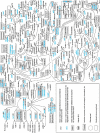
Reconstruction of a metabolic network required for growth in an engineered nutrient niche. Whole-genome comparison of 5 genomes representing extraintestinal Salmonella serovars and 10 genomes representing gastrointestinal Salmonella serovars reveals metabolic pathwaysgastrointestinal pathogens use to fuel their growth in the inflamed gut (213). The graphic shows pathways that are degrading in genomes of extraintestinal Salmonella serovars (genes in blue font) but are intact in genomes of gastrointestinal Salmonella serovars. The metabolic network predicted by this in silico analysis provides a window into the nutrient niche that is engineered by S. Typhimurium virulence factors in the gut. (Adapted from reference .)

References

    1. Bordenave G. 2003. Louis Pasteur (1822–1895). Microbes Infect 5:553–560. doi:10.1016/S1286-4579(03)00075-3. - DOI - PubMed
    1. Koch R. 1882. Die Aetiologie der Tuberkulose. Berliner Klin Wochenschr 15:221–230.
    1. Roux EYA. 1888. Contribution à l’étude de la diphtérie. Ann Inst Pasteur 2:421–499.
    1. Neisser M, Shiga K. 1903. Ueber freie Receptoren von Typhus- und Dysenteriebazillen und über das Dysenterietoxin. Dtsch Med Wochenschr 29:61–62. doi:10.1055/s-0028-1138255. - "V体育2025版" DOI
    1. Conradi H. 1903. Über lösliche, durch aseptische Autolyse erhaltene Giftstoffe von Ruhr- und Typhus-Bazillen. Dtsch Med Wochenschr 29:26–28. doi:10.1055/s-0028-1138228. - DOI

Publication types

MeSH terms