"VSports最新版本" Skip to main page content
U.S. flag

An official website of the United States government

Dot gov

The . gov means it’s official. Federal government websites often end in . gov or . mil VSports app下载. Before sharing sensitive information, make sure you’re on a federal government site. .

Https

The site is secure. The https:// ensures that you are connecting to the official website and that any information you provide is encrypted and transmitted securely. V体育官网.

. 2021 Jan;27(1):82-91.
doi: 10.1111/cns.13496. Epub 2020 Dec 6.

"V体育2025版" The novel Nrf2 activator CDDO-EA attenuates cerebral ischemic injury by promoting microglia/macrophage polarization toward M2 phenotype in mice

Affiliations

V体育官网 - The novel Nrf2 activator CDDO-EA attenuates cerebral ischemic injury by promoting microglia/macrophage polarization toward M2 phenotype in mice

Xia Lei et al. CNS Neurosci Ther. 2021 Jan.

Abstract

The aim of present study was to explore whether 2-cyano-3, 12-dioxooleana-1, 9-dien-28-oic acid (CDDO)-ethylamide (CDDO-EA) attenuates cerebral ischemic injury and its possible mechanisms using a middle cerebral artery occlusion (MCAO) model in C57BL/6 mice. Our results showed that intraperitoneal injection (i. p. ) of CDDO-EA (2 and 4 mg/kg) augmented NFE2-related factor 2 (Nrf2) and heme oxygenase-1 (HO-1) expression in ischemic cortex after MCAO. Moreover, CDDO-EA (2 mg/kg, i. p. ) significantly enhanced Nrf2 nuclear accumulation, associated with increased cytosolic HO-1 expression, reduced neurological deficit and infarct volume as well as neural apoptosis, and shifted polarization of microglia/macrophages toward an antiinflammatory M2 phenotype in ischemic cortex after MCAO. Using an in vitro model, we confirmed that CDDO-EA (100 μg/mL) increased HO-1 expression and primed microglial polarization toward M2 phenotype under inflammatory stimulation in BV2 microglial cells. These findings suggest that a novel Nrf2 activator CDDO-EA confers neuroprotection against ischemic injury. VSports手机版.

Keywords: CDDO-EA; HO-1; Nrf2; cerebral ischemia; microglia/macrophage V体育安卓版. .


Conflict of interest statement (VSports最新版本)

The author(s) declared no potential conflicts of interest for the research, authorship, and publication of this article.

Figures

FIGURE 1
FIGURE 1
CDDO‐EA increases Nrf2 and HO‐1 expression in cortex after transient MCAO. A, Chemical structure of CDDO‐EA. B, Scheme for the experimental design. C, Nrf2 and HO‐1 protein expression in the ischemic cortex was detected by Western blot 48 h after MCAO. Mice were injected with CDDO‐EA (0, 1, 2, and 4 mg/kg, i.p.) 30 min after MCAO surgery. Cortexes were collected at 48 h after MCAO, and homogenates were blotted with anti‐Nrf2, anti‐HO‐1, and anti‐β‐actin. Quantification of optical density was normalized to sham controls. D, Mice were injected with CDDO‐EA (2 mg/kg, i.p.) followed by MCAO. Ischemic cortexes were collected at 48 h after MCAO and subjected to subcellular fractionation. Nuclear Nrf2 and cytosolic HO‐1 were detected by Western blot, and Histone H3 and β‐actin were used as loading controls for nuclear and total protein, respectively. Data are mean ± SD. (ns denotes not significant, * P < 0.05, n = 3)
FIGURE 2
FIGURE 2
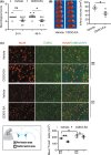
CDDO‐EA reduces neurological deficit, infarct volume, and neuronal death following ischemic injury in mice. A, Assessment of neurological deficits in mice using a modified Longa score 24 and 48 h following MCAO. B, Representative images of TTC‐stained brain coronal sections obtained 48 h following transient MCAO with vehicle or CDDO‐EA for the determination of total infarct volume. C, Coronal sections from MCAO + Vehicle and MCAO + CDDO‐EA group on 48 h after MCAO were subjected to TUNEL staining (green) and immunostaining for the NeuN (red) in center lesion area and peri‐lesion area of ischemic cortexes. Quantification was performed by counting the TUNEL+/NeuN+ neurons in the indicated region and expressed as cells per mm2. Scale bars = 20 μm. Data are mean ± SD. Data were analyzed using an unpaired t test with Welch correction (ns denotes not significant, * P < 0.05, ** P < 0.01, n = 8 in A, n = 4 in B and C)
FIGURE 3
FIGURE 3
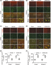
CDDO‐EA is associated with decreased M1 phenotype expression and increased M2 phenotype expression in microglia/macrophages within the ischemic cortex after MCAO. A–D, Coronal sections from MCAO + Vehicle and MCAO + CDDO‐EA cortex and striatum 48 h after MCAO were subjected to immunostaining for either CD16 or CD206 (red) and for Iba 1 (green) 48 h after MCAO. E, F, Quantification was performed by counting the CD16+/ or CD206+/Iba1+ microglia/macrophages in the region (per mm2). Scale bars = 50 or 10 μm. Data are mean ± SD (* P < 0.05, n = 3)
FIGURE 4
FIGURE 4
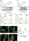
CDDO‐EA increases expression of HO‐1, CD206, and CCL22, reduces CD16 and CD11b expression, and enhances microglial phagocytosis in LPS‐activated BV2 microglial cells. A, B, Cells were treated with CDDO‐EA (0, 50, 100, or 200 μg/mL), LPS (100 ng/mL), LPS (100 ng/mL) + CDDO‐EA (100 μg/mL), or LPS (100 ng/mL) + PPIX (10 μg/mL) for 24 h, lysates collected, and homogenates were immunoblotted with anti‐HO‐1 and anti‐β‐actin. C, D, BV2 cells were treated with LPS (100 ng/mL), LPS (100 ng/mL) + CDDO‐EA (100 μg/mL), or LPS + CDDO‐EA + PPIX (10 μg/mL) for 24 h, and the level of CD16, CD11b, CD206, and CCL22 mRNA were detected with real‐time quantitative PCR. E, BV2 cells were treated with LPS (100 ng/mL), LPS (100 ng/mL) + CDDO‐EA (100 μg/mL), or LPS + CDDO‐ EA + PPIX (10 μg/mL) for 21 h, fluorescent microspheres were added into the medium for 3 h, and then, the cells were stained with phalloidin to visualize F‐actin. The left panel is representative images of intra‐microglia fluorescence 3 h after fluorescent microspheres uptake. Scale bars = 50 μm (ns denotes not significant, * P < 0.05, ** P < 0.01, n = 3‐6)

VSports最新版本 - References

    1. Lapchak PA, Zhang JH. The high cost of stroke and stroke cytoprotection research. Transl Stroke Res. 2017;8(4):307‐317. - PMC - PubMed
    1. Hafez S, Coucha M, Bruno A, Fagan SC, Ergul A. Hyperglycemia, acute ischemic stroke, and thrombolytic therapy. Transl Stroke Res. 2014;5(4):442‐453. - PMC - PubMed
    1. An C, Shi Y, Li P, et al. Molecular dialogs between the ischemic brain and the peripheral immune system: dualistic roles in injury and repair. Prog Neurogibol. 2014;115:6‐24. - PMC - PubMed
    1. Brown CM, Bushnell CD, Samsa GP, Goldstein LB, Colton CA. Chronic systemic immune dysfunction in African‐Americans with small vessel‐type ischemic stroke. Transl Stroke Res. 2015;6(6):430‐436. - PMC - PubMed
    1. Hu X, Leak RK, Shi Y, et al. Microglial and macrophage polarization‐new prospects for brain repair. Nat Rev Neurol. 2015;11(1):56‐64. - PMC - PubMed

Publication types

V体育安卓版 - MeSH terms