Skip to main page content
U.S. flag

An official website of the United States government

Dot gov

The . gov means it’s official. Federal government websites often end in . gov or . mil. Before sharing sensitive information, make sure you’re on a federal government site. VSports app下载.

Https

The site is secure. The https:// ensures that you are connecting to the official website and that any information you provide is encrypted and transmitted securely V体育官网. .

. 2020 Sep 29;39(1):202.
doi: 10.1186/s13046-020-01677-w.

FadA promotes DNA damage and progression of Fusobacterium nucleatum-induced colorectal cancer through up-regulation of chk2

Affiliations

FadA promotes DNA damage and progression of Fusobacterium nucleatum-induced colorectal cancer through up-regulation of chk2

VSports注册入口 - Pin Guo et al. J Exp Clin Cancer Res. .

Abstract

Background: Globally, colorectal cancer (CRC) affects more than 1 million people each year VSports手机版. In addition to non-modifiable and other environmental risk factors, Fusobacterium nucleatum infection has been linked to CRC recently. In this study, we explored mechanisms underlying the role of Fusobacterium nucleatum infection in the progression of CRC in a mouse model. .

Methods: C57BL/6 J-Adenomatous polyposis coli (APC) Min/J mice [APC (Min/+)] were treated with Fusobacterium nucleatum (109 cfu/mL, 0 V体育安卓版. 2 mL/time/day, i. g. , 12 weeks), saline, or FadA knockout (FadA-/-) Fusobacterium nucleatum. The number, size, and weight of CRC tumors were determined in isolated tumor masses. The human CRC cell lines HCT29 and HT116 were treated with lentiviral vectors overexpressing chk2 or silencing β-catenin. DNA damage was determined by Comet assay and γH2AX immunofluorescence assay and flow cytometry. The mRNA expression of chk2 was determined by RT-qPCR. Protein expression of FadA, E-cadherin, β-catenin, and chk2 were determined by Western blot analysis. .

Results: Fusobacterium nucleatum treatment promoted DNA damage in CRC in APC (Min/+) mice. Fusobacterium nucleatum also increased the number of CRC cells that were in the S phase of the cell cycle. FadA-/- reduced tumor number, size, and burden in vivo. FadA-/- also reduced DNA damage, cell proliferation, expression of E-cadherin and chk2, and cells in the S phase V体育ios版. Chk2 overexpression elevated DNA damage and tumor growth in APC (Min/+) mice. .

Conclusions: In conclusion, this study provided evidence that Fusobacterium nucleatum induced DNA damage and cell growth in CRC through FadA-dependent activation of the E-cadherin/β-catenin pathway, leading to up-regulation of chk2 VSports最新版本. .

Keywords: Colorectal cancer; DNA damage; E-cadherin/β-catenin pathway; FadA, chk2; Fusobacterium nucleatum. V体育平台登录.


Conflict of interest statement

The author declares no competing interest exists.

Figures
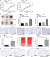
Fig. 1
Fig. 1
Fusobacterium nucleatum contributes to CRC. Male BALB/C nude mice were subcutaneously injected with HCT16 and HT29 cells treated with PBS, Escherichia coli, or Fusobacterium nucleatum to establish xenograft tumor animal models. a Tumor volume in HT29 and HCT116 cells. b Weight of tumors in nude mice formed by cells with different treatments. c Representative micrographs showing Ki-67 immunohistochemistry in xenograft tumor tissues (400 ×). d Number of tumors and tumor load in APC (Min/+) mice (n = 10 per group). e Representative micrographs showing PCNA (400 ×) and γH2AX (400 ×) immunohistochemistry. *p < 0.05 vs. PBS or Escherichia coli. Data are expressed as mean ± s.d. Data from multiple groups were compared using one-way analysis of variance (ANOVA) and Tukey’s post hoc test. Data comparison at different time points was performed using repeated measures ANOVA, followed by Bonferroni post hoc test. Non-parametric Mann-Whitney U test was used for comparison of data that were not normally distributed
Fig. 2
Fig. 2
Fusobacterium nucleatum leads to DNA damage in CRC cells. HCT16 and HT29 cells were treated with PBS, Escherichia coli, or Fusobacterium nucleatum with different MOI. a The number of cells counted by hemocytometry at hour 6, 24 and 48 after treatment to detect cell proliferation. b Representative micrographs (400 ×) showing Comet assay. c Representative micrographs (400 ×) showing γH2AX immunofluorescence assay. d γH2AX formation determined by flow cytometry. e The number of cells in different cell cycle phases. *p < 0.05 vs. PBS or Escherichia coli. Data are expressed as mean ± s.d. Data from multiple groups were compared using one-way analysis of variance (ANOVA) and Tukey’s post hoc test. Data comparison at different time points was performed using repeated measures ANOVA, followed by Bonferroni post hoc test. Each experiment was repeated three times
Fig. 3
Fig. 3
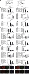
Fusobacterium nucleatum induces DNA damage in CRC cells via FadA. HCT16 and HT29 cells were treated with WT or FadA−/− Fusobacterium nucleatum (MOI: 1000). a The number of cells counted by hemocytometry at at hour 6, 24 and 48 after treatment to detect cell proliferation. b Representative micrographs showing Comet analysis. c Representative micrographs (400 ×) depicting the γH2AX immunofluorescence assay. d γH2AX formation as determined by flow cytometry. e The number of cells in different cell cycle phases *p < 0.05 vs. WT Fusobacterium nucleatum. Data are expressed as mean ± s.d. Data from two groups were compared using the unpaired t test. Data comparison at different time points was performed using repeated measures ANOVA, followed by Bonferroni post hoc test. Each experiment was repeated three times
Fig. 4
Fig. 4
FadA regulates E-cadherin, β-catenin, and chk2 expression in CRC cells. a Expression of chk2 in HCT116 and HT29 cells after treatment with WT or FadA−/− Fusobacterium nucleatum (MOI: 1000) for different time periods. b Protein expression of FadA, E-cadherin, β-catenin, and chk2 in HCT116 and HT29 cells treated with WT or FadA−/− Fusobacterium nucleatum (MOI: 1000) for 2 h. c Protein expression of FadA, E-cadherin, β-catenin, and chk2 in HCT116 and HT29 cells treated with genistein for 1 h and then with WT or FadA−/− Fusobacterium nucleatum (MOI: 1000) for 2 h. PTK inhibitor genistein inhibits all FadAc-activated functions. d Protein expression of FadA, E-cadherin, β-catenin, and chk2 in β-catenin-knockdown HCT116 and HT29 cells treated with WT or FadA−/− Fusobacterium nucleatum (MOI: 1000) for 2 h. e β-catenin nucleation and chk2 expression in HCT116 cells after different treatments determined by a confocal microscopy (400 ×). * p < 0.05 or ** p < 0.01 vs. WT Fusobacterium nucleatum. Data are expressed as mean ± s.d. and n.d. stands for no data. Data comparison at different time points was performed using repeated measures ANOVA, followed by Bonferroni post hoc test. Non-parametric Mann-Whitney U test was used for data that were not normally distributed
Fig. 5
Fig. 5
Overexpression of chk2 induces tumor growth in nude mice. Male BALB/C nude mice were subcutaneously injected with chk2-overexpressed HCT16 and HT29 cells to establish xenograft tumor animal models (n = 5 per group). a Tumor volume in nude mice. b Tumor weight in nude mice. * p < 0.05 vs. oe-NC. Data are expressed as mean ± s.d. Data from two groups were compared using the unpaired t test. Data comparison at different time points was performed using repeated measures ANOVA, followed by Bonferroni post hoc test. Each experiment was repeated three times
Fig. 6
Fig. 6
FadA up-regulates E-cadherin/β-catenin activation and chk2 to induce DNA damage in CRC cells. a Tumor number, size, and load in APCMin/+ mice on 12th week after treatment with WT or FadA−/− Fusobacterium nucleatum. b Protein expression of FadA, E-cadherin/β-catenin, and chk2 in CRC tissue determined by Western blot analysis. c Protein expression of β-catenin in the nucleus. d Representative micrographs showing β-catenin (400 ×), chk2 protein (400×), and γH2AX (400 ×) immunohistochemistry in CRC tissues. Data are expressed as mean ± s.d. Data from two groups were compared using the unpaired t test
Fig. 7
Fig. 7
Schematic diagrams showing the proposed molecular pathway of Fusobacterium nucleatum-induced FadA-dependent DNA damage and development of CRC

References

    1. Sillo TO, Beggs AD, Morton DG, Middleton G. Mechanisms of immunogenicity in colorectal cancer. Br J Surg. 2019;106:1283–1297. doi: 10.1002/bjs.11204. - DOI - PMC - PubMed
    1. Newton PT. New insights into niclosamide action: autophagy activation in colorectal cancer. Biochem J. 2019;476:779–781. doi: 10.1042/BCJ20190020. - "V体育官网" DOI - PubMed
    1. Antoni S, Soerjomataram I, Moller B, Bray F, Ferlay J. An assessment of GLOBOCAN methods for deriving national estimates of cancer incidence. Bull World Health Organ. 2016;94:174–184. doi: 10.2471/BLT.15.164384. - DOI (VSports最新版本) - PMC - PubMed
    1. Bray F, Ferlay J, Soerjomataram I, Siegel RL, Torre LA, Jemal A. Global cancer statistics 2018: GLOBOCAN estimates of incidence and mortality worldwide for 36 cancers in 185 countries. CA Cancer J Clin. 2018;68:394–424. doi: 10.3322/caac.21492. - DOI - PubMed
    1. Moghimi-Dehkordi B, Safaee A. An overview of colorectal cancer survival rates and prognosis in Asia. World J Gastrointest Oncol. 2012;4:71–75. doi: 10.4251/wjgo.v4.i4.71. - DOI - PMC - PubMed

MeSH terms