Skip to main page content
U.S. flag

An official website of the United States government

Dot gov

The . gov means it’s official. Federal government websites often end in . gov or . mil VSports app下载. Before sharing sensitive information, make sure you’re on a federal government site. .

Https

The site is secure. The https:// ensures that you are connecting to the official website and that any information you provide is encrypted and transmitted securely. V体育官网.

. 2020 Jul 7;9(13):e015640.
doi: 10.1161/JAHA.119.015640. Epub 2020 Jun 15.

Sacubitril/Valsartan Improves Cardiac Function and Decreases Myocardial Fibrosis Via Downregulation of Exosomal miR-181a in a Rodent Chronic Myocardial Infarction Model

Affiliations

Sacubitril/Valsartan Improves Cardiac Function and Decreases Myocardial Fibrosis Via Downregulation of Exosomal miR-181a in a Rodent Chronic Myocardial Infarction Model (VSports在线直播)

V体育ios版 - Evgeniya Vaskova et al. J Am Heart Assoc. .

Abstract

Background Exosomes are small extracellular vesicles that function as intercellular messengers and effectors. Exosomal cargo contains regulatory small molecules, including miRNAs, mRNAs, lncRNAs, and small peptides that can be modulated by different pathological stimuli to the cells. One of the main mechanisms of action of drug therapy may be the altered production and/or content of the exosomes. Methods and Results We studied the effects on exosome production and content by neprilysin inhibitor/angiotensin receptor blockers, sacubitril/valsartan and valsartan alone, using human-induced pluripotent stem cell-derived cardiomyocytes under normoxic and hypoxic injury model in vitro, and assessed for physiologic correlation using an ischemic myocardial injury rodent model in vivo. We demonstrated that the treatment with sacubitril/valsartan and valsartan alone resulted in the increased production of exosomes by induced pluripotent stem cell-derived cardiomyocytes in vitro in both conditions as well as in the rat plasma in vivo. Next-generation sequencing of these exosomes exhibited downregulation of the expression of rno-miR-181a in the sacubitril/valsartan treatment group. In vivo studies employing chronic rodent myocardial injury model demonstrated that miR-181a antagomir has a beneficial effect on cardiac function. Subsequently, immunohistochemical and molecular studies suggested that the downregulation of miR-181a resulted in the attenuation of myocardial fibrosis and hypertrophy, restoring the injured rodent heart after myocardial infarction VSports手机版. Conclusions We demonstrate that an additional mechanism of action of the pleiotropic effects of sacubitril/valsartan may be mediated by the modulation of the miRNA expression level in the exosome payload. .

Keywords: exosomes; mechanism of action; miRNAs; sacubitril/valsartan V体育安卓版. .


Figures

Figure 1
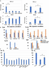
Figure 1. Exosome production in prehypoxia and posthypoxia conditions by induced pluripotent stem cell‐derived cardiomyocytes in vitro.
A, Size distribution of the induced pluripotent stem cell‐derived cardiomyocytes derived exosomes in prehypoxia and posthypoxia conditions. B, A significant increase in the exosome production (particles/mL) was seen in the LBQ657/valsartan (LBQ/val), valsartan (val), and LBQ657 treatment groups vs the control arms in comparison with both prehypoxic and posthypoxic control arms (*P<0.05; **P<0.01).
Figure 2
Figure 2. In vitro LBQ657/valsartan (LBQ/val), valsartan (val), LBQ657 (LBQ), and exosome effects on human‐induced pluripotent stem cell‐derived cardiomyocytes viability.
A, qRT (quantitative reverse transcription)–polymerase chain reaction demonstrated no significant difference in apoptosis‐associated gene expressions between the drug treatment vs untreated (negative) control arms while the exosome treatment arm significantly reversed the expression of the Bcl‐2 and Bax genes. B, FACS (fluorescence‐activated cell sorting) analysis of induced pluripotent stem cell‐derived cardiomyocytes viability measured live, apoptotic, and necrotic cells after 6 and 18 hours of hypoxia. C, The survival rate of induced pluripotent stem cell‐derived cardiomyocytes after hypoxic insult measured by MTT (3‐(4,5‐dimethylthiazol‐2‐yl)‐2,5‐diphenyltetrazolium bromide) analysis is shown. The viability values were represented as a percent of the untreated cells cultured under normoxic condition (regarded as 100% viability). The bars demonstrate the mean of triplicate experiments with SD. The exosome treatment group of the hypoxia‐injured induced pluripotent stem cell‐derived cardiomyocytes demonstrated significantly improved viability (*P<0.05). hyp indicates hypoxic; and norm, normoxic.
Figure 3
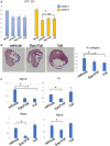
Figure 3. The effect of sacubitril/valsartan (sac/val) and valsartan (val) on cardiac function and remodeling.
A, Left ventricular ejection fraction (LVEF) at weeks 2 and 8. B, Representative Masson‐Trichrome stain images of cardiac tissues in rat model of myocardial infarction. The myocardial fibers and collagen are colored blue and indicated by white lines. Scale bar=300 μm. Quantitative analysis demonstrated significant reduction of % collagen in the sacubitril/valsartan and valsartan treatment groups when compared with the vehicle group. C, Quantitative polymerase chain reaction analysis for the gene expression of rat hearts related to myocardial fibrosis and hypertrophy, demonstrating the significant effects of sacubitril/valsartan vs the vehicle and valsartan treatment groups (*P<0.05).
Figure 4
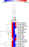
Figure 4. Heatmap of hierarchical clustering of the top 15 differentially expressed miR in the sacubitril/valsartan (sac/val), valsartan (val), and vehicle treatment groups.
rno‐miR‐181a‐1, rno‐miR‐181a‐2, and rno‐miR‐181a‐5p demonstrate the highest expression difference between the exosomes derived from the plasma of sacubitril/valsartan–treated vs valsartan‐treated and vehicle‐treated rats. Red denotes high and blue denotes low expression levels.
Figure 5
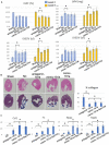
Figure 5. Downregulation of rno‐miR‐181a (mir‐181a antagomir) improves cardiac function and myocardial remodeling processes.
A, Left ventricular ejection fraction (LVEF), left ventricular end‐diastolic volume (LVEDV), left ventricular end systolic volume (LVESV), left ventricular mass (LVM) at weeks 2 and 8. The mir‐181a antagomir treatment group demonstrates significant salutary effects vs the negative control (NC) group. B, Representative images of cardiac tissues in myocardial infarction rats stained with hematoxylin‐eosin (upper panel) and Masson trichrome (lower panel). The myocardial fibers and collagen are colored blue and indicated by white lines. Scale bar=300 μm. Quantitative analysis demonstrated a significant reduction in the percentage of collagen in the miR‐181a antagomir treatment group when compared with NC and the other treatment groups. C, Quantitative polymerase chain reaction analysis for myocardial hypertrophy and fibrosis gene expression in the mir‐181a antagomir treatment group *P<0.05.

References

    1. Benjamin EJ, Muntner P, Alonso A, Bittencourt MS, Callaway CW, Carson AP, Chamberlain AM, Chang AR, Cheng S, Das SR, et al. Heart disease and stroke statistics‐2019 update: a report from the American Heart Association. Circulation. 2019;139:e56–e66. - "V体育官网" PubMed
    1. Ziaeian B, Fonarow GC. Epidemiology and aetiology of heart failure. Nat Rev Cardiol. 2016;13:368–378. - PMC - PubMed
    1. Savarese G, Lund LH. Global public health burden of heart failure. Card Fail Rev. 2017;03:7–11. - PMC - PubMed
    1. Khder Y, Shi V, McMurray JJV, Lefkowitz MP. Sacubitril/valsartan (LCZ696) in heart failure In: Bauersachs J, Butler J, Sandner P, eds. Heart Failure. Cham: Springer International Publishing; 2017:133–165.
    1. Karch R, Neumann F, Ullrich R, Neumüller J, Podesser BK, Neumann M, Schreiner W. The spatial pattern of coronary capillaries in patients with dilated, ischemic, or inflammatory cardiomyopathy. Cardiovasc Pathol. 2005;14:135–144. - PubMed

Publication types

MeSH terms