<var date-time="kdIn8hxD"></var> Skip to main page content
U.S. flag

An official website of the United States government

Dot gov

The VSports app下载. gov means it’s official. Federal government websites often end in . gov or . mil. Before sharing sensitive information, make sure you’re on a federal government site. .

Https

The site is secure. The https:// ensures that you are connecting to the official website and that any information you provide is encrypted and transmitted securely. V体育官网.

. 2020 Oct 1;116(12):1981-1994.
doi: 10.1093/cvr/cvaa008.

"V体育平台登录" The LINC00961 transcript and its encoded micropeptide, small regulatory polypeptide of amino acid response, regulate endothelial cell function

Affiliations

The LINC00961 transcript and its encoded micropeptide, small regulatory polypeptide of amino acid response, regulate endothelial cell function

Helen L Spencer et al. Cardiovasc Res. .

Abstract

Aims: Long non-coding RNAs (lncRNAs) play functional roles in physiology and disease, yet understanding of their contribution to endothelial cell (EC) function is incomplete. We identified lncRNAs regulated during EC differentiation and investigated the role of LINC00961 and its encoded micropeptide, small regulatory polypeptide of amino acid response (SPAAR), in EC function. VSports手机版.

Methods and results: Deep sequencing of human embryonic stem cell differentiation to ECs was combined with Encyclopedia of DNA Elements (ENCODE) RNA-seq data from vascular cells, identifying 278 endothelial enriched genes, including 6 lncRNAs V体育安卓版. Expression of LINC00961, first annotated as an lncRNA but reassigned as a protein-coding gene for the SPAAR micropeptide, was increased during the differentiation and was EC enriched. LINC00961 transcript depletion significantly reduced EC adhesion, tube formation, migration, proliferation, and barrier integrity in primary ECs. Overexpression of the SPAAR open reading frame increased tubule formation; however, overexpression of the full-length transcript did not, despite production of SPAAR. Furthermore, overexpression of an ATG mutant of the full-length transcript reduced network formation, suggesting a bona fide non-coding RNA function of the transcript with opposing effects to SPAAR. As the LINC00961 locus is conserved in mouse, we generated an LINC00961 locus knockout (KO) mouse that underwent hind limb ischaemia (HLI) to investigate the angiogenic role of this locus in vivo. In agreement with in vitro data, KO animals had a reduced capillary density in the ischaemic adductor muscle after 7 days. Finally, to characterize LINC00961 and SPAAR independent functions in ECs, we performed pull-downs of both molecules and identified protein-binding partners. LINC00961 RNA binds the G-actin sequestering protein thymosin beta-4x (Tβ4) and Tβ4 depletion phenocopied the overexpression of the ATG mutant. SPAAR binding partners included the actin-binding protein, SYNE1. .

Conclusion: The LINC00961 locus regulates EC function in vitro and in vivo. The gene produces two molecules with opposing effects on angiogenesis: SPAAR and LINC00961. V体育ios版.

Keywords: Angiogenesis; Endothelial cell; Hind limb ischaemia; LncRNA; Micropeptide VSports最新版本. .


Figures

Graphical Abstract
Graphical Abstract
Figure 1
Figure 1
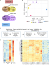
Identification of endothelial cell enriched genes. (A) Schematic representation of the RNA-seq samples: day 0 H9 hESC (ESC); Day 3 mesodermal population CD326lowCD56+ (MP); Day 3 remaining population (non-MP); Day 7 EC CD144+CD31+(EC); Day 7 remaining population (non-EC); HSVEC. (B) PCA of the RNA-seq samples. The plot was generated on the regularized log-transformed data using DESEq2. (C) Summary of the selection of candidates to identify genes enriched in ‘immature’ and ‘mature’ ECs. (D) Heatmap showing the expression data [as row z-score of the Log2(FPKM + 1)] during differentiation of the 278 EC-enriched genes. (E) Heatmap showing the expression data [as row z-score of the Log2 (FPKM + 1)] of the 278 EC-enriched genes in ENCODE RNA-seq samples.
Figure 2
Figure 2
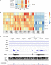
LINC00961 is enriched in immature and mature ECs. (A) Heatmap of the six lncRNAs identified in our EC differentiation protocol in each of the isolated cell populations. (B) Heatmap of these six lncRNAs in ENCODE RNA-seq samples including various types of EC lineages such as, venous, arterial, and lympthatic ECs. (C) Genomic organization of the LINC00961 gene, read profile from the ESC to EC RNA-seq, and conservation track based on UCSC alignment and PhyloP score.
Figure 3
Figure 3
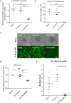
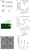
Functional impact of LINC00961/SPAAR depletion in ECs. (A) Confirmation of the dsiRNA-mediated depletion of LINC00961 transcript in HUVECs by qRT-PCR (n = 4, unpaired t-test). (B) Network formation assay in LINC00961 depleted HUVECs. Branch length assessed by Image J Angiogenesis plugin (n = 3, unpaired t-test). (C) Representative phase contrast and Calcein AM staining of network formation assay of LINC00961 depleted and control HUVECs. Phase Scale bar = 0.5 mm. Calcein AM Scale bar = 0.1 mm. (D) Impact of LINC00961 depletion on HUVEC adhesion (n = 3). (E) Analysis of average barrier resistance, expressed as Rb [Ohm × cm2], across a 10 h time course (n = 4 except for mock n = 3, one-way ANOVA). For data represented as fold change, the statistical analysis was done on the Log2 fold change using an one sample t-test. On the graphs, *P < 0.05 **P < 0.01 ***P < 0.001.
Figure 4
Figure 4
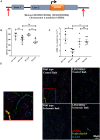
LINC00961/SPAAR KO mice have a reduced adductor muscle capillary density following HLI at 7 days. (A) Schematic representation of the deleted region of the LINC00961 mouse locus using CRISPR/Cas9 technology by Taconic©. Red arrows indicate the position of the guide RNA strands utilized to delete the whole locus. (B) Capillary density per sample. Five random regions of interest from three sections per sample were counted (n = 4 WT mice/6 KO mice, one-way ANOVA, ** P < 0.01, ns, not significant). (C) αSMA positive vessel density per sample. (D) Representative adductor muscle immunofluorescent images: Isolectin b4 (IB4) capillary/endothelium, αSMA, and nuclear DAPI, scale bar 50 µm. Zoomed panel on left corresponds to red box on area of WT control limb image.
Figure 5
Figure 5
Impact of LINC00961 transcript and SPAAR micropeptide overexpression in in vitro angiogenic assays. (A) Schematic representation of LINC00961 LV constructs with transcript length in base pairs (bp) and encoded peptide length in amino acids (aa). (B) qRT-PCR validation of the LV constructs overexpression in HUVECs using primers targeting the ORF sequence. Unpaired t-test, comparison test vs. LV-EMPTY (n = 4). (C) Representative western blot of SPAAR micropeptide and β-actin in HUVECs infected with the LV constructs. (D) Network formation assay comparing HUVECs transfected with LV constructs. Branch length assessed by Image J Angiogenesis plugin. Unpaired t-test vs. LV-EMPTY (n = 3). (E) Representative Phase contrast of network formation assay of HUVECs transfected with LV constructs. (F) Analysis of average barrier resistance, expressed as Rb [Ohm × cm2], across a 10 h time course (n = 4, one-way ANOVA). Scale bar =0.5 mm. On the graphs, *P < 0.05, **P < 0.01, ***P < 0.001.
Figure 6
Figure 6
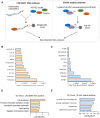
LINC00961 and SPAAR both bind to actin-binding proteins. (A) Schematic of the LINC00961 RNA and SPAAR peptide pull-down experiments in HUVECs. (B) List of the top 10 proteins identified in LINC00961 RNA pull-down (ranked on label-free quantification value). (C) List of the top 10 proteins identified in HA-SPAAR peptide pull-down (ranked on label-free quantification value). (D) GO analysis on enriched proteins from LINC00961 immunoprecipitation. (E) GO analysis on enriched proteins from SPAAR immunoprecipitation.
Figure 7
Figure 7
Thymosin beta 4-x KD in HUVECS has a similar phenotype to LV-ΔΔATG961 overexpression on tubule formation. (A) Network formation assay in dsiRNA-mediated TMSB4X depleted HUVECs. Branch length assessed by Image J Angiogenesis plugin, n = 5, unpaired t-test. (B) Representative phase contrast and Calcein AM staining of network formation assay of depleted HUVECs. Phase contrast Scale bar = 0.5 mm. Calcein AM Scale bar = 0.1 mm. On the graphs, *P < 0.05, **P < 0.01, ***P < 0.001.

V体育ios版 - Comment in

References

    1. Toborek M, Kaiser S.. Endothelial cell functions. Relationship to atherogenesis. Basic Res Cardiol 1999;94:295–314. - PubMed
    1. De Vries MR, Simons KH, Jukema JW, Braun J, Quax PH.. Vein graft failure: from pathophysiology to clinical outcomes. Nat Rev Cardiol 2016;13:451–470. - PubMed
    1. Pearson J. Normal endothelial cell function. Lupus 2000;9:183–188. - PubMed (VSports在线直播)
    1. Su JB. Vascular endothelial dysfunction and pharmacological treatment. World J Cardiol 2015;7:719. - PMC - PubMed
    1. Zhao H, Zhao Y, Li Z, Ouyang Q, Sun Y, Zhou D, Xie P, Zeng S, Dong L, Wen H.. FLI1 and PKC co-activation promote highly efficient differentiation of human embryonic stem cells into endothelial-like cells. Cell Death Dis 2018;9:131. - PMC - PubMed

Publication types

MeSH terms