Skip to main page content
U.S. flag

An official website of the United States government

Dot gov

The . gov means it’s official. Federal government websites often end in . gov or . mil. Before sharing sensitive information, make sure you’re on a federal government site. VSports app下载.

Https

The site is secure V体育官网. The https:// ensures that you are connecting to the official website and that any information you provide is encrypted and transmitted securely. .

. 2020 Apr;34(4):1017-1026.
doi: 10.1038/s41375-019-0639-x. Epub 2019 Nov 18.

V体育官网 - RIG-I-based immunotherapy enhances survival in preclinical AML models and sensitizes AML cells to checkpoint blockade

Affiliations

RIG-I-based immunotherapy enhances survival in preclinical AML models and sensitizes AML cells to checkpoint blockade

Michael Ruzicka et al. Leukemia. 2020 Apr.

Abstract

Retinoic acid-inducible gene-I (RIG-I) is a cytoplasmic immune receptor sensing viral RNA. It triggers the release of type I interferons (IFN) and proinflammatory cytokines inducing an adaptive cellular immune response. We investigated the therapeutic potential of systemic RIG-I activation by short 5'-triphosphate-modified RNA (ppp-RNA) for the treatment of acute myeloid leukemia (AML) in the syngeneic murine C1498 AML tumor model. ppp-RNA treatment significantly reduced tumor burden, delayed disease onset and led to complete remission including immunological memory formation in a substantial proportion of animals. Therapy-induced tumor rejection was dependent on CD4+ and CD8+ T cells, but not on NK or B cells, and relied on intact IFN and mitochondrial antiviral signaling protein (MAVS) signaling in the host. Interestingly, ppp-RNA treatment induced programmed death ligand 1 (PD-L1) expression on AML cells and established therapeutic sensitivity to anti-PD-1 checkpoint blockade in vivo. In immune-reconstituted humanized mice, ppp-RNA treatment reduced the number of patient-derived xenografted (PDX) AML cells in blood and bone marrow while concomitantly enhancing CD3+ T cell counts in the respective tissues. Due to its ability to establish a state of full remission and immunological memory, our findings show that ppp-RNA treatment is a promising strategy for the immunotherapy of AML. VSports手机版.


Conflict of interest statement

The authors declare no competing financial interests. Parts of this work have been performed for the doctoral theses of MR, EH, HM, and MZ at the LMU Munich V体育安卓版.

Figures

Fig. 1
Fig. 1
Systemic ppp-RNA treatment in C1498-GFP tumor bearing mice. a Therapy scheme for in vivo experiments in the syngeneic C1498-GFP AML model. AML was induced by injecting 106 C1498-GFP AML cells into the tail vein. On days 3, 7, 10 and 14, mice were treated with 50 µg ppp-RNA i.v. b ppp-RNA treated (bar charts indicate mean of n = 3 with SEM of a single experiment) and untreated (bar charts indicate mean of n = 5 with SEM) C57BL/6 mice were sacrificed on day 17. Single cell suspensions of blood, bone marrow, livers, lungs, ovaries, and spleens were analyzed by flow cytometry determining the fraction of GFP+ cells (AML cells). Statistical significance was determined using the Student’s t test with comparisons indicated by brackets. c C1498-GFP AML was induced in C57BL/6 mice (n = 16 per group derived from three independent experiments) and ppp-RNA therapy was applied according to the scheme in a. Survival data were plotted in a Kaplan–Meier survival curve and statistical significance was calculated with the log-rank test
Fig. 2
Fig. 2
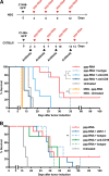
ppp-RNA induced tumor rejection is mediated by cellular immunity. a NSG mice and C57BL/6 mice (a, b) were inoculated with C1498-GFP AML cells and therapy was applied according to the schemes depicted in a. NSG mice (n = 5 for ppp-RNA treated, n = 4 for untreated, both derived from one experiment), C57BL/6 mice (n = 8 per group for ppp-RNA treated, untreated and ppp-RNA treated + NK1.1 depleted C57BL/6 mice; n = 7 for ppp-RNA treated+ CD4/CD8 depleted C57BL/6 mice. Data derive from two independent experiments; n = 4 for ppp-RNA treated + CD19 depleted C57BL/6 mice, these data derive from a single experiment). Depleting antibodies were administered as described in the materials and methods section. Corresponding isotype controls were tested in a total of n = 4 mice per group in a single experiment. Survival data were plotted in two Kaplan–Meier survival curves. P values of immune cell depleted groups compared to respective isotype controls were calculated using the log-rank test: p = 0.018 for CD4, p = 0.003 for CD8, p = 0.376 for NK1.1, p = 0.401 for CD19 depletion
Fig. 3
Fig. 3
ppp-RNA treatment depends on intact IFN alpha signaling. a As depicted in the schematic C57BL/6 WT, Mavs/ and Ifnar1/ mice were treated with 50 μg of ppp-RNA on days 3, 7, 10, and 14 after inoculation with C1498-GFP AML cells. Blood was drawn after the first (day 3) and fourth (day 14) treatment, and levels of murine CXCL10 were measured via ELISA. Each symbol represents a single mouse and error bars indicate SD. Statistical differences between genotypes at one time point were determined by one-way ANOVA with Tukey’s post-hoc test. b Ifnar1/ mice (n = 4 per group) were inoculated with C1498-GFP AML cells and treated according to the scheme depicted in a. Survival data were plotted in a Kaplan–Meier survival curve. p = 0.073 for ppp-RNA versus untreated mice. c C1498-GFP AML-bearing C57BL/6 wild type mice (n = 10) were randomized into two groups, of which one received 5 × 104 IU murine IFN alpha (mIFNα) i.p. on days 3, 7, 10, and 14 as depicted in the schematic in c. Survival data were plotted in a Kaplan–Meier survival curve. p = 0.352 for mIFNα treated versus untreated mice. d C57BL/6 Mavs/ (n = 9 per group) were treated with 50 μg of ppp-RNA on days 3, 7, 10, and 14 or left untreated according to the scheme depicted in a. Survival data were plotted in a Kaplan–Meier survival curve. The data shown are derived from one (b, c) or two independent (d) experiments
Fig. 4
Fig. 4
Tumor rechallenge of C1498-GFP AML surviving mice and adoptive transfer of CD8+ T cells. a C1498-GFP AML surviving C57BL/6 mice (n = 7) were rechallenged with 106 C1498-GFP AML cells. No further treatment was applied. Tumor-naive C57BL/6 mice (n = 10) served as controls. Survival data derived from three independent experiments were plotted in a Kaplan–Meier survival curve. b As depicted in the schematic C57BL/6 mice (n = 4) were treated with CD8+ T cells from C1498-GFP AML surviving (T cells (survivor), n = 3) or tumor-naive mice (T cells (ctrl), n = 5), respectively. Untreated mice (n = 8) served as controls. C1498-GFP AML was induced 12 h later in all three groups. T cells were isolated as described in the “Materials and methods” section. Survival data derived from two independent experiments were plotted in a Kaplan–Meier survival curve. Significance was calculated using log-rank test
Fig. 5
Fig. 5
ppp-RNA treatment primes AML cells for anti-PD-1 checkpoint inhibition. a 2.5 × 105 C1498-GFP AML cells were seeded in six-well format and treated with interferon gamma. PD-L1 expression was determined by flow cytometry 72 h after stimulation. b C1498-GFP AML-bearing C57BL/6 mice received three treatments of 50 μg ppp-RNA on days 8, 11, and 14 after tumor induction. Twelve hours upon the last treatment (day 15), mice were sacrificed and single cell suspensions of lung tissue were analyzed by flow cytometry, determining PD-L1 expression on GFP+ AML cells. c, d C57BL/6 mice (n = 13 per group derived from two independent experiments) were inoculated with C1498-GFP AML cells on day 0 and treated with 25 μg of ppp-RNA on days 3, 7, 10, and 14. Hundred micrograms of anti-PD-1 antibody was injected i.p. on days 6, 9, and 13. Levels of murine CXCL10 were determined by ELISA in blood serum obtained 4 h after the first treatment with 25 μg of ppp-RNA (n = 8 per group) c. Survival data were plotted in a Kaplan–Meier survival curve (e). Statistical significance was determined by the Student’s t test (a, b), one-way ANOVA with the Tukey’s post-hoc test (c) and the log-rank test (e)
Fig. 6
Fig. 6
Efficacy of ppp-RNA in a humanized mouse model of AML. Twelve NSG mice were inoculated with 4.5 × 105 PDX AML-491 cells i.v. on day 0 and 107 human PBMCs were injected on day 52. ppp-RNA treatment was given on days 53, 56, and 59. Mice were sacrificed 12 h after the last treatment on day 60. Single cell suspensions of peripheral blood (a) and bone marrow (b) were analyzed by flow cytometry, determining levels of murine CD45 positive (mCD45+) and human CD3 positive cells (hCD3+) relating to murine plus human CD45 positive (pan hCD45+) cells. Tumor burdens were determined by the detection of mCherry positive cells. Bar charts depict mean values plus SEM of n = 6 (a) and n = 3 (b). Statistical significance was determined using a Student’s t test with comparisons indicated by brackets

References (VSports)

    1. Reinisch A, Chan SM, Thomas D, Majeti R. Biology and clinical relevance of acute myeloid leukemia stem cells. Semin Hematol. 2015;52:150–64. doi: 10.1053/j.seminhematol.2015.03.008. - DOI - PMC - PubMed
    1. Kassim AA, Savani BN. Hematopoietic stem cell transplantation for acute myeloid leukemia: a review. Hematol/Oncol Stem Cell Ther. 2017;10:245–51. doi: 10.1016/j.hemonc.2017.05.021. - DOI - PubMed
    1. Lichtenegger FS, Krupka C, Haubner S, Köhnke T, Subklewe M. Recent developments in immunotherapy of acute myeloid leukemia. J Hematol Oncol. 2017;10:1–20. doi: 10.1186/s13045-017-0505-0. - DOI - PMC - PubMed
    1. Poeck H, Besch R, Maihoefer C, Renn M, Tormo D, Morskaya SS, et al. 5′-Triphosphate-siRNA: turning gene silencing and Rig-I activation against melanoma. Nat Med. 2008;14:1256–63. doi: 10.1038/nm.1887. - V体育2025版 - DOI - PubMed
    1. Tormo D, Checińska A, Alonso-Curbelo D, Pérez-Guijarro E, Cañón E, Riveiro-Falkenbach E, et al. Targeted activation of innate immunity for therapeutic induction of autophagy and apoptosis in melanoma cells. Cancer Cell. 2009;16:103–14. doi: 10.1016/j.ccr.2009.07.004. - V体育安卓版 - DOI - PMC - PubMed

Publication types

MeSH terms

VSports手机版 - LinkOut - more resources