Skip to main page content
U.S. flag

An official website of the United States government

Dot gov

The . gov means it’s official. Federal government websites often end in . gov or . mil. Before sharing sensitive information, make sure you’re on a federal government site VSports app下载. .

Https

The site is secure V体育官网. The https:// ensures that you are connecting to the official website and that any information you provide is encrypted and transmitted securely. .

. 2019 May;23(5):3451-3463.
doi: 10.1111/jcmm.14241. Epub 2019 Feb 26.

Nrf2 promotes breast cancer cell migration via up-regulation of G6PD/HIF-1α/Notch1 axis

Affiliations

Nrf2 promotes breast cancer cell migration via up-regulation of G6PD/HIF-1α/Notch1 axis

Hong-Sheng Zhang et al. J Cell Mol Med. 2019 May.

"V体育ios版" Abstract

Abnormal metabolism of tumour cells is closely related to the occurrence and development of breast cancer, during which the expression of NF-E2-related factor 2 (Nrf2) is of great significance. Metastatic breast cancer is one of the most common causes of cancer death worldwide; however, the molecular mechanism underlying breast cancer metastasis remains unknown VSports手机版. In this study, we found that the overexpression of Nrf2 promoted proliferation and migration of breast cancers cells. Inhibition of Nrf2 and overexpression of Kelch-like ECH-associated protein 1 (Keap1) reduced the expression of glucose-6-phosphate dehydrogenase (G6PD) and transketolase of pentose phosphate pathway, and overexpression of Nrf2 and knockdown of Keap1 had opposite effects. Our results further showed that the overexpression of Nrf2 promoted the expression of G6PD and Hypoxia-inducing factor 1α (HIF-1α) in MCF-7 and MDA-MB-231 cells. Overexpression of Nrf2 up-regulated the expression of Notch1 via G6PD/HIF-1α pathway. Notch signalling pathway affected the proliferation of breast cancer by affecting its downstream gene HES-1, and regulated the migration of breast cancer cells by affecting the expression of EMT pathway. The results suggest that Nrf2 is a potential molecular target for the treatment of breast cancer and targeting Notch1 signalling pathway may provide a promising strategy for the treatment of Nrf2-driven breast cancer metastasis. .

Keywords: G6PD; HIF-1α; Notch1; Nrf2; breast cancer V体育安卓版. .


Conflict of interest statement

The authors declare that there are no conflicts of interest.

Figures

Figure 1
Figure 1
Nrf2 overexpression and Kelch‐like ECH‐associated protein 1 (Keap1) knockdown promote proliferation of breast cancers cells. A, Expression of Nrf2 mRNA in MCF‐7 and MBA‐DA‐231 cells was examined by qRT‐PCR. GADPH was used as a reference for RNA. B, Expression of Nrf2 protein in MCF‐7 and MBA‐DA‐231 cells was performed by western blotting analysis. The signal obtained with a β‐actin antibody served as loading control. Blots were the representative of three independent experiments. C, Quantitative analysis of Nrf2 protein in MCF‐7 and MBA‐DA‐231 cells was shown. D, Cell viability was analysed by MTS assay. Results were means ± SEM of three independent experiments. **P < 0.01 compared to untreated cells
Figure 2
Figure 2
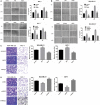
Nrf2 overexpression and Kelch‐like ECH‐associated protein 1 (Keap1) knockdown promote migration and invasion of breast cancers cells. A, Wound healing assay with MCF‐7 cells expressing shNrf2, shKeap1, Nrf2 or Keap1. B, Quantitative analysis of wound healing assay with MDA‐MB‐231 cells expressing shNrf2, shKeap1, Nrf2 or Keap1. Images were captured 24, 48, 72 h after wound was formed. The percentage of migration was assigned as 100% when complete fusion occurred, and 0% at t = 0 h. Relative migratory rate was shown in the graph. C, Transwell invasion assay of MCF‐7 and MDA‐MB‐231 cells expressing Nrf2 or Keap1 for 24 h. D, Quantitative analysis of transwell invasion assay of MCF‐7 and MDA‐MB‐231 cells expressing shNrf2 or shKeap1 for 24 h. Representative figures from three independent experiments were shown. Results were means ± SEM of three independent experiments. **P < 0.01 compared to untreated cells
Figure 3
Figure 3
Effect of Nrf2 on pentose‐phosphate pathway in breast cancer cells. A, Expression of glucose‐6‐phosphate dehydrogenase (G6PD) and TKT mRNA in MCF‐10A, MCF‐7 and MBA‐DA‐231 cells was examined by qRT‐PCR. GADPH was used as a reference for RNA. (B) Expression of G6PD and TKT mRNA in MCF‐7 cells was examined by qRT‐PCR. GADPH was used as a reference for RNA. (C) Expression of G6PD and TKT mRNA in MBA‐DA‐231 cells was examined by qRT‐PCR. GADPH was used as a reference for RNA. (D) Expression of G6PD and TKT protein in MCF‐7 and MBA‐DA‐231 cells was performed by western blotting analysis. The signal obtained with a β‐actin antibody served as loading control. Blots were the representative of three independent experiments. (E) Quantitative analysis of G6PD and TKT protein in MCF‐7 and MBA‐DA‐231 cells was shown. Results were means ± SEM of three independent experiments. **P < 0.01 compared to untreated cells
Figure 4
Figure 4
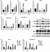
Nrf2 regulated Notch signalling in breast cancer cells. A, Expression of Notch1‐4, Dll1, Dll3, Dll4, Jagged1 and Jagged2 mRNA in MCF‐7 and MBA‐DA‐231 cells was examined by qRT‐PCR. GADPH was used as a reference for RNA. B, Expression of Notch1‐4, Dll1, Dll3, Dll4, Jagged1 and Jagged2 protein in MCF‐7 and MBA‐DA‐231 cells was performed by western blotting analysis. The signal obtained with a β‐actin antibody served as loading control. Blots were the representative of three independent experiments. C, Quantitative analysis of Notch1‐4, Dll1, Dll3, Dll4, Jagged1 and Jagged2 protein in MCF‐7 and MBA‐DA‐231 cells was shown. Results were means ± SEM of three independent experiments. **P < 0.01 compared to untreated cells
Figure 5
Figure 5
Nrf2 regulated Notch signalling via glucose‐6‐phosphate dehydrogenase (G6PD) in breast cancer cells. A, Relative luciferase activity was assayed in MCF‐7 and MBA‐DA‐231 cells. B, Expression of G6PD and Notch1 mRNA in MCF‐7 and MBA‐DA‐231 cells was examined by qRT‐PCR. GADPH was used as a reference for RNA. C, Expression of G6PD and Notch1 protein in MCF‐7 and MBA‐DA‐231 cells was performed by western blotting analysis. The signal obtained with a β‐actin antibody served as loading control. Blots were the representative of three independent experiments. D, Quantitative analysis of G6PD and Notch1 protein in MCF‐7 and MBA‐DA‐231 cells was shown. Results were means ± SEM of three independent experiments. **P < 0.01 compared to untreated cells
Figure 6
Figure 6
Nrf2 regulated Notch signalling via HIF‐1α in breast cancer cells. A, Expression of HIF‐1α and Notch1 mRNA in MCF‐7 and MBA‐DA‐231 cells was examined by qRT‐PCR. GADPH was used as a reference for RNA. B, Expression of HIF‐1α and Notch1 protein in MCF‐7 and MBA‐DA‐231 cells was performed by western blotting analysis. The signal obtained with a β‐actin antibody served as loading control. Blots were the representative of three independent experiments. C, Quantitative analysis of HIF‐1α and Notch1 protein in MCF‐7 and MBA‐DA‐231 cells was shown. D, Expression of HIF‐1α and Notch1 mRNA in Nrf2 overexpression or knockdown MCF‐7 and MBA‐DA‐231 cells was examined by qRT‐PCR. GADPH was used as a reference for RNA. E, Expression of HIF‐1α and Notch1 protein in Nrf2 overexpression or knockdown MCF‐7 and MBA‐DA‐231 cells was performed by western blotting analysis. The signal obtained with a β‐actin antibody served as loading control. Blots were the representative of three independent experiments. F, Quantitative analysis of HIF‐1α and Notch1 protein in Nrf2 overexpression or knockdown MCF‐7 and MBA‐DA‐231 cells was shown. Results were means ± SEM of three independent experiments. **P < 0.01 compared to untreated cells
Figure 7
Figure 7
Nrf2 can affect breast cancer cell proliferation and migration by influencing the expression of Notch1. A, Expression of Notch1 and Hes1 mRNA in MCF‐7 and MBA‐DA‐231 cells was examined by qRT‐PCR. GADPH was used as a reference for RNA. B, Expression of Notch1 and Hes1 protein in MCF‐7 and MBA‐DA‐231 cells was performed by western blotting analysis. The signal obtained with a β‐actin antibody served as loading control. Blots were the representative of three independent experiments. C, Quantitative analysis of Notch1 and Hes1 protein in MCF‐7 and MBA‐DA‐231 cells was shown. D, Cell viability was analysed by MTS assay. E, Wound healing assay with MCF‐7 and MBA‐DA‐231 cells expressing shNrf2, shKeap1, Nrf2 or Kelch‐like ECH‐associated protein 1 (Keap1). Images were captured 24, 48, 72 h after wound was created. The percentage of migration was assigned as 100% when complete fusion occurred, and 0% at t = 0 h. Relative migratory rate was shown in the graph. F, Transwell invasion assay of MCF‐7 and MBA‐DA‐231 cells expressing shNrf2, shKeap1, Nrf2 or Keap1 for 24 h. Representative figures from three independent experiments were shown. G, Expression of Notch1, E‐cadherin, N‐cadherin and Snail1 protein in MCF‐7 and MBA‐DA‐231 cells was performed by western blotting analysis. The signal obtained with a β‐actin antibody served as loading control. Blots were the representative of three independent experiments. H, Quantitative analysis of Notch1, E‐cadherin, N‐cadherin and Snail1 protein in MCF‐7 and MBA‐DA‐231 cells was shown. Results were means ± SEM of three independent experiments. **P < 0.01 compared to untreated cells
Figure 8
Figure 8
A model of Nrf2‐mediated Notch1 activation via glucose‐6‐phosphate dehydrogenase (G6PD)/HIF‐1α axis in breast cancer cells

References

    1. Hu ZI, McArthur HL. Immunotherapy in breast cancer: the New Frontier. Curr Breast Cancer Rep. 2018;10:35‐40. - PMC (VSports最新版本) - PubMed
    1. Walker A, Singh A, Tully E, et al. Nrf2 signaling and autophagy are complementary in protecting breast cancer cells during glucose deprivation. Free Radic Biol Med. 2018;120:407‐413. - "V体育2025版" PMC - PubMed
    1. Carlisi D, De Blasio A, Drago‐Ferrante R, et al. Parthenolide prevents resistance of MDA‐MB231 cells to doxorubicin and mitoxantrone: the role of Nrf2. Cell Death Discov. 2017;3:17078. - PMC - PubMed
    1. Yamamoto M, Kensler TW, Motohashi H. The KEAP1‐NRF2 system: a thiol‐based sensor‐effector apparatus for maintaining redox homeostasis. Physiol Rev. 2018;98:1169‐1203. - PMC - PubMed
    1. Raghunath A, Sundarraj K, Nagarajan R, et al. Antioxidant response elements: discovery, classes, regulation and potential applications. Redox Biol. 2018;17:297‐314. - PMC - PubMed

VSports app下载 - Publication types

MeSH terms

Substances

Associated data