Skip to main page content
U.S. flag

An official website of the United States government

Dot gov

The . gov means it’s official. Federal government websites often end in VSports app下载. gov or . mil. Before sharing sensitive information, make sure you’re on a federal government site. .

Https

The site is secure. The https:// ensures that you are connecting to the official website and that any information you provide is encrypted and transmitted securely. V体育官网.

. 2018 May;6(10):e13683.
doi: 10.14814/phy2.13683.

Matrix metalloproteinase (MMP)-7 in Barrett's esophagus and esophageal adenocarcinoma: expression, metabolism, and functional significance (V体育平台登录)

Affiliations

Matrix metalloproteinase (MMP)-7 in Barrett's esophagus and esophageal adenocarcinoma: expression, metabolism, and functional significance (VSports app下载)

Hanan M Garalla et al. Physiol Rep. 2018 May.

Abstract (V体育平台登录)

Matrix metalloproteinase (MMP)-7, unlike many MMPs, is typically expressed in epithelial cells. It has been linked to epithelial responses to infection, injury, and tissue remodeling including the progression of a number of cancers. We have now examined how MMP-7 expression changes in the progression to esophageal adenocarcinoma (EAC), and have studied mechanisms regulating its expression and its functional significance. Immunohistochemistry revealed that MMP-7 was weakly expressed in normal squamous epithelium adjacent to EAC but was abundant in epithelial cells in both preneoplastic lesions of Barrett's esophagus and EAC particularly at the invasive front. In the stroma, putative myofibroblasts expressing MMP-7 were abundant at the invasive front but were scarce or absent in adjacent tissue. Western blot and ELISA revealed high constitutive secretion of proMMP-7 in an EAC cell line (OE33) that was inhibited by the phosphatidylinositol (PI) 3-kinase inhibitor LY294002 but not by inhibitors of protein kinase C, or MAP kinase activation. There was detectable proMMP-7 in cultured esophageal myofibroblasts but it was undetectable in media. Possible metabolism of MMP-7 by myofibroblasts studied by proteomic analysis indicated degradation via extensive endopeptidase, followed by amino- and carboxpeptidase, cleavages. Myofibroblasts exhibited increased migration and invasion in response to conditioned media from OE33 cells that was reduced by MMP-7 knockdown and immunoneutralization VSports手机版. Thus, MMP-7 expression increases at the invasive front in EAC which may be partly attributable to activation of PI 3-kinase. Secreted MMP-7 may modify the tumor microenvironment by stimulating stromal cell migration and invasion. .

Keywords: Barrett's esophagus; MMP-7; PI3-kinase; myofibroblast; proteomic. V体育安卓版.


"V体育2025版" Figures

Figure 1
Figure 1
Expression of MMP‐7 in EAC and preneoplastic tissue. (A) epithelial cells in control squamous tissue (n = 9); (B) epithelial cells in Barrett's metaplasia (n = 8); (C) epithelial cells in dysplasia (n = 11); (D) epithelial cells in EAC (n = 14); (E) epithelial cells at the invasive front in EAC (n = 13); (F) stromal cells in dysplasia (n = 4); (G) stromal cells in EAC (n = 14); (H) stromal cells at the invasive front in EAC (n = 13). In each case, upper part of the panel shows a ×40 image with arrows indicating staining, and lower part shows the proportion of cells in that compartment assigned to four categories of staining intensity (0–3: 0,open bars; 1, stippling on white background; 2, cross hatching; 3, stippling on black background). Results from 14 surgically resected tumors; not all compartments were represented in all tumors, hence some “n” values less than 14; too few stromal cells were identified in macroscopically normal tissue, Barrett's tissue, and all but 4 of the dysplastic tissues, to be scored;. Means ± S.E.
Figure 2
Figure 2
Expression of MMP‐7 in Barrett's esophagus biopsies. (A) epithelial cells in Barrett's intestinal metaplasia (n = 17); (B) epithelial cells in low‐grade dysplasia (n = 17); (C) epithelial cells in high‐grade dysplasia (n = 12). In each case, upper panel shows a ×40 image and the lower panel shows the proportion of cells in that compartment assigned to categories of staining intensity 0–3 (see Fig 1 legend for details). Means ± S.E.
Figure 3
Figure 3
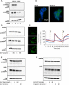
Expression of MMP‐7 in OE33 cells is insensitive to PMA and gastrin. (A) OE33, but not OE19, cell media contains abundant proMMP‐7 and a minor band corresponding to MMP‐7; there is also abundant proMMP‐7 in OE33 cell extracts and just detectable amounts in OE19 cell extracts: GAPDH in cell extracts confirms equal loading; PMA (100 nmol/L) does not stimulate proMMP‐7 expression. (B) Immunocytochemistry using CCK2R antibody confirms expression of CCK2R in OE33‐Gr cells (which have been stably transfected with CCK2R) but not wild‐type OE33 cells (green CCK2R staining; blue, nuclear staining with DAPI). (C) Heptadecapeptide gastrin (G17, 0.1–10 nmol/L) did not influence proMMP‐7 abundance in OE33‐Gr cells or media. (D) Left, representative fluorescence images of OE33‐Gr cells (top, control), treated with G17 (10 nmol/L, center), or ionomycin (bottom); right, traces from four cells, two responded to G17 (10 nmol/L, applied at I) and all responded to ionomycin (applied at II). (E) Gly‐extended G17 (G17‐Gly, 1 and 10 nmol/L) had no effect on proMMP‐7 abundance in media or cells of OE33 cells. (F) G17 extended to include the C‐terminal flanking peptide of progastrin (G17‐CFP, 1 and 10 nmol/L) and intact progastrin had no effect on proMMP‐7 abundance in OE33 media or cells.
Figure 4
Figure 4
Role of PI 3‐kinase in MMP‐7 expression in OE33 cells. (A) Western blot showing suppression of proMMP‐7 in media of OE33 cells following treatment with brefeldin A (BFA, 10 μg/mL) and LY294002 (LY, 50 μmol/L) but not Ro32‐0432 (Ro, 2 μmol/L) or UO126 (UO, 10 μmol/L). (B) Similar data obtained using an ELISA for MMP‐7 in media. (C) Inhibition of PI 3‐kinase with LY294002 suppressed phosphorylation (p‐, phospho; t‐, total) of the downstream mediator Akt in cell extracts. In each case, experiments were performed in serum‐free medium added together with relevant drugs at the start of the experiment, the duration of the experiment was 6 h. Horizontal bars, P < 0.05.
Figure 5
Figure 5
Expression of MMP‐7 in myofibroblasts. (A) In cell extracts of three different CAM lines (CAM‐1, ‐2 and ‐3) there was a band corresponding to proMMP‐7 (28 kD) in western blots; in some cases, and in OE33 cells run as a positive control, there was a minor band at 26 kD. (B) Immunocytochemistry of MMP‐7 in myofibroblasts revealed expression (green) in a high proportion of cells (left), and at a higher power (right) localization is confirmed to vesicular organelles. (C) In cell extracts of myofibroblasts treated with BFA to arrest secretion there was an increase in intracellular proMMP‐7. (D) However, in the media of three different CAM lines proMMP‐7 or related proteins were undetectable, although in the same experiments there was abundant proMMP‐7 in OE33 cell media run as a positive control, and there was abundant MMP‐1 in both CAM and OE33 cell media.
Figure 6
Figure 6
Proteolysis of MMP‐7 by myofibroblasts. (A) In the medium of myofibroblasts incubated (37°C, 30 min) with recombinant human MMP‐7 (2 μg), western blot reveals both the starting material (19 kD, see control samples of MMP‐7 incubated in medium in the absence of cells, ‘Control’) and two bands corresponding to degradation products at 14 and 11 kD. (B) Label‐free quantification of MMP‐7 in the medium, based on the summed intensity of 5 unique tryptic peptides shows substantial loss of protein by 24 h compared with 30 min. (C) Analysis of MMP‐7 fragments after 30 min incubation revealed numerous fragments attributable to endopeptidase cleavage followed by aminopeptidase or carboxypeptidase trimming of the cleavage products, separately or in concert; blue, tryptic peptides; red, semi‐tryptic on the N‐terminal side of Lys or Arg; green, semi‐tryptic on the C‐terminal side of Lys or Arg; black, non‐tryptic peptides. Inset is a schematic representation of the distribution and relative abundance of cleavage products averaged over three biological replicates using Peptigram (Manguy et al. 2017). For each residue, a green bar is drawn if this position is covered by at least one peptide in the sample; height of the bars is proportional to the count of peptides overlapping this position; colour intensity is proportional to the summed ion intensities of peptides overlapping this position, with dark green indicating high peptide intensity and light green indicating low peptide intensity. No fragments are observed before residue 95 which is the start of the mature form of MMP‐7 used in this study.
Figure 7
Figure 7
MMP‐7 mediates cancer cell stimulation of myofibroblast migration. (A) recombinant MMP‐7 (2 μg/mL) stimulates the migration (left) and invasion (right) of EAC myofibroblasts in Boyden chambers. (B) siRNA knockdown of MMP‐7 expression in OE33 cells reduces expression by 80% while expression of MMP‐1 is preserved. (C) representative images of myofibroblasts in migration (top) and invasion (bottom) assays using Boyden chamber chemotaxis assays in response to conditioned medium from control (left), sham (center), and siRNA‐treated (right) OE33 cells; scale bar 20 μm. (D) Conditioned medium (hatched bars) from sham and MMP‐7 siRNA‐treated OE33 cells stimulates migration (left), invasion (center), and proliferation (right) of EAC myofibroblasts and siRNA treatment significantly reduces the migration and invasion responses. Representative results are shown for migration studies in two different CAM lines. Horizontal bars, P < 0.05.
Figure 8
Figure 8
Schematic representation of the role of MMP‐7 in the progression from Barrett's esophagus to esophageal adenocarcinoma. Differences in font size and line thickness indicate the magnitude of effects. It is suggested that there is progressively increased expression of proMMP‐7 in progress to EAC at least in part due to activation of PI 3‐kinase, that following secretion conversion of proMMP‐7 to MMP‐7 activates stromal cells stimulating myofibroblast migration and invasion and remodeling of the cancer cell microenvironment.

References

    1. Adachi, Y. , Itoh F., Yamamoto H., Matsuno K., Arimura Y., Kusano M., et al. 1998. Matrix metalloproteinase matrilysin (MMP‐7) participates in the progression of human gastric and esophageal cancers. Int. J. Oncol. 13:1031–1035. - PubMed
    1. Adachi, Y. , Li R., Yamamoto H., Min Y., Piao W., Wang Y., et al. 2009. Insulin‐like growth factor‐I receptor blockade reduces the invasiveness of gastrointestinal cancers via blocking production of matrilysin. Carcinogenesis 30:1305–1313. - PubMed
    1. Balabanova, S. , Holmberg C., Steele I., Ebrahimi B., Rainbow L., Burdyga T., et al. 2014. The neuroendocrine phenotype of gastric myofibroblasts and its loss with cancer progression. Carcinogenesis 35:1798–1806. - PMC - PubMed
    1. Beales, I. L. , and Ogunwobi O. O.. 2009. Glycine‐extended gastrin inhibits apoptosis in Barrett's oesophageal and oesophageal adenocarcinoma cells through JAK2/STAT3 activation. J. Mol. Endocrinol. 42:305–318. - PubMed
    1. Beales, I. L. , Ogunwobi O., Cameron E., El‐Amin K., Mutungi G., and Wilkinson M.. 2007. Activation of Akt is increased in the dysplasia‐carcinoma sequence in Barrett's oesophagus and contributes to increased proliferation and inhibition of apoptosis: a histopathological and functional study. BMC Cancer 7:97. - PMC - PubMed

Publication types

MeSH terms

LinkOut - more resources