Skip to main page content
U.S. flag

An official website of the United States government

Dot gov

The . gov means it’s official. Federal government websites often end in . gov or . mil. Before sharing sensitive information, make sure you’re on a federal government site. VSports app下载.

Https

The site is secure V体育官网. The https:// ensures that you are connecting to the official website and that any information you provide is encrypted and transmitted securely. .

. 2018 Aug;67(8):1493-1504.
doi: 10.1136/gutjnl-2017-315193. Epub 2018 Feb 6.

CPT2 downregulation adapts HCC to lipid-rich environment and promotes carcinogenesis via acylcarnitine accumulation in obesity

Affiliations

CPT2 downregulation adapts HCC to lipid-rich environment and promotes carcinogenesis via acylcarnitine accumulation in obesity

Naoto Fujiwara (V体育官网) et al. Gut. 2018 Aug.

"VSports app下载" Abstract

Objective: Metabolic reprogramming of tumour cells that allows for adaptation to their local environment is a hallmark of cancer. Interestingly, obesity-driven and non-alcoholic steatohepatitis (NASH)-driven hepatocellular carcinoma (HCC) mouse models commonly exhibit strong steatosis in tumour cells as seen in human steatohepatitic HCC (SH-HCC), which may reflect a characteristic metabolic alteration VSports手机版. .

Design: Non-tumour and HCC tissues obtained from diethylnitrosamine-injected mice fed either a normal or a high-fat diet (HFD) were subjected to comprehensive metabolome analysis, and the significance of obesity-mediated metabolic alteration in hepatocarcinogenesis was evaluated. V体育安卓版.

Results: The extensive accumulation of acylcarnitine species was seen in HCC tissues and in the serum of HFD-fed mice. A similar increase was found in the serum of patients with NASH-HCC. The accumulation of acylcarnitine could be attributed to the downregulation of carnitine palmitoyltransferase 2 (CPT2), which was also seen in human SH-HCC V体育ios版. CPT2 downregulation induced the suppression of fatty acid β-oxidation, which would account for the steatotic changes in HCC. CPT2 knockdown in HCC cells resulted in their resistance to lipotoxicity by inhibiting the Src-mediated JNK activation. Additionally, oleoylcarnitine enhanced sphere formation by HCC cells via STAT3 activation, suggesting that acylcarnitine accumulation was a surrogate marker of CPT2 downregulation and directly contributed to hepatocarcinogenesis. HFD feeding and carnitine supplementation synergistically enhanced HCC development accompanied by acylcarnitine accumulation in vivo. .

Conclusion: In obesity-driven and NASH-driven HCC, metabolic reprogramming mediated by the downregulation of CPT2 enables HCC cells to escape lipotoxicity and promotes hepatocarcinogenesis. VSports最新版本.

Keywords: CPT2; acylcarnitine; hepatocellular carcinoma; metabolic reprograming; metabolome V体育平台登录. .


Conflict of interest statement

Competing interests: None declared.

Figures
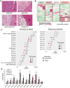
Figure 1
Figure 1. Acylcarnitine species markedly accumulate in obesity-driven HCC tissues
(A) Hematoxylin and eosin (H&E)-stained images of livers from mice with obesity- and NASH-driven HCC (scale bar, 100 μm). NT and T indicate non-tumor and tumor areas, respectively. (B) Hierarchical clustering analyses of liquid chromatography-mass spectrometry data were performed using NT and HCC tissues obtained from DEN-injected 8-month-old mice fed a ND or a HFD (ND-NT, n = 3; ND-HCC, n = 3; HFD-NT, n = 3; HFD-HCC, n = 4). (C) Significantly accumulated metabolites in HFD-HCC tissues compared with ND-NT or ND-HCC tissues (false discovery rate < 0.25). Size of the circles indicates fold changes and color of the circles indicates types of metabolites (acylcarnitine species or others). AC, acylcarnitine; FA, fatty acid. (D) The bar graph shows the relative amounts of acylcarnitine species of various carbon chain lengths. The data are presented as the fold change relative to the average amount in ND-NT tissues (means ± SEM); * p < 0.05 vs. ND-NT tissues, p < 0.05 vs. ND-HCC tissues, p < 0.05 vs. HFD-NT tissues.
Figure 2
Figure 2. Expression of acylcarnitine-metabolism-related genes in obesity-driven HCC
(A) The relative expression levels of the indicated genes determined using real-time PCR in NT and HCC tissues obtained from DEN-injected, ND- or HFD-fed mice (means ± SEM, n = 5 per group); * p < 0.05 vs. ND-NT tissues, p < 0.05 vs. HFD-NT tissues. (B) The relative expression levels of the indicated genes determined using real-time PCR in NT and tumor tissues obtained from HFD-fed MUP-uPA mice (n = 6) and PIK3CA Tg mice (n = 5). The data are presented as the means ± SEM; * p < 0.05 vs. NT tissues. (C) WB analysis of CPT1A and CPT2 proteins in NT and HCC tissues obtained from DEN-injected, ND- or HFD-fed mice. Data were quantified using ImageJ software (means ± SEM, n = 5 per group); *p < 0.05 vs. ND-NT tissues, p < 0.05 vs. HFD-NT tissues. (D) Representative IHC image of CPT2 in the liver obtained from DEN-injected HFD-fed mice (scale bar, 50 μm). Bar graph shows relative CPT2 expression levels in ND-HCC, HFD-NT, and HFD-HCC tissues compared with ND-NT tissues based on IF staining analysis (n = 7 per group); *p < 0.05 vs. ND-NT tissues, p < 0.05 vs. ND-HCC tissues, p < 0.05 vs. HFD-NT tissues. (E) Acylcarnitine concentrations in culture supernatants of control- and CPT2 KD-Dih10 cells (means ± SEM, n = 3 per group); *p < 0.05. (F) Relative PPARα mRNA expression levels determined using real-time PCR in tumor and non-tumor tissues obtained from DEN+HFD (n = 5 per group), MUP-uPA+HFD (n = 6), and PIK3CA Tg mice models (n = 5). The data are presented as the means ± SEM. DEN+HFD model; *p < 0.05 vs. ND-NT tissues, p < 0.05 vs. HFD-NT tissues. MUP-uPA+HFD, and PIK3CA Tg mice models; *p < 0.05 vs. NT tissues. (G) Relative CPT2 mRNA expression levels determined using real-time PCR in Dih10 cells 24 h after incubation with fenofibrate (50 μM) or vehicle control (DMSO). The data are presented as the fold change relative to the average level in vehicle-treated control Dih10 cells (means ± SEM, n = 3 per group); *p < 0.05.
Figure 3
Figure 3. Serum acylcarnitine levels are elevated in patients with NAFLD-related HCC
(A) Serum levels of total carnitine, free carnitine, and acylcarnitine in DEN-injected ND- or HFD-fed mice and non-injected age-matched ND- or HFD-fed mice (means ± SEM, n = 5 per group); *p < 0.05 vs. ND group, †p < 0.05 vs. ND+DEN group, ‡p < 0.05 vs. HFD group. (B) Violin plots of acylcarnitine and free carnitine serum levels in patients with NAFLD with or without HCC. (C) ROC curves of serum acylcarnitine, AFP, and DCP for the presence of HCC and their AUROCs. (D) Multivariable analysis for the presence of HCC using the logistic regression model. (E) Representative images of CPT2 immunostaining using human SH-HCC and adjacent non-tumor liver tissue (scale bar, 50 μm). (F) Relative CPT2 expression levels in conventional HCC and SH-HCC tissues compared with paired adjacent non-tumor liver tissues were analyzed using IF staining (n = 20 per group); *p < 0.05.
Figure 4
Figure 4. CE–MS analyses of obesity-driven HCC tissues
(A) Concentration of β-hydroxybutyrate in NT and HCC tissues obtained from DEN-injected, ND- or HFD-fed mice, and liver tissues obtained from age-matched ND-fed control mice measured using the β-hydroxybutyrate assay kit. Data are the means ± SEM (n = 6 per group); *p < 0.05. (B) CE-MS analyses of NT and HCC tissues obtained from DEN-injected, ND- or HFD-fed, 8-month-old mice (ND-NT, n = 3; ND-HCC, n = 3; HFD-NT, n = 3; HFD-HCC, n = 4). Representative metabolites in the glycolytic pathway and TCA cycle are shown; *p < 0.05 vs. ND-NT tissues, p < 0.05 vs. HFD-NT tissues.
Figure 5
Figure 5. CPT2 down-regulation enables HCC cells to adapt to a lipid-rich environment
(A) Growth curve of control, CPT2 knockdown1 (KD1), and CPT2 KD2 Dih10 cells (means ± SEM, n = 3 per group). (B) Control, CPT2 KD1, and CPT2 KD2 Dih10 cells were incubated with PA and SA at the indicated concentrations for 24 h, after which cell viability was assessed (means ± SEM, n = 3 per group); *p < 0.05 vs. control Dih10 cells incubated with 100 μM PA or SA, p < 0.05 vs. control Dih10 cells incubated with 300 μM PA or SA. (C) Control, CPT2 KD1, and CPT2 KD2 Dih10 cells were incubated with 200 μM PA and the expression levels of the indicated proteins were then assessed using WB at the indicated time points. (D) Relative expression levels of Puma were determined using real-time PCR in control, CPT2 KD1, and CPT2 KD2 Dih10 cells 10 h after their incubation with 200 μM PA or the vehicle control (means ± SEM, n = 3 per group); *p < 0.05. (E) PA-resistant Dih10 cells were established by their chronic exposure to 50% of the lethal concentration of PA (75 μM) for 1 week. Control and PA-resistant Dih10 cells were incubated with 100 μM PA or control media for 48 h, after which cell numbers were assessed (means ± SEM, n = 3 per group); *p < 0.05. (F) Relative expression levels of CPT2 were determined using real-time PCR in control and PA-resistant Dih10 cells (means ± SEM, n = 3 per group); *p < 0.05.
Figure 6
Figure 6. AC18:1 enhances the self-renewal of HCC cells via STAT3 activation
(A) Growth curve of Dih10 cells incubated with AC16:0 or AC18:1 at the indicated concentrations. (B) Sphere formation assays of Dih10 cells with 5 μM AC16:0, 5 μM AC18:1, or control medium. Bar graph shows the total number of spheres per well (means ± SEM from 8 wells per group); *p < 0.05 vs. the control. Representative images are shown (scale bar, 200 μm). (C) Relative expression levels of stem cell markers as determined by real-time PCR in Dih10 cells incubated for 24 h with 5 μM AC 18:1 or the vehicle control (means ± SEM from 3 wells per group); *p < 0.05. (D) Dih10 cells were incubated with 5 μM AC18:1 for 24 h, after which the expression levels of the indicated proteins were assessed by WB. Data were quantified using the ImageJ software (means ± SEM, n = 3 per group); *p < 0.05. (E, F) Effects of the STAT3 inhibitor WP1066 (2 μM) on AC18:1-induced promotion of sphere formation (E) and the enhanced expression of stem cell marker mRNAs in Dih10 cells (F). Data are the means ± SEM (n = 3 per group); *p < 0.05.
Figure 7
Figure 7. HFD feeding and carnitine supplementation synergistically promote hepatocarcinogenesis
(A) Protocol for L-carnitine supplementation in combination with HFD feeding of DEN-injected mice. The mice were randomly assigned to the ND (n = 12), ND + carnitine (n = 12), HFD (n = 15), and HFD + carnitine groups (n = 14). (B, C) Effects of L-carnitine supplementation on HCC development. Representative images of the livers (B) and tumor numbers (C) from each group of mice are shown. The data are presented as the means ± SEM; *p < 0.05 vs. the HFD group. (D, E) Effects of L-carnitine supplementation on HFD-induced body weight gain, elevation of ALT levels, and liver steatosis. (D) Bar graphs show the body weight and serum ALT levels in the HFD and HFD + carnitine groups. The data are presented as the means ± SEM. (E) Representative H&E-stained images of the non-tumor area of the livers from mice in the HFD and HFD + carnitine groups (scale bar: 100 μm). (F) Relative amounts of acylcarnitine species of various carbon chain lengths in HCC tissues from the HFD and HFD + carnitine groups. Three tumors from each group were pooled and the amounts of acylcarnitine species measured. The data are presented as the fold change relative to the level in the HFD group. (G) WB analysis of STAT3 phosphorylation in HCC tissues from the HFD and HFD + carnitine groups. (H) Proposed mechanisms of obesity- and NASH-driven enhanced carcinogenesis.

Comment in

References

    1. El-Serag HB. Hepatocellular carcinoma. The New England journal of medicine. 2011;365(12):1118–27. - PubMed
    1. Calle EE, Rodriguez C, Walker-Thurmond K, et al. Overweight, obesity, and mortality from cancer in a prospectively studied cohort of U.S. adults. The New England journal of medicine. 2003;348(17):1625–38. - PubMed
    1. Fujiwara N, Nakagawa H, Kudo Y, et al. Sarcopenia, intramuscular fat deposition, and visceral adiposity independently predict the outcomes of hepatocellular carcinoma. J Hepatol. 2015;63(1):131–40. - V体育官网入口 - PubMed
    1. Park EJ, Lee JH, Yu GY, et al. Dietary and genetic obesity promote liver inflammation and tumorigenesis by enhancing IL-6 and TNF expression. Cell. 2010;140(2):197–208. - "V体育官网" PMC - PubMed
    1. Tilg H, Hotamisligil GS. Nonalcoholic fatty liver disease: Cytokine-adipokine interplay and regulation of insulin resistance. Gastroenterology. 2006;131(3):934–45. - PubMed (VSports注册入口)

Publication types

MeSH terms

Grants and funding (V体育ios版)