Skip to main page content
U.S. flag

An official website of the United States government

Dot gov

The . gov means it’s official. Federal government websites often end in . gov or . mil VSports app下载. Before sharing sensitive information, make sure you’re on a federal government site. .

Https

The site is secure V体育官网. The https:// ensures that you are connecting to the official website and that any information you provide is encrypted and transmitted securely. .

. 2017 Dec 14;8(70):115526-115545.
doi: 10.18632/oncotarget.23308. eCollection 2017 Dec 29.

Curcumol potentiates celecoxib-induced growth inhibition and apoptosis in human non-small cell lung cancer

Affiliations

"VSports app下载" Curcumol potentiates celecoxib-induced growth inhibition and apoptosis in human non-small cell lung cancer

Fangfang Cai et al. Oncotarget. .

Abstract

Combinatorial therapies that target multiple signaling pathways may provide improved therapeutic responses over monotherapies. Celecoxib and curcumol are two highly hydrophobic drugs which show bioavailability problems due to their poor aqueous solubility. In the present study, we evaluated the effects of celecoxib and curcumol alone and in combination on cell proliferation, invasion, migration, cell cycle and apoptosis induction in non-small cell lung cancer (NSCLC) cells using in vitro and in vivo experiments. Our data showed that the sensitivity of a combined therapy using low concentration of celecoxib and curcumol was higher than that of celecoxib or curcumol alone. Suppression of NF-κB transcriptional activity, activation of caspase-9/caspase-3, cell cycle G1 arrest, and inhibition of survival MAPK and PI3K/AKT signaling pathway contributed to the synergistic effects of this combination therapy for induction of apoptosis. Additionally, either celecoxib alone or in combination with curcumol inhibited NSCLC cell migration and invasion by suppressing FAK and matrix metalloproteinase-9 activities. Furthermore, the combined treatment reduced tumor volume and weight in xenograft mouse model, and significantly decreased tumor metastasis nodules in lung tissues by tail vein injection. Our results confirm and provide mechanistic insights into the prominent anti-proliferative activities of celecoxib and/or curcumol on NSCLC cells, which provide a rationale for further detailed preclinical and potentially clinical studies of this combination for the therapy of lung cancer. VSports手机版.

Keywords: apoptosis; celecoxib; curcumol; synergism; tumor metastasis V体育安卓版. .


"VSports" Conflict of interest statement

CONFLICTS OF INTEREST The authors declare no conflicts of interest.

Figures

Figure 1
Figure 1. Effect of celecoxib and curcumol on the growth of tumor cells in vitro
(A) Chemical structure of celecoxib and curcumol. (B) A549 cells, H1299 cells and BEAS-2B cells were treated with curcumol at different concentrations (0, 10, 20, 30, 40, 50, 60 μM) for 24 h, and the cell viability was assessed by MTT assay. (C) A549 cells, H1299 cells and BEAS-2B cells were treated with indicated concentrations (0, 5, 10, 20, 40, 80, 160 μM) of celecoxib for 24 h in the absence or presence of 30 μM curcumol. The cell viability was assessed by MTT assay. Data are represented as mean ± SD. *p < 0.05, **p < 0.01.
Figure 2
Figure 2. Curcumol enhances celecoxib-induced cell apoptosis and their combination suppresses the clonogenic growth of NSCLC cells
(A) A549 and H1299 cells were exposed to celecoxib (30 μM) and/or curcumol (30 μM). 18 h later, all cells were harvested for flow cytometry analysis. Annexin V/PI-stained cells were analyzed and the percentage of apoptotic cells was determined. The experiments were carried out independently in triplicate; representative data are shown. Data are represented as mean ± SD. *p < 0.05, **p < 0.01. Annexin V/PI double staining profile of A549 cells is also included. (B) A549 and H1299 cells were exposed to celecoxib (30 μM) and/or curcumol (30 μM) for 18 h. TUNEL assays were performed according to the manufacturer’s instructions. The rate of apoptosis was expressed as the percentage of total cells counted. Each bar shows the mean ± SD of three independent experiments, performed in triplicate. *p < 0.05, **p < 0.01. TUNEL staining profile of A549 cells is also shown. A dark brown DAB signal indicates positive staining, while shades of blue-green to greenish tan signifies a non-reactive cell. (C) Colony formation ability of NSCLC cells treated with celecoxib (30 μM) and/or curcumol (30 μM). The experiments were repeated three times (n = 3); representative data are shown. Data are represented as mean ± SD. *p < 0.05, **p < 0.01. Representative dishes of A549 cells evaluated by colony-forming assay are also included.
Figure 3
Figure 3. Celecoxib and curcumol-induced apoptosis is mediated through the caspase-dependent mitochondrial pathway in NSCLC cells
(A) Caspase-8, caspase-9, caspase-3, and PARP expression levels in A549 cells under different treatment conditions. All gels run under the same experimental conditions and the representative images of three different experiments were cropped and shown. (B) Activity of caspase-3 and caspase-9 in A549 cells treated with celecoxib and curcumol alone or in combination for 24 h. Data are presented as fold increases as determined by quantitative analysis. *p < 0.05. (C) Viability of A549 cells after treatment with caspase inhibitors. Cells were treated with inhibitors for 2 h before the 24 h treatments, after which cell viability was determined by MTT assay. Data are representative of three independent experiments and are represented as mean ± SD. **p < 0.01. (D) Expressions of the Bcl-2 family proteins, Bcl-2, Bcl-xl, Bax, Bad, in A549 cells under different treatment conditions. All gels run under the same experimental conditions and the representative images of three different experiments were cropped and shown. (E) Band intensity shown in (D) was quantified by Image J software. The ratio of Bax:Bcl-2 was shown. The results shown are representative of three different experiments. Data are represented as mean ± SD, *p < 0.05, **p < 0.01, ***p < 0.001.
Figure 4
Figure 4. Effects of celecoxib and curcumol on NF-κB, PI3K/AKT and MAPKs signaling pathway
(A) A549 and H1299 were treated with celecoxib (30 μM) and/or curcumol (30 μM) for 24 h. Nuclear proteins were extracted and subjected to Western blotting for p65 detection. Lamin B was used as loading control. Additionally, the whole cell extracts with the same treatment were prepared and analyzed for IκB-αexpression. (B) A549 and H1299 were treated with celecoxib (30 μM) and/or curcumol (30 μM) for 24 h. AKT, p-AKT, PI3K, and p-PI3K proteins in whole cell lysates were determined with specific antibodies. GAPDH was used as loading control. (C) A549 and H1299 were treated with celecoxib (30 μM) and/or curcumol (30 μM) for 24 h. Western blotting was performed to detect the levels of p-p38, p38, ERK and p-ERK respectively. Densitometric quantification of the immunoblot data in (A-C) is also shown and data are represented as mean ± SD. *p < 0.05, **p < 0.01. (D) The MEK inhibitor PD98059 and PI3K inhibitor LY294002 were used to evaluate whether ERK phosphorylation and AKT inactivation, respectively, are required for apoptosis. The percentages of apoptotic cell death were measured via Annexin V/PI staining followed by flow cytometry analysis. Data are represented as mean ± SD, *p < 0.05.
Figure 5
Figure 5. Effect of celecoxib and curcumol on cell cycle distribution
(A) A549 and H1299 were treated with celecoxib (30 μM) and/or curcumol (30 μM) for 24 h. The cells were then fixed with 70% ethanol and stained with PI. The cell cycle distribution (G0/G1, G2/M and S) was determined by flow cytometry. (B) Western blotting was also performed to detect the levels of cell cycle regulators (cyclin D1, cyclin E, cdk2 and p21). Band intensity was quantified by Image J software. The results shown are representative of three different experiments. Data are represented as mean ± SD, *p < 0.05, **p < 0.01.
Figure 6
Figure 6. Effect of celecoxib and curcumol on cell migration
(A) Wound healing assays. A549 and H1299 cells were treated with celecoxib (30 μM) and/or curcumol (30 μM). Photographs were taken immediately and after 24 h of creating the scratch. Images shown are representative of three independent experiments. (B) Transwell assay. A549 and H1299 cells were treated with celecoxib (30 μM) and/or curcumol (30 μM). After 16 h pretreatment and 9 h incubation in the upper chamber, the cells migrating to the lower membrane were stained and counted in five fields with a magnification of × 100. N = 3, bar = 50 μm. The experiments were carried out in triplicate and representative data are shown. (C) Effects of celecoxib and curcumol on FAK, p-FAK, MMP-2 and MMP-9 protein expression. A549 and H1299 cells were treated with celecoxib (30 μM) and/or curcumol (30 μM). 24 h later, cells were harvested for Western blotting analysis using indicated antibodies. The level of GAPDH served as the loading control. Band intensities were calculated using software Image J. Relative intensities are also shown. Data represent mean values of triplicate samples. (D) A549 and H1299 cells were treated with celecoxib (30 μM) and/or curcumol (30 μM). 24 h later, cells were harvested for RNA extraction and quantitative real time PCR using primers specific for human MMP-2, MMP-9, and GAPDH (internal control). Data represent mean values of triplicate samples. *p < 0.05, **p < 0.01.
Figure 7
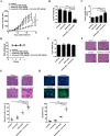
Figure 7. Celecoxib and curcumol combined therapy inhibits in vivo tumor xenograft growth in a subcutaneous tumor model
A549 cells were injected subcutaneously into the dorsal flanks of athymic nude mice. When tumors reached a size of approximately 50 mm3, mice were i.g. with celecoxib and i.p. with curcumol or the combination of two drugs every other day for a total of 25 days. (A) The tumor growth inhibitory effects of different treatments were compared. (B) At the end of the study, the excised tumors from each group were weighed. (C) Tumor double time of each group. (D) The weight of nude mice from each group did not change significantly during the experiment. (E) Liver weight of mice at the end of the experiment show in (A). (F) Representative photomicrographs of liver sections stained with H&E of mice treated with PBS, celecoxib, curcumol, or celecoxib + curcumol. (G) Determination of tumor necrosis after combined treatment with celecoxib and curcumol. Tumor necrosis areas are shown by H&E staining and observed under light microscope (×100). The viable tumor cells are indicated by a blue arrow. Tumor necrosis was determined by software Image J. Two sections/mouse and three mice were prepared. (H) Determination of apoptosis after combined treatment with celecoxib and curcumol. TUNEL assay was used to detect apoptotic cells (stained green and indicated with blank arrows) (× 200). The ratio of apoptotic cells to total cells: TUNEL positive cells were counted from three fields of the highest density of positive-stained cells in each section to determine the percentage of apoptotic cells. All data are shown as mean ± SD. *p < 0.05, **p < 0.01.
Figure 8
Figure 8. Celecoxib and curcumol combined therapy inhibits tumor metastasis in tail vein injection mouse model
A549 cells-xenografted nude mice (n = 8 per group) were i.g. with celecoxib and i.p. with curcumol or the combination of two drugs five times a week for a total of 6 weeks. (A) Bioluminescence imaging of whole bodies of nude mice after injection of A549-C8-luc for 35 days using the IVIS system. (B) Representative whole organ imaging and H&E tissue staining of nude mice injected with A549 cells. Lung (left) and liver (right) images with corresponding pathological analyses after tail vein injection for 6 weeks.
Figure 9
Figure 9. A working model for the synergistic effects of celecoxib and curcumol on NSCLC cells
Celecoxib and curcumol cooperatively induce apoptosis through caspase-dependent pathway and cell cycle arrest. In NSCLC cells, combined treatment with celecoxib and curcumol at a low dosage inhibits AKT phosphorylation via the PI3K inhibition, which contributes to inhibition of IκBα phosphorylation and degradation, suppresses the nuclear translocation of p65, and, in turn, decreases the expression of NF-κB target genes, such as Bcl-2 and Bcl-xl. Thus, the increase in the Bax:Bcl-2 ratio induces the depolarization of the mitochondrial membrane with the release of cytochrome c and the consequent activation of caspase-9 and caspase-3, resulting in apoptosis of NSCLC cells. Inhibition of NF-κB also results in aberrant expression of cell cycle regulators, such as cyclin D1, cyclin E, cdk2 and p21, which leads to cell cycle G1 arrest and finally promotes cell apoptosis. In addition, the suppression of NF-κB leads to decreased expression of MMP-9 and phospho-FAK, thereby inhibiting tumor cell migration and invasion. Moreover, the combination of celecoxib and curcumol also suppresses the activation of MAPK pathway. All of the above may account for the synergistic effects of celecoxib and curcumol on NSCLC cells.

References

    1. Siegel RL, Miller KD, Jemal A. Cancer statistics, 2016. CA Cancer J Clin. 2016;66:7–30. - PubMed
    1. Bakhle YS. COX-2 and cancer: a new approach to an old problem. Br J Pharmacol. 2001;134:1137–1150. - PMC - PubMed
    1. Cianchi F, Cortesini C, Bechi P, Fantappie O, Messerini L, Vannacci A, Sardi I, Baroni G, Boddi V, Mazzanti R, Masini E. Up-regulation of cyclooxygenase 2 gene expression correlates with tumor angiogenesis in human colorectal cancer. Gastroenterology. 2001;121:1339–1347. - "V体育安卓版" PubMed
    1. Dempke W, Rie C, Grothey A, Schmoll HJ. Cyclooxygenase-2: a novel target for cancer chemotherapy? J Cancer Res Clin Oncol. 2001;127:411–417. - PubMed
    1. Hida T, Yatabe Y, Achiwa H, Muramatsu H, Kozaki K, Nakamura S, Ogawa M, Mitsudomi T, Sugiura T, Takahashi T. Increased expression of cyclooxygenase 2 occurs frequently in human lung cancers, specifically in adenocarcinomas. Cancer Res. 1998;58:3761–3764. - PubMed