Skip to main page content
U.S. flag

An official website of the United States government

Dot gov

The . gov means it’s official. Federal government websites often end in . gov or VSports app下载. mil. Before sharing sensitive information, make sure you’re on a federal government site. .

Https

The site is secure V体育官网. The https:// ensures that you are connecting to the official website and that any information you provide is encrypted and transmitted securely. .

. 2017 Oct 18;7(1):13429.
doi: 10.1038/s41598-017-12868-5.

Cdk2 strengthens the intra-S checkpoint and counteracts cell cycle exit induced by DNA damage

Affiliations

Cdk2 strengthens the intra-S checkpoint and counteracts cell cycle exit induced by DNA damage

Katarina Bačević et al. Sci Rep. .

V体育安卓版 - Abstract

Although cyclin-dependent kinase 2 (Cdk2) controls the G1/S transition and promotes DNA replication, it is dispensable for cell cycle progression due to redundancy with Cdk1. Yet Cdk2 also has non-redundant functions that can be revealed in certain genetic backgrounds and it was reported to promote the G2/M DNA damage response checkpoint in TP53 (p53)-deficient cancer cells. However, in p53-proficient cells subjected to DNA damage, Cdk2 is inactivated by the CDK inhibitor p21. We therefore investigated whether Cdk2 differentially affects checkpoint responses in p53-proficient and deficient cell lines VSports手机版. We show that, independently of p53 status, Cdk2 stimulates the ATR/Chk1 pathway and is required for an efficient DNA replication checkpoint response. In contrast, Cdk2 is not required for a sustained DNA damage response and G2 arrest. Rather, eliminating Cdk2 delays S/G2 progression after DNA damage and accelerates appearance of early markers of cell cycle exit. Notably, Cdk2 knockdown leads to down-regulation of Cdk6, which we show is a non-redundant pRb kinase whose elimination compromises cell cycle progression. Our data reinforce the notion that Cdk2 is a key p21 target in the DNA damage response whose inactivation promotes exit from the cell cycle in G2. .


Conflict of interest statement

The authors declare that they have no competing interests.

Figures
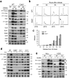
Figure 1
Figure 1
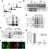
Cdk2 is required for efficient Chk1 activation and G1 arrest upon exposure to HU. (a) Immunoblots showing phosphorylation of Chk1, Mcm2 and p53 as well as the expression of indicated cell cycle regulators after 20 h in hydroxyurea (HU-0h) and 6 hrs after release, in wild-type (WT) and Cdk2−/− HCT-116 cells. NT, non treated cells. Arrow shows hyper-phosphorylated Cdk1. LC, loading control. (b) Flow cytometry analysis showing cell cycle progression after release from HU block in WT and Cdk2−/− HCT-116 cells. Arrows show accelerated appearance of G1 (2 N) cell population in Cdk2−/− cells. See Supplementary Fig. S3 for quantification. (c) Video-microscopy data showing fraction of mitotic cells after the release of HU block in WT and Cdk2−/− HCT-116 cells. Mean and standard deviation of three separate experiments are given. (d) Immunoblots showing phosphorylation of Chk1 and p53 as well as the expression of Cdc6 in the presence of 2 mM hydroxyurea (HU-0h, 20 h) in WT, Cdk2−/− and two clones (clone 7 and 11) expressing Cdk2 in Cdk2−/− HCT-116 cells. NT, non-treated cells. LC, loading control. (e) Immunoblots showing Chk1 phosphorylation in non-treated (NT) and HU-arrested control (Ctl) and Cdk2 knockdown (siCdk2) HeLa and U2OS cells. Arrows, phosphorylation-dependent SDS-PAGE mobility shift. LC, loading control. See Supplementary Fig. S4 for FACS analysis. Uncropped images of blots are shown in Supplementary Fig. S17.
Figure 2
Figure 2
CycA-Cdk1/2 complexes are major DNA damage-induced p21 targets in HCT-116 cells. (a) Flow cytometry analysis of non-treated (NT) and HCT-116 cells exposed to bleomycin or ICRF-193 (ICRF) for 24 h. (b) Immunoblot analysis showing pRb and p130 pocket proteins in WT and p21-/- HCT-116 cells exposed to bleomycin or ICRF for 12 and 24h. Arrows show hypo-phosphorylated pRb and p130. NT, non-treated cells. LC, loading control. (c) Immunoblot analysis of p21 immunoprecipitates (IP) from HCT-116 cells exposed to bleomycin or ICRF (24 h). Numbers 1, 2 and 3 indicate different Cdk1 phospho-isoforms. (d) Immunoblot analysis of CycA and CycB1 immunoprecipitates (IP) from HCT-116 cells exposed to bleomycin or ICRF for indicated times. (e) p21 immunodepletion (ID) experiment. Immunoblot analysis of CycA and CycB1 immunoprecipitates before (mock-treated: -) and after p21 immunodepletion (+) of cell extracts from non-treated (NT) or HCT-116 cells exposed to bleomycin or ICRF for 24 h. Numbers 1, 2 and 3 indicate different Cdk1 phospho-isoforms. (f) Immunofluorescence showing CycB1 and p21 localization in HCT-116 cells arrested in G2 with ICRF (24 h). Arrow shows a cell in G2 with high p21 content and nuclear CycB1. Bar, 10 mM. (g) CycB1 localization in non-treated (NT) and HCT-116 cells exposed to bleomycin (24 h). Cytoplasmic (Cyt), mainly or partly nuclear (Nuc) or mitotic (Mit) CycB1-positive cells were scored in five independent experiments (more than 1000 cells). Error bars indicate standard errors. Asterisks indicate that the effect of bleomycin is statistically significant (paired t-test) at the 0.01 level (p = 0.0089). Uncropped images of blots are shown in Supplementary Fig. S17.
Figure 3
Figure 3
Cdk2 is not required for Chk1 activation upon DNA damage by bleomycin, and its absence slows S/G2 progression. (a) Immunoblots showing DNA damage response in WT, Cdk2−/− and p53−/− HCT-116 cells exposed to bleomycin for 24 h and 48 h. NT, non-treated cells. (b) FACS analysis of non-treated (NT) and WT, Cdk2−/− and p53−/− HCT-116 cells exposed to bleomycin 24 h and 48 h. Arrow shows a G1 population that escaped from G2 arrest. (c) Immunoblot analysis of CycA and CycB1 immunoprecipitates from WT, Cdk2−/− and p53−/− HCT-116 cells exposed to bleomycin. 1, 2 and 3 indicate different Cdk1 phospho-isoforms. (d) Immunoblots showing CycB1 and Wee1 phosphorylation (PS642) in non-treated (−) and WT, Cdk2−/− and p53−/− HCT-116 cells exposed to bleomycin for 24 h and 48 h. Arrow indicates slow SDS-PAGE mobility band that co-migrates with hyper-phosphorylated Wee1 (PS642-Wee1) band. LE, longer ECL exposure to appreciate SDS-PAGE shift of different Wee1 phospho-isoforms. Bar on the right indicates migration of 100 kDa molecular mass marker. LC, loading control. Uncropped images of blots are shown in Supplementary Fig. S17.
Figure 4
Figure 4
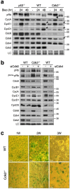
Cdk2 downregulation accelerates DNA damage-induced cell cycle exit by reducing Cdk6. (a) Immunoblot analysis showing indicated cell cycle regulators in wild-type (WT), Cdk2−/− and p53−/− HCT-116 cells exposed to bleomycin. Arrow indicates hypo-phosphorylated pRb accumulation. Arrowheads show increase of CycE1 and CycD1 after 48 h in WT cells. LC, loading control. (b) Immunoblot analysis of indicated cell cycle regulators in proliferating wild-type (WT) and Cdk2−/− HCT-116 cells that were previously depleted for Cdk6 (siCdk6, left panel) or Cdk4 (only WT, right panel). Cells were harvested and lysed 48 h after transfection with Ctl-, Cdk6- or Cdk4-specific siRNA. LC, loading control. (c) β-galactosidase staining of WT and Cdk2−/− cells 1 week (W), 2 weeks and 3 weeks after 24 h treatment with bleomycin. Bar, 10 μM. Uncropped images of blots are shown in Supplementary Fig. S17.
Figure 5
Figure 5
CycE1-Cdk2, and not CycE1-Cdk1, is preferential p21 target in senescence. (a) Immunofluorescence showing localization of CycE1 and p21 in proliferating WT and Cdk2−/− HCT-116 cells. Arrow shows the cell with elevated levels of both CycE1 and p21. Bar, 10 μM. See Supplementary Fig. S10 for larger fields. (b) Cells expressing high CycE1 levels were scored for p21 expression. Mean and standard deviation of three separate experiments are given. For more detailed explanation of the criteria used for quantification see Materials & Methods. (c) Immunofluorescence showing co-localization of CycE1 and Ki-67 in proliferating WT HCT-116 cells. Arrow shows the cell with elevated levels of CycE1 with low Ki-67 signal. Bar, 10 μM. See Supplementary Fig. S12 for larger fields. (d) Cells expressing high CycE1 levels were scored for Ki-67 expression. Mean and standard deviation of three separate experiments are given. For more detailed explanation of the criteria used for quantification see Materials & Methods. (e) Immunofluorescence showing proliferating HCT-116 cultures with subpopulation of cells with strong γH2AX and p21 signals. Bar, 10 μM. See Supplementary Fig. S13 for larger fields. (f) Immunofluorescence showing proliferating cultures of Cdk2−/− HCT-116 cells with ectopic expression of Cdk2 (clone 7) that restored accumulation of p21 and CycE1. Bar, 10 mM.
Figure 6
Figure 6
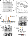
Cdk2 KD strengthens the G2/M checkpoint and promotes DNA damage-induced cell cycle exit in U2OS cells by reducing Cdk6. (a) Immunoblots showing effects of KU-55933 (KU) and caffeine (Caf) on Chk1 phosphorylation in ICRF- and bleomycin-treated U2OS cells. Arrow, ATM-mediated Chk2 mobility shift; NT- non-treated cells. (b) Occurrence of mitoses based on video-microscopy data of non-treated (Ctl) and U2OS cells exposed to ICRF (top) and bleomycin (below) in the absence (−) or presence of Caffeine (+Caf) or KU-55933 (+KU). Mitoses were counted at 2-hour intervals for 30 hours and normalized to the total cell number, as described in Methods. (c) Video-microscopy data showing mitoses in ICRF-treated control (siCtl), Cdk2 KD (siCdk2) and Chk1 KD (siChk1) U2OS cells. Mitoses were counted at 8-hour intervals. Mean and standard deviation of three separate experiments are given. See also Supplementary Fig. S15. (d) Immunoblots showing effects of Control (Ct), Chk1 (Ch1), Chk2 (Ch2) and Cdk2 depletion on pRb pathway in ICRF-treated U2OS cells. Arrow shows hypo-phosphorylated pRb. LC, loading control. (e) Immunoblots showing effects of Control (Ct), Chk1 (Ch1), Chk2 (Ch2) and Cdk2 depletion on DNA damage response regulators in ICRF-treated U2OS cells. LC, loading control. (f) Model: How Cdk2 inhibition promotes implementation of G2 exit program after DNA damage. See Supplementary Fig. S16 for more details. Uncropped images of blots are shown in Supplementary Fig. S17.

References (V体育安卓版)

    1. Santamaria D, et al. Cdk1 is sufficient to drive the mammalian cell cycle. Nature. 2007;448:811–815. doi: 10.1038/nature06046. - "VSports在线直播" DOI - PubMed
    1. Tetsu O, McCormick F. Proliferation of cancer cells despite CDK2 inhibition. Cancer Cell. 2003;3:233–245. doi: 10.1016/S1535-6108(03)00053-9. - DOI - PubMed
    1. Hochegger H, et al. An essential role for Cdk1 in S phase control is revealed via chemical genetics in vertebrate cells. J Cell Biol. 2007;178:257–268. doi: 10.1083/jcb.200702034. - V体育ios版 - DOI - PMC - PubMed
    1. Berthet C, Aleem E, Coppola V, Tessarollo L, Kaldis P. Cdk2 knockout mice are viable. Curr Biol. 2003;13:1775–1785. doi: 10.1016/j.cub.2003.09.024. - DOI - PubMed
    1. Ortega S, et al. Cyclin-dependent kinase 2 is essential for meiosis but not for mitotic cell division in mice. Nat Genet. 2003;35:25–31. doi: 10.1038/ng1232. - "V体育平台登录" DOI - PubMed

Publication types

MeSH terms

Substances