Skip to main page content
U.S. flag

An official website of the United States government

Dot gov

The . gov means it’s official VSports app下载. Federal government websites often end in . gov or . mil. Before sharing sensitive information, make sure you’re on a federal government site. .

Https

The site is secure V体育官网. The https:// ensures that you are connecting to the official website and that any information you provide is encrypted and transmitted securely. .

. 2017 Oct 3;114(40):10713-10718.
doi: 10.1073/pnas.1711235114. Epub 2017 Sep 11.

Gut bacteria from multiple sclerosis patients modulate human T cells and exacerbate symptoms in mouse models

Affiliations

Gut bacteria from multiple sclerosis patients modulate human T cells and exacerbate symptoms in mouse models

Egle Cekanaviciute et al. Proc Natl Acad Sci U S A. .

"V体育官网" Erratum in

Abstract

The gut microbiota regulates T cell functions throughout the body VSports手机版. We hypothesized that intestinal bacteria impact the pathogenesis of multiple sclerosis (MS), an autoimmune disorder of the CNS and thus analyzed the microbiomes of 71 MS patients not undergoing treatment and 71 healthy controls. Although no major shifts in microbial community structure were found, we identified specific bacterial taxa that were significantly associated with MS. Akkermansia muciniphila and Acinetobacter calcoaceticus, both increased in MS patients, induced proinflammatory responses in human peripheral blood mononuclear cells and in monocolonized mice. In contrast, Parabacteroides distasonis, which was reduced in MS patients, stimulated antiinflammatory IL-10-expressing human CD4+CD25+ T cells and IL-10+FoxP3+ Tregs in mice. Finally, microbiota transplants from MS patients into germ-free mice resulted in more severe symptoms of experimental autoimmune encephalomyelitis and reduced proportions of IL-10+ Tregs compared with mice "humanized" with microbiota from healthy controls. This study identifies specific human gut bacteria that regulate adaptive autoimmune responses, suggesting therapeutic targeting of the microbiota as a treatment for MS. .

Keywords: autoimmunity; microbiome; multiple sclerosis. V体育安卓版.


V体育ios版 - Conflict of interest statement

The authors declare no conflict of interest.

"VSports注册入口" Figures

Fig. 1.
Fig. 1.
MS patient microbiota alter self-Treg differentiation despite similar alpha and beta diversity to control microbiota. (A) Quantification of CD25+FoxP3+ lymphocytes within the CD3+CD4+ population in MS patients (n = 7) and healthy controls (n = 9), run in triplicate and results averaged, in response to extracts from total bacteria isolated from the stool samples of the same subjects who were PBMC donors (thus, self bacteria). CD25+FoxP3+ lymphocyte induction is expressed as the fold difference over no-bacteria control. **P < 0.01, two-tailed Mann–Whitney test. (BD) Comparison of microbial community composition of untreated MS patients (n = 71) and healthy controls (n = 71). (B) Chao1 metric of alpha diversity. Data are presented as mean ± SEM. (C) Principal coordinate analysis (PCoA) plot of beta diversity (unweighted UniFrac). (D) Mean relative abundance of microbial genera. Akkermansia and Parabacteroides are outlined. Colors represent bacterial genera.
Fig. 2.
Fig. 2.
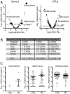
Relative abundances of individual microbial genera differ between MS patients and controls. (A) Volcano plots of the relative abundance distribution of microbial genera (Left) and OTUs (Right). The x axes show log twofold of relative abundance ratio between MS patients (n = 71) and controls (n = 71) after variance-stabilizing transformation. The y axes show negative log10 of P value (negative binomial Wald test with Benjamini–Hochberg correction for multiple comparisons). (B) Summary of taxonomic differences between MS and control microbiomes. (C) Relative abundance plots of selected microbial genera (highlighted in A) that were found to be significantly different between MS and controls. Data are shown as mean ± SEM.
Fig. 3.
Fig. 3.
A. calcoaceticus inhibits Treg differentiation and stimulates Th1 differentiation in vitro. (A and B) Representative flow cytometry plots (A) and quantification (B) of CD25+FoxP3+ cell differentiation within the CD3+CD4+ population in response to A. calcoaceticus (A. calc) (n = 6 PBMC donors). (C and D) Representative flow cytometry plots (C) and quantification (D) of IFNγ+ Th1 lymphocytes within the CD3+CD4+ population in response to A. calcoaceticus (A. calc) (n = 11 PBMC donors). *P < 0.05, **P < 0.01, two-tailed repeated-measures t test. Data are shown as mean ± SEM.
Fig. 4.
Fig. 4.
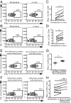
A. muciniphila increases Th1 lymphocyte differentiation in vitro. (AD) Representative flow cytometry plots (A and B) and quantification (C and D) of IFNγ+ and of Tbet+ Th1 lymphocytes within the CD3+CD4+ population in response to A. muciniphila (A. muci). n = 6 PBMC donors for the IFNγ experiment; n = 7 PBMC donors for the Tbet experiment. *P < 0.05, two-tailed repeated-measures t test. (EH) Representative flow cytometry plots (E and F) and quantification (G and H) of IFNγ+ Th1 lymphocytes within the CD3+CD4+ population in response to nonself or self bacteria from subjects with or without detected A. muciniphila. n = 6 subjects without A. muciniphila; n = 12 subjects with A. muciniphila. *P < 0.05, two-tailed t test; **P < 0.01, two-way ANOVA for repeated measures. Data are shown as mean ± SEM.
Fig. 5.
Fig. 5.
P. distasonis stimulates IL-10+ Treg differentiation in vitro. (AD) Representative flow cytometry plots (A and B) and quantification (C and D) of CD25+ and CD25+IL-10+ lymphocytes within the CD3+CD4+ population in response to P. distasonis (P. dist). n = 6 PBMC donors. *P < 0.05, **P < 0.01, two-tailed repeated measures t test. Data are shown as mean ± SEM.
Fig. 6.
Fig. 6.
Monocolonization of GF mice with MS-associated bacteria mediates T lymphocyte differentiation. (AC) Representative flow cytometry plots (A) and quantification (B and C) of CD4+IFNγ+ lymphocytes (B) and CD4+IL-10+ lymphocytes (C) within the live cell population in GF mice colonized with A. calcoaceticus, A. muciniphila, and P. distasonis. GF mice and specific pathogen-free (SPF) mice are used as controls. n = 3–8 mice per group. *P < 0.05, **P < 0.01, ***P < 0.001, ****P < 0.0001, one-way ANOVA with Tukey adjustment for multiple comparisons. Data are shown as mean ± SEM. CLN, cervical lymph nodes; MLN, mesenteric lymph nodes.
Fig. 7.
Fig. 7.
Transfer of healthy control microbiota protects against EAE and mediates Treg induction in mouse mesenteric lymph nodes compared with transfer of MS patient microbiota. (A) Clinical EAE scores of mice that had been colonized with healthy control or MS patient microbiota for at least 5 wk or kept GF before the induction of EAE at 9–10 wk of age. Asterisks indicate significance between both the MS vs. control and the MS vs. GF groups. n = 6–8 mice per group. ****P < 0.0001, two-way ANOVA with Tukey adjustment for multiple comparisons. Data are shown as mean ± SEM. (B) PCoA of mouse microbiota at different time points after colonization with fecal microbiota from donor pair #1. n = 3–5 mice per group. EAE induction occurs at 35 d after transplantation. PC1, -2, -3, principal components 1, 2, and 3. (C and E) Representative flow cytometry plots of FoxP3+ lymphocytes within CD4+ populations and IL-10+ lymphocytes within CD4+FoxP3+ populations before EAE induction (C) and at peak of EAE disease (E). (D and F) Frequencies of IL-10+ lymphocytes within CD4+FoxP3+ populations in mesenteric lymph nodes of mice killed before EAE induction (D) and at peak of EAE progression (22 d after immunization) (F). *P < 0.05, **P < 0.01, ****P < 0.0001, one-way ANOVA with Tukey adjustment for multiple comparisons. Data are shown as mean ± SEM.

Comment in

References

    1. Lee YK, Mazmanian SK. Has the microbiota played a critical role in the evolution of the adaptive immune system? Science. 2010;330:1768–1773. - V体育官网入口 - PMC - PubMed
    1. Faith JJ, Ahern PP, Ridaura VK, Cheng J, Gordon JI. Identifying gut microbe-host phenotype relationships using combinatorial communities in gnotobiotic mice. Sci Transl Med. 2014;6:220ra11. - PMC - PubMed
    1. Round JL, Mazmanian SK. Inducible Foxp3+ regulatory T-cell development by a commensal bacterium of the intestinal microbiota. Proc Natl Acad Sci USA. 2010;107:12204–12209. - PMC - PubMed
    1. Lozupone CA, et al. Alterations in the gut microbiota associated with HIV-1 infection. Cell Host Microbe. 2013;14:329–339. - PMC - PubMed
    1. Chu H, et al. Gene-microbiota interactions contribute to the pathogenesis of inflammatory bowel disease. Science. 2016;352:1116–1120. - PMC - PubMed

V体育安卓版 - Publication types

"V体育官网" MeSH terms