"V体育2025版" Skip to main page content
U.S. flag

An official website of the United States government

Dot gov

The . gov means it’s official. Federal government websites often end in . gov or . mil. Before sharing sensitive information, make sure you’re on a federal government site VSports app下载. .

Https

The site is secure. The https:// ensures that you are connecting to the official website and that any information you provide is encrypted and transmitted securely V体育官网. .

. 2017 Dec;67(6):1232-1242.
doi: 10.1016/j.jhep.2017.08.010. Epub 2017 Aug 23.

Macrophage heme oxygenase-1-SIRT1-p53 axis regulates sterile inflammation in liver ischemia-reperfusion injury (V体育平台登录)

Affiliations

Macrophage heme oxygenase-1-SIRT1-p53 axis regulates sterile inflammation in liver ischemia-reperfusion injury

Kojiro Nakamura et al. J Hepatol. 2017 Dec.

V体育平台登录 - Abstract

Background & aims: Hepatic ischemia-reperfusion injury (IRI), characterized by exogenous antigen-independent local inflammation and hepatocellular death, represents a risk factor for acute and chronic rejection in liver transplantation VSports手机版. We aimed to investigate the molecular communication involved in the mechanism of liver IRI. .

Methods: We analyzed human liver transplants, primary murine macrophage cell cultures and IR-stressed livers in myeloid-specific heme oxygenase-1 (HO-1) gene mutant mice, for anti-inflammatory and cytoprotective functions of macrophage-specific HO-1/SIRT1 (sirtuin 1)/p53 (tumor suppressor protein) signaling. V体育安卓版.

Results: Decreased HO-1 expression in human post-reperfusion liver transplant biopsies correlated with a deterioration in hepatocellular function (serum ALT; p<0. 05) and inferior patient survival (p<0. 05). In the low HO-1 liver transplant biopsy group, SIRT1/Arf (alternative reading frame)/p53/MDM2 (murine double minute 2) expression levels decreased (p<0. 05) while cleaved caspase 3 and frequency of TUNEL+cells simultaneously increased (p<0. 05). Immunofluorescence showed macrophages were the principal source of HO-1 in human and mouse IR-stressed livers. In vitro macrophage cultures revealed that HO-1 induction positively regulated SIRT1 signaling, whereas SIRT1-induced Arf inhibited ubiquitinating activity of MDM2 against p53, which in turn attenuated macrophage activation V体育ios版. In a murine model of hepatic warm IRI, myeloid-specific HO-1 deletion lacked SIRT1/p53, exacerbated liver inflammation and IR-hepatocellular death, whereas adjunctive SIRT1 activation restored p53 signaling and rescued livers from IR-damage. .

Conclusion: This bench-to-bedside study identifies a new class of macrophages activated via the HO-1-SIRT1-p53 signaling axis in the mechanism of hepatic sterile inflammation. This mechanism could be a target for novel therapeutic strategies in liver transplant recipients. VSports最新版本.

Lay summary: Post-transplant low macrophage HO-1 expression in human liver transplants correlates with reduced hepatocellular function and survival. HO-1 regulates macrophage activation via the SIRT1-p53 signaling network and regulates hepatocellular death in liver ischemia-reperfusion injury V体育平台登录. Thus targeting this pathway in liver transplant recipients could be of therapeutic benefit. .

Keywords: Heme oxygenase-1; Innate immunity; Ischemia-reperfusion injury; Liver transplantation; Myeloid-specific mutant mice; P53; Sirtuin 1 VSports注册入口. .


Conflict of interest statement (VSports app下载)

Conflict of interest

The authors who have taken part in this study declared that they do not have anything to disclose regarding funding or conflict of interest with respect to this manuscript.

Please refer to the accompanying ICMJE disclosure forms for further details.

Figures (V体育2025版)

Fig. 1
Fig. 1. Post-transplant hepatic HO-1 levels and OLT patient outcomes
(A) Based on Western blot-assisted relative protein expression, twenty-one human liver transplant biopsies were identified as HO-1 “high” (n = 11) vs. “low” (n = 10). Representative HO-1 expression is shown in the bottom panel. (B–D) Serum ALT, AST and total bilirubin (T-Bil) levels at post-operative day 1–14 (POD 1–14). Solid line indicates high while dotted line low HO-1 OLT groups. Data are shown as mean ± SEM. #p <0.05 vs. high HO-1 (Mann-Whitney U test). (E) The cumulative probability of OLT survival (Kaplan-Meier method). Solid line indicates high and dotted line low HO-1 group. #p <0.05 vs. high HO-1 group (Log-rank test). (This figure appears in colour on the web.)
Fig. 2
Fig. 2. HO-1 levels in human OLT correlate with SIRT1, p53, cleaved caspase 3, MDM2, and p14Arf levels
Twenty-one human liver transplant biopsies were divided, based on the relative HO-1 expression, into “high” (n = 11) and “low” (n = 10) groups (Fig. 1A). (A–E) Relative expression of SIRT1 (A; mean: 2.78 vs. 1.09), p53 (B, mean: 1.11 vs. 0.63), cleaved caspase 3 (C; mean: 0.66 vs. 1.13), MDM2 (D; mean: 0.80 vs. 0.46) and p14Arf (E; mean: 0.69 vs. 0.36). Data are shown in dot plots; bars indicate mean ± SEM. #p <0.05 high (n = 11) vs. low (n = 10) HO-1 (Mann-Whitney U test). (F) Representative Western blot-assisted OLT expression of HO-1, SIRT1, p53, cleaved caspase 3, MDM2 and p14Arf (A/B: high HO-1; P/Q: low HO-1).
Fig. 3
Fig. 3. Frequency of TUNEL + cells in human OLT is negatively correlated with macrophage-expressing HO-1
(A) Immunofluorescence staining of CD68 (blue, macrophage), HO-1 (red), TUNEL (green) and merged image in high vs. low HO-1 human OLT. Representative of three in each group is shown. (B) Immunofluorescence staining of CD68 (red), HO-1 (green), DAPI (blue) and merged image in IR-stressed mouse livers. Upper panels: 6 h reperfusion/OLT (18 h cold storage). Lower panels: 6 h reperfusion/90 min warm ischemia. Representative of three in each group is shown. (This figure appears in colour on the web.)
Fig. 4
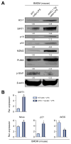
Fig. 4. HO-1 upregulates SIRT1–p53 signaling in BMDM
BMDM from WT or mHO-1 Tg mice were stimulated with LPS (100 ng/ml, 6 h). (A) Western blot-assisted detection of HO-1, SIRT1, p19, p53, MDM2, PUMA and p-Stat1 (Tyr701). β-actin expression served as an internal control for normalization. The values under the bands represent relative ratios of normalized intensity compared to WT/cells + LPS. Representative of three experiments is shown. (B) Quantitative RT-PCR-assisted detection of mRNA coding for SIRT1, Noxa, p21 and iNOS. Data normalized to B2M gene (n = 4/group) are presented as mean ± SD. *p <0.05 vs. WT/cells + LPS (Student t test).
Fig. 5
Fig. 5. SIRT1 depresses macrophage activation and upregulates p53 signaling in vitro
LPS-stimulated BMDM (100 ng/ml, 6 h) with/without Res (SIRT1 inducer, 100 μM, 12 h) or siRNA SIRT1 were screened for activation markers. (A and B) Western blot-assisted detection of SIRT1, p-Stat1 (Tyr701), p-IkBα (Ser32), IkBα, iNOS, p19, p53, Ac-p53 (Lys379), MDM2, and PUMA. Band intensities were quantified with ImageJ and normalized by dividing target band intensity by that of housekeeping β-actin. The values under the bands represent relative ratios of normalized intensity, compared to cells + LPS. Representative of three experiments is shown. (C) Quantitative RT-PCR-assisted detection of mRNA coding for SIRT1, Noxa, p21, iNOS, IL-1β, MCP1, CCL5, CXCL10 and GzmB. Data normalized to B2M gene expression (n = 4/group) are presented as the mean ± SD. *p <0.05 vs. cells + LPS (one-way ANOVA).
Fig. 6
Fig. 6. SIRT1 inhibits macrophage activation through p19/p53 signaling
(A–C) BMDM stimulated with LPS (100 ng/ml, 6 h) were treated with Res (100 μM, 12 h), siRNA (p53) or siRNA (p19). (A) 25 μg of protein lysate from BMDM were divided by immunoprecipitation into ubiquitinated (UB-) and non-ubiquitinated (nonUB-) proteins and analyzed by Western blotting. Representative images (upper panel) and densitometry quantification (lower panel) are shown. *p <0.05 vs. cells + LPS, #p <0.05 vs. cells + LPS + Res, n = 3/group (one-way ANOVA). (B) Western blot-assisted detection of SIRT1, p53, PUMA, p-IkBα (Ser32) and IkBα in LPS-stimulated BMDM. Band intensities were normalized by dividing target band intensity by that of β-actin. The values under the bands represent relative ratios of normalized intensity compared to that of cells + LPS. Representative of three experiments is shown. (C) Quantitative RT-PCR-assisted detection of mRNA coding for SIRT1, Noxa, p21, iNOS, IL-1β, MCP1 and CCL5. Data were normalized to B2M gene expression (*p <0.05 vs. cells + LPS, #p <0.05 vs. cells + LPS + Res, n = 4/group, one-way ANOVA). (D and E) Livers in WT mice underwent 90 min of warm ischemia followed by 6 h reperfusion. (D) Western blot-assisted detection of SIRT1, p19, p53, MDM2 and PUMA. The values under the bands represent relative ratios of normalized intensity compared to that of IR. Representative of three experiments is shown. (E) Quantitative RT-PCR-assisted detection of mRNA coding for Noxa. Data normalized to HPRT gene expression (*p <0.05 vs. IR, #p <0.05 vs. IR + VHC, n = 4/group, one-way ANOVA) are presented as mean ± SD.
Fig. 7
Fig. 7. Macrophage HO-1 deficiency depresses SIRT1–p53 signaling and augments inflammation, while SIRT1 activation restores p53/anti-inflammatory phenotype in BMDM
BMDM were harvested from HO-1 proficient (WT) or myeloid-specific HO-1 deficient (mHO-1 KO) mice and activated with LPS (100 ng/ml, 6 h). In some experiments, cells were pretreated with Res (100 μM) or VHC for 12 h. (A) Western blot-assisted detection of HO-1, SIRT1, p19, p53, Ac-p53 (Lys379), MDM2, and p-Stat1 (Tyr701). β-actin expression as internal control was used for normalization. The values under the bands represent the relative ratio of normalized intensity compared to WT/cells + LPS. Representative of three experiments is shown. (B) Quantitative RT-PCR-assisted detection of SIRT1, Noxa, p21, iNOS, TNFα, MCP1, IL-1β, GzmB and IL-12p40. Data normalized to B2M gene expression (n = 4/group). (C) ELISA-assisted detection of MCP1 in BMDM culture medium (pg/μl, n = 4/group). Data are presented as mean ± SD. *p <0.05 vs. WT/cells + LPS, #p <0.05 vs. HO-1 deficient/cells + LPS + Res (one-way ANOVA).
Fig. 8
Fig. 8. Myeloid-specific HO-1 deficiency depresses SIRT1–p53 and augments IRI, while SIRT1 activation restores p53 and attenuates liver IRI/inflammation in mHO-1 KO mice
Groups of control and mHO-1 KO mice were subjected to warm liver IRI. Some mice were treated with Res (25 mg/kg i.p. at −1 h) or VHC. (A) Representative H&E staining (original magnification, 100×; n = 4–6/group). (B) Suzuki’s histological grading of liver IRI and sALT/AST [IU/L] (n = 4–6/group). (C) Western blot-assisted detection of SIRT1, p19, p53, Ac-p53 (Lys379) and MDM2. β-actin expression serves as an internal control. The values under the bands represent the relative ratio of normalized intensity compared to IR/Control. Representative of three experiments is shown. (D) ELISA-assisted detection of serum MCP1 (pg/ml, n = 3–4/group) (E) Quantitative RT-PCR-assisted detection of mRNA coding for SIRT1, Noxa, TNFα and MCP1. Data normalized to HPRT gene expression (n = 4/group) are presented as mean ± SD. *p <0.05 vs. IR/Control, #p <0.05 vs. IR/mHO-1 KO + Res (one-way ANOVA). (F) Schematic illustration of macrophage regulation by HO-1–SIRT1–p53 axis. HO-1 upregulates SIRT1 (#1), whereas SIRT1-induced Arf (p19 in mouse, p14 in human) (#2) inhibits MDM2 E3 ligase activity to stabilize p53 (#3). Consequently, p53 tumor suppressor protein upregulated by HO1/SIRT1/p19 axis (#1–3) attenuates macrophage activation (#4). DEG: ubiquitin-degradation of p53 by MDM2. (This figure appears in colour on the web.)

Comment in

References

    1. Zhai Y, Petrowsky H, Hong JC, Busuttil RW, Kupiec-Weglinski JW. Ischaemia-reperfusion injury in liver transplantation-from bench to bedside. Nat Rev Gastroenterol Hepatol. 2013;10:79–89. - PMC - PubMed
    1. Zhai Y, Shen XD, O’Connell R, Gao F, Lassman C, Busuttil RW, et al. Cutting edge: TLR4 activation mediates liver ischemia/reperfusion inflammatory response via IFN regulatory factor 3-dependent MyD88-independent pathway. J Immunol. 2004;173:7115–7119. - PubMed
    1. Sosa RA, Zarrinpar A, Rossetti M, Lassman CR, Naini BV, Datta N, et al. Early cytokine signatures of ischemia/reperfusion injury in human orthotopic liver transplantation. JCI Insight. 2016;1:e89679. - PMC (V体育官网入口) - PubMed
    1. Maines MD. The heme oxygenase system: a regulator of second messenger gases. Annu Rev Pharmacol Toxicol. 1997;37:517–554. - "V体育ios版" PubMed
    1. Amersi F, Buelow R, Kato H, Ke B, Coito AJ, Shen XD, et al. Upregulation of heme oxygenase-1 protects genetically fat Zucker rat livers from ischemia/reperfusion injury. J Clin Invest. 1999;104:1631–1639. - PMC - PubMed

Publication types (VSports)