Skip to main page content
U.S. flag

An official website of the United States government

Dot gov

The . gov means it’s official. Federal government websites often end in VSports app下载. gov or . mil. Before sharing sensitive information, make sure you’re on a federal government site. .

Https

The site is secure. The https:// ensures that you are connecting to the official website and that any information you provide is encrypted and transmitted securely V体育官网. .

. 2017 Aug 17;130(7):933-942.
doi: 10.1182/blood-2017-01-762658. Epub 2017 Jun 12.

Loss of thymic innate lymphoid cells leads to impaired thymopoiesis in experimental graft-versus-host disease

Affiliations

"VSports app下载" Loss of thymic innate lymphoid cells leads to impaired thymopoiesis in experimental graft-versus-host disease

Jarrod A Dudakov (VSports在线直播) et al. Blood. .

V体育2025版 - Abstract

Graft-versus-host disease (GVHD) and posttransplant immunodeficiency are frequently related complications of allogeneic hematopoietic transplantation. Alloreactive donor T cells can damage thymic epithelium, thus limiting new T-cell development. Although the thymus has a remarkable capacity to regenerate after injury, endogenous thymic regeneration is impaired in GVHD. The mechanisms leading to this regenerative failure are largely unknown. Here we demonstrate in experimental mouse models that GVHD results in depletion of intrathymic group 3 innate lymphoid cells (ILC3s) necessary for thymic regeneration. Loss of thymic ILC3s resulted in deficiency of intrathymic interleukin-22 (IL-22) compared with transplant recipients without GVHD, thereby inhibiting IL-22-mediated protection of thymic epithelial cells (TECs) and impairing recovery of thymopoiesis. Conversely, abrogating IL-21 receptor signaling in donor T cells and inhibiting the elimination of thymic ILCs improved thymopoiesis in an IL-22-dependent fashion. We found that the thymopoietic impairment in GVHD associated with loss of ILCs could be improved by restoration of IL-22 signaling VSports手机版. Despite uninhibited alloreactivity, exogenous IL-22 administration posttransplant resulted in increased recovery of thymopoiesis and development of new thymus-derived peripheral T cells. Our study highlights the role of innate immune function in thymic regeneration and restoration of adaptive immunity posttransplant. Manipulation of the ILC-IL-22-TEC axis may be useful for augmenting immune reconstitution after clinical hematopoietic transplantation and other settings of T-cell deficiency. .


VSports最新版本 - Conflict of interest statement

Conflict-of-interest disclosure: The authors declare no competing financial interests. A patent application has been filed on the use of IL-22 as a thymopoietic growth factor (US 61/487 517) with J. A. D. , A. M. H. , and M. R. M. v. d V体育ios版. B. listed as inventors.

"V体育2025版" Figures

Figure 1.
Figure 1.
Thymic IL-22 levels are reduced in mice with GVHD. (A-B) BALB/c (H-2d) recipients were transplanted with 5 × 10⁶ TCD bone marrow (BM) cells from B6 mice (H-2b) with or without 1 × 10⁶ BALB/c T cells to induce GVHD. (A) Total thymic cellularity on day 7 after transplant. (B) Thymic cellularity on day 7 and day 21 after transplant, analyzed by linear regression with slope comparison (1/slope for each line displayed on the graph). (C) B6 (H-2b) recipients were transplanted with 5 × 10⁶ TCD BM cells from B10.BR mice (H-2k) with or without 2 × 10⁶ B10.BR T cells to induce GVHD. Absolute levels of IL-22 were measured on day 7 post-BMT in B6 recipients or normal non-BMT controls (n = 10 per group). (D) BALB/c (H-2d) recipients were transplanted with 5 × 10⁶ TCD BM cells from B6 mice (H-2b) with or without 1 × 10⁶ BALB/c T cells to induce GVHD. Absolute levels of IL-22 were measured on day 7 post-BMT in BALB/c recipients or normal non-BMT controls (n = 9-10 per group). (E) Correlation of thymic cellularity and IL-22 levels in BMT recipients or normal controls from panels C and D. Left panel shows values from allo-BMT without T cells (blue circles and squares), indicating that after TCD BMT, the thymic cellularity and intrathymic IL-22 levels have a strong negative correlation. Right panel shows values from mice transplanted with allogeneic T cells (gray circles and squares), indicating that GVHD causes a loss of the correlation between thymic cellularity and the compensatory IL-22 response. Untransplanted normal controls are the same in both panels (white circles and squares). Bar graphs represent mean ± standard error of the mean (SEM) of at least 2 independent experiments. *P < .05; **P < .01; ***P < .001.
Figure 2.
Figure 2.
Intrathymic ILCs are eliminated and their production of IL-22 is reduced in mice with GVHD. (A-G) BALB/c recipients (H-2d) were transplanted with 5 × 10⁶ TCD BM cells with or without 1 × 10⁶ T cells from B6 donors (H-2b) to induce GVHD and were analyzed on day 7 after transplant (n = 7-10 mice per group). (A) Total number and (B) frequency of cTECs, mTECs, and DP thymocytes. (C) Back-gating analysis of thymic IL-22+ cells after TCD allo-BMT; thymic harvest and incubation in vitro with brefeldin A for 5 hours, followed by intracellular and surface antibody staining. Representative flow cytometry plots were concatenated from 5 independent observations with isotype antibody (RORγt, IL-7Rα) or CD45 (CD4, CCR6, CD8, CD3, NKp46) negative controls or whole thymus positive controls (CD45). (D) Total number of intrathymic CD45+CD3CD8CD4+IL7R+RORγt+ ILC3s in BMT recipients or normal untransplanted controls. (E-F) Thymocytes were harvested and incubated for 5 hours with brefeldin A. Intracellular IL-22 was assessed by flow cytometry, demonstrating the frequency (E) and absolute number (F) of IL-22+ ILC3s in BMT recipients or normal untransplanted controls. (G) Absolute levels of intrathymic IL-23 on day 7 after BMT in BALB/c recipients or normal untransplanted controls. (H) B6 (CD45.2) recipients were transplanted with 10 000 LineageSca1+ckit+ BM cells from congenic CD45.1+ donors, and BM, spleen, and thymus were analyzed on day 4 after transplant for ILC3 chimerism and absolute numbers of donor and host ILC3s (n = 5-15 mice per group). (I) Absolute number of host-derived (CD45.2+H-2d+) and donor BM–derived (CD45.1+H-2b+) thymic ILCs 7 days after B6→BALB/c transplant (performed as in panels A-G), gated on CD45+CD3CD8CD4+ cells (n = 9-10 per group). Bar graphs represent mean ± SEM of at least 2 independent experiments.*P < .05; **P < .01; ***P < .001. FSC, forward scatter; ns, not significant.
Figure 3.
Figure 3.
IL-22 deficiency exacerbates thymic GVHD. WT or Il22−/− BALB/c recipients (H-2d) were transplanted with 5 × 10⁶ B6 TCD BM (H-2b) cells and 1 × 10⁶ WT B6 T cells. (A) Total thymic cellularity (n = 10-14 per group) and (B) number of CD4+CD8+ donor BM-derived DP cells (n = 5 per group) 21 days post-BMT. (C) Proportion of thymocyte subsets shown on the left, and MHCII+EpCAM+ TECs gated on viable CD45 cells shown on the right. (D) Total number of cTECs and mTECs (n = 5-9 per group). Bar graphs represent mean ± SEM of at least 2 independent experiments. **P < .01.
Figure 4.
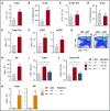
Figure 4.
Abrogation of IL-21 signaling in donor T cells maintains intrathymic ILC3s and ameliorates thymic GVHD in an IL-22–dependent fashion. (A-D) WT BALB/c recipients (H-2d) transplanted with 5 × 10⁶ CD45.1 B6 TCD BM (H-2b) cells and 1 × 10⁶ CD45.2 B6 T cells from either WT or Il21r−/− B6 donors to induce GVHD. Thymus harvested on day 7 post-BMT. (A) Absolute number of intrathymic CD45+CD3CD8CD4+IL7R+RORγt+ ILC3s (n = 10 per group). (B) Absolute amount of thymic IL-22 measured by enzyme-linked immunosorbent assay (ELISA) (n = 9 per group). (C) ILC3s were isolated from thymus and incubated for 4 hours in the presence of brefeldin A and then examined for intracellular expression of IL-22 (n = 10 per group). (D) Absolute amount of thymic IL-23 measured by ELISA (n = 9 per group). (E-H) WT BALB/c recipients were transplanted with CD45.1 B6 TCD BM cells and WT or Il21r−/− CD45.2 B6 donor T cells as above; thymus was harvested 21 days post-BMT. (E) Total thymus cellularity (n = 40-44 per group). (F) Absolute number of CD45EpCAM+MHCII+Ly51hiUEA-1lo cTECs and CD45EpCAM+MHCII+Ly51loUEA-1hi mTECs (n = 12-14 per group). (G) Proportion of thymocyte subsets by flow cytometry gated on CD45+ cells. (H) Absolute number of BM-derived (H-2b+CD45.1+) CD4+CD8+ DP thymocytes (n = 9 per group). (I-J) B6→BALB/c BMT with WT B6 marrow, WT or Il21r−/− B6 donor T cells, and WT or Il22−/− BALB/c recipients (n = 10 per group). (I) Total thymus cellularity and (J) absolute number of CD4+CD8+ DP thymocytes at 21 days post-BMT. (K) Total thymic cellularity and absolute number of CD4+CD8+ DP thymocytes 21 days after B6→LP BMT (H-2b→H-2b) with WT B6 TCD BM and either WT or Il21r−/− B6 T cells (n = 17 per group). Bar graphs represent mean ± SEM of at least 2 independent experiments. *P < .05; **P < .01; ***P < .001.
Figure 5.
Figure 5.
Deletion of IL-21 signaling increases the number of donor graft–derived Tregs in the thymus. (A-E) WT BALB/c (H-2d) recipients were transplanted with 5 × 10⁶ TCD BM cells from CD45.1+ B6 (H-2b) mice and 1 × 10⁶ T cells derived from either WT (n = 10 recipients) or Il21r−/− (n = 10 recipients) CD45.2+ B6 mice to induce GVHD. (A) Concatenated flow cytometry plots showing transplanted donor graft–derived T cells on day 7 post-BMT. (B) Proportion of transplanted donor T cells of all thymic T cells on day 7 post-BMT. (C) Absolute number of transplanted donor graft–derived T cells in the thymus on day 7 post-BMT. (D) Absolute number of total CD3+CD8CD25+CD4+FoxP3+ cells in the thymus on day 21 post-BMT (n = 9 per group). (E) Proportion of host-derived, graft T-cell–derived, and marrow-derived Tregs of all Tregs in the thymus on day 21 post-BMT, showing transplant with WT T cells in the left column and with IL-21R knockout (KO) T cells in right column; also shown are representative flow cytometry plots of Tregs derived from the donor marrow or from the T cells present in the donor graft. (F) Absolute number of donor BM–derived (H-2b+CD45.1+) or donor T-cell–derived (H-2b+CD45.2+) Tregs 21 days post-BMT (n = 9 per group). Dot plots concatenated from 5 independent observations from 1 experiment and gated on donor-derived (H-2b+) Tregs. Bar graphs represent mean ± SEM, combined from at least 2 independent experiments. *P < .05; **P < .01; ***P < .001.
Figure 6.
Figure 6.
Exogenous administration of IL-22 improves thymopoiesis and T-cell reconstitution after allo-BMT. (A) WT B6 recipients (H-2b) were transplanted with LP TCD BM (H-2b; 5 × 10⁶) and WT LP T cells (4 × 10⁶) (n = 9-10 recipients per group). GVHD mice were treated once per day with either phosphate-buffered saline (PBS) or rmIL-22 (200 mg/kg) starting on day 7 post-BMT; CD4+CD8+ DP thymocytes were assessed 3 weeks post-BMT. (B) Experimental model of FVB→BALB/c BMT (H-2q→H-2d) with RAG2-GFP marrow to evaluate thymic export of donor marrow–derived new peripheral T cells after IL-22 treatment. (C) BALB/c mice were transplanted with 5 × 10⁶ RAG2-GFP (FVB background) TCD BM cells and 0.1 × 10⁶ FVB T cells. Recipient mice (n = 5 per group) were treated once per day with either PBS or rmIL-22 (200 mg/kg) starting on day 7 post-BMT, and GFP+ T cells were enumerated in the spleen at 28 days post-BMT. (D) Thymic injury and depletion of thymocytes trigger ILC3 production of IL-22, which acts directly on TECs to induce their proliferation and survival. Through this trophic effect on TECs, IL-22 can promote endogenous immune regeneration. During GVHD, alloreactive T cells eliminate ILCs, thereby abrogating their production of IL-22 and impairing IL-22–mediated recovery of thymic epithelium. Administration of exogenous recombinant IL-22 can circumvent the loss of ILCs to enhance thymopoiesis and improve immune reconstitution after allo-BMT. Bar graphs represent mean ± SEM. *P < .05.

VSports - Comment in

  • Innately interesting interactions.
    Komanduri KV. Komanduri KV. Blood. 2017 Aug 17;130(7):844-845. doi: 10.1182/blood-2017-06-791848. Blood. 2017. PMID: 28818978 No abstract available.

References

    1. van den Brink MR, Alpdogan O, Boyd RL. Strategies to enhance T-cell reconstitution in immunocompromised patients. Nat Rev Immunol. 2004;4(11):856-867. - PubMed
    1. Ramadan A, Paczesny S. Various forms of tissue damage and danger signals following hematopoietic stem-cell transplantation. Front Immunol. 2015;6:14. - PMC - PubMed
    1. Na IK, Lu SX, Yim NL, et al. . The cytolytic molecules Fas ligand and TRAIL are required for murine thymic graft-versus-host disease. J Clin Invest. 2010;120(1):343-356. - VSports在线直播 - PMC - PubMed
    1. Krenger W, Holländer GA. The immunopathology of thymic GVHD. Semin Immunopathol. 2008;30(4):439-456. - PubMed (VSports)
    1. Krenger W, Rossi S, Holländer GA. Apoptosis of thymocytes during acute graft-versus-host disease is independent of glucocorticoids. Transplantation. 2000;69(10):2190-2193. - PubMed

V体育官网入口 - Substances