Skip to main page content
U.S. flag

An official website of the United States government

Dot gov

The VSports app下载. gov means it’s official. Federal government websites often end in . gov or . mil. Before sharing sensitive information, make sure you’re on a federal government site. .

Https

The site is secure. The https:// ensures that you are connecting to the official website and that any information you provide is encrypted and transmitted securely. V体育官网.

. 2016 Apr 26:6:25017.
doi: 10.1038/srep25017.

Enterobacter cloacae inhibits human norovirus infectivity in gnotobiotic pigs

Affiliations

"VSports" Enterobacter cloacae inhibits human norovirus infectivity in gnotobiotic pigs

"V体育ios版" Shaohua Lei et al. Sci Rep. .

Abstract

Human noroviruses (HuNoVs) are the leading cause of epidemic gastroenteritis worldwide. Study of HuNoV biology has been hampered by the lack of an efficient cell culture system. Recently, enteric commensal bacteria Enterobacter cloacae has been recognized as a helper in HuNoV infection of B cells in vitro VSports手机版. To test the influences of E. cloacae on HuNoV infectivity and to determine whether HuNoV infects B cells in vivo, we colonized gnotobiotic pigs with E. cloacae and inoculated pigs with 2. 74 × 10(4) genome copies of HuNoV. Compared to control pigs, reduced HuNoV shedding was observed in E. cloacae colonized pigs, characterized by significantly shorter duration of shedding in post-inoculation day 10 subgroup and lower cumulative shedding and peak shedding in individual pigs. Colonization of E. cloacae also reduced HuNoV titers in intestinal tissues and in blood. In both control and E. cloacae colonized pigs, HuNoV infection of enterocytes was confirmed, however infection of B cells was not observed in ileum, and the entire lamina propria in sections of duodenum, jejunum, and ileum were HuNoV-negative. In summary, E. cloacae inhibited HuNoV infectivity, and B cells were not a target cell type for HuNoV in gnotobiotic pigs, with or without E. cloacae colonization. .


Figures

Figure 1
Figure 1. Fecal E. cloacae shedding.
Colonization of E. cloacae in the Gn pigs throughout the HuNoV infection study was confirmed by monitoring fecal shedding. The colony forming unit (CFU) was measured in serial dilutions of fecal samples by enumeration of colonies grown on agar plates of culture media. Data shown are pooled from independent experiments performed on each day, with individual animal data points.
Figure 2
Figure 2. Lower HuNoV shedding in E. cloacae colonized Gn pigs.
(a) Fecal HuNoV shedding was monitored daily from PID1 to PID10 by qRT-PCR to quantify viral genomes in feces. Dashed line indicates 5000 HuNoV genomes. (b) Cumulative virus shedding was shown as area under curve calculated for individual pigs based on (a). (c) Peak shedding titers from PID1 to PID3, PID7, or PID10 in each pig was presented. Sample sizes are shown in Table 1. Data are presented as mean with individual animal data points (b,c). Statistical significance was determined by Student’s t-test. NS, not significant, *P < 0.05.
Figure 3
Figure 3. Lower HuNoV titers in small intestine and blood in E. cloacae colonized Gn pigs.
HuNoV genomes in duodenum, jejunum, and ileum in pigs euthanized on PID3 (a) and PID10 (b) were measured by qRT-PCR. HuNoV genomes in plasma (c) and whole blood cells (d) were measured by qRT-PCR. Sample sizes in control groups, PID3 n = 3, PID10 n = 3; in E. cloacae groups, PID3 n = 4, PID10 n = 4. Data are presented as mean with individual animal data points. Statistical significance was determined by Student’s t-test. NS, not significant, *P < 0.05.
Figure 4
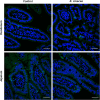
Figure 4. HuNoV infection of enterocytes in Gn pigs.
HuNoV capsid in duodenum (top panel) and jejunum (bottom panel) of Gn pigs euthanized on PID3 was detected by immunohistochemistry. Cell nuclei (blue) were counterstained with HuNoV capsid (bright green). Representative images showing HuNoV was detected in enterocytes in control and E. cloacae colonized Gn pigs. Scale bar, 50 μm.
Figure 5
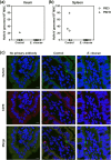
Figure 5. HuNoV infection of B cells was not observed in Gn pigs.
MNC from ileum (a) and spleen (b) were isolated and measured for HuNoV titers by qRT-PCR. Data are presented as individual animal data points. (c) Ileum tissue sections were stained to detect HuNoV capsid protein (bright green) and B cells (CD79+, red) with counterstain of cell nuclei (DAPI, blue). Representative images showing that HuNoV was observed in enterocytes but not in B cells in control and E. cloacae colonized Gn pigs. Scale bar, 10 μm.
Figure 6
Figure 6. Colonization of E. cloacae in Gn pigs stimulated the development of IPP.
(a) Representative H&E staining images of ileum showing more developed IPP in E. cloacae colonized Gn pigs compared to that of control pigs on both 6 days (top panel) and 13 days (bottom panel) post E. cloacae inoculation, Scale bar, 0.5 mm. (b) 12 random locations including all pigs in each group were measured to characterize the IPP size, including width from muscularis mucosae to muscularis (mm to m) associated with gut associated lymphoid tissue (GALT), width of GALT, height and width of follicles. Statistical significance was determined by Mann-Whitney test. *P < 0.05; **P < 0.01; ***P < 0.001. Error bars denote SEM.

"V体育官网" References

    1. Ahmed S. M. et al. Global prevalence of norovirus in cases of gastroenteritis: a systematic review and meta-analysis. Lancet Infect Dis 14, 725–30 (2014). - PMC - PubMed
    1. Karst S. M. Pathogenesis of noroviruses, emerging RNA viruses. Viruses 2, 748–81 (2010). - PMC - PubMed
    1. Zheng D. P. et al. Norovirus classification and proposed strain nomenclature. Virology 346, 312–23 (2006). - "V体育安卓版" PubMed
    1. Kroneman A. et al. Proposal for a unified norovirus nomenclature and genotyping. Arch Virol 158, 2059–68 (2013). - PMC - PubMed
    1. Bull R. A., Eden J. S., Rawlinson W. D. & White P. A. Rapid evolution of pandemic noroviruses of the GII.4 lineage. PLoS Pathog 6, e1000831 (2010). - PMC - PubMed

Publication types

MeSH terms

VSports注册入口 - Grants and funding

LinkOut - more resources