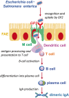
"VSports最新版本" Intestinal M cells
- PMID: 26634447
- PMCID: V体育官网 - PMC4892784
- DOI: 10.1093/jb/mvv121
VSports app下载 - Intestinal M cells
Abstract (V体育官网入口)
We have an enormous number of commensal bacteria in our intestine, moreover, the foods that we ingest and the water we drink is sometimes contaminated with pathogenic microorganisms. The intestinal epithelium is always exposed to such microbes, friend or foe, so to contain them our gut is equipped with specialized gut-associated lymphoid tissue (GALT), literally the largest peripheral lymphoid tissue in the body. GALT is the intestinal immune inductive site composed of lymphoid follicles such as Peyer's patches. M cells are a subset of intestinal epithelial cells (IECs) residing in the region of the epithelium covering GALT lymphoid follicles. Although the vast majority of IEC function to absorb nutrients from the intestine, M cells are highly specialized to take up intestinal microbial antigens and deliver them to GALT for efficient mucosal as well as systemic immune responses. I will discuss recent advances in our understanding of the molecular mechanisms of M-cell differentiation and functions. VSports手机版.
Keywords: M cell; Spi-B; antigen uptake; enteroid; gut-associated lymphoid tissue (GALT) V体育安卓版. .
© The Authors 2015. Published by Oxford University Press on behalf of the Japanese Biochemical Society. All rights reserved. V体育ios版.
Figures


References
-
- Bianconi E., Piovesan A., Facchin F., Beraudi A., Casadei R., Frabetti F., Vitale L., Pelleri M.C., Tassani S., Piva F., Perez-Amodio S., Strippoli P., Canaider S. (2013) An estimation of the number of cells in the human body. Ann. Hum. Biol. 40, 463–471 - PubMed
-
- Peterson C.T., Sharma V., Elmén L., Peterson S.N. (2015) Immune homeostasis, dysbiosis and therapeutic modulation of the gut microbiota. Clin. Exp. Immunol. 179, 363–377 - "VSports注册入口" PMC - PubMed
-
- Qin J. Li R. Raes J. Arumugam M. Burgdorf K.S. Manichanh C. Nielsen T. Pons N. Levenez F. Yamada T. Mende D.R. Li J. Xu J. Li S. Li D. Cao J. Wang B. Liang H. Zheng H. Xie Y. Tap J. Lepage P. Bertalan M. Batto J.M. Hansen T. Le Paslier D. Linneberg A. Nielsen H.B. Pelletier E. Renault P. Sicheritz-Ponten T. Turner K. Zhu H. Yu C. Li S. Jian M. Zhou Y. Li Y. Zhang X. Li S. Qin N. Yang H. Wang J. Brunak S. Doré J. Guarner F. Kristiansen K. Pedersen O. Parkhill J. Weissenbach J MetaHIT Consortium, Bork P. Ehrlich S.D. Wang J. (2010) A human gut microbial gene catalogue established by metagenomic sequencing. Nature 464, 59–65 - PMC - PubMed
-
- International Human Genome Sequencing Consortium. (2004) Finishing the euchromatic sequence of the human genome. Nature 431, 931–945 - PubMed
-
- Revy P., Muto T., Levy Y., Geissmann F., Plebani A., Sanal O., Catalan N., Forveille M., Dufourcq-Labelouse R., Gennery A., Tezcan I., Ersoy F., Kayserili H., Ugazio A.G., Brousse N., Muramatsu M., Notarangelo L.D., Kinoshita K., Honjo T., Fischer A., Durandy A. (2000) Activation-induced cytidine deaminase (AID) deficiency causes the autosomal recessive form of the Hyper-IgM syndrome (HIGM2). Cell 102, 565–575 - PubMed
Publication types
MeSH terms
- "V体育官网" Actions
- "VSports在线直播" Actions
- "V体育安卓版" Actions
- V体育2025版 - Actions
- Actions (VSports)
- "V体育2025版" Actions
- Actions (VSports)
Substances
- V体育2025版 - Actions
- Actions (V体育2025版)
- "V体育官网" Actions
LinkOut - more resources
Full Text Sources (VSports手机版)
VSports - Other Literature Sources

