Skip to main page content
U.S. flag

An official website of the United States government

Dot gov

The . gov means it’s official VSports app下载. Federal government websites often end in . gov or . mil. Before sharing sensitive information, make sure you’re on a federal government site. .

Https

The site is secure. The https:// ensures that you are connecting to the official website and that any information you provide is encrypted and transmitted securely. V体育官网.

. 2016 Jan 15;310(2):G81-92.
doi: 10.1152/ajpgi.00065.2015. Epub 2015 Nov 25.

"V体育ios版" Bile acids regulate intestinal cell proliferation by modulating EGFR and FXR signaling

Affiliations

Bile acids regulate intestinal cell proliferation by modulating EGFR and FXR signaling

Avafia Y Dossa et al. Am J Physiol Gastrointest Liver Physiol. .

Abstract

Bile acids (BAs) are synthesized in the liver and secreted into the intestine. In the lumen, enteric bacteria metabolize BAs from conjugated, primary forms into more toxic unconjugated, secondary metabolites. Secondary BAs can be injurious to the intestine and may contribute to disease. The epidermal growth factor receptor (EGFR) and the nuclear farnesoid X receptor (FXR) are known to interact with BAs. In this study we examined the effects of BAs on intestinal epithelial cell proliferation and investigated the possible roles for EGFR and FXR in these effects. We report that taurine-conjugated cholic acid (TCA) induced proliferation, while its unconjugated secondary counterpart deoxycholic acid (DCA) inhibited proliferation VSports手机版. TCA stimulated phosphorylation of Src, EGFR, and ERK 1/2. Pharmacological blockade of any of these pathways or genetic ablation of EGFR abrogated TCA-stimulated proliferation. Interestingly, Src or EGFR inhibitors eliminated TCA-induced phosphorylation of both molecules, suggesting that their activation is interdependent. In contrast to TCA, DCA exposure diminished EGFR phosphorylation, and pharmacological or siRNA blockade of FXR abolished DCA-induced inhibition of proliferation. Taken together, these results suggest that TCA induces intestinal cell proliferation via Src, EGFR, and ERK activation. In contrast, DCA inhibits proliferation via an FXR-dependent mechanism that may include downstream inactivation of the EGFR/Src/ERK pathway. Since elevated secondary BA levels are the result of specific bacterial modification, this may provide a mechanism through which an altered microbiota contributes to normal or abnormal intestinal epithelial cell proliferation. .

Keywords: bile acid; intestine; necrotizing enterocolitis; proliferation V体育安卓版. .


Figures

Fig. 1.
Fig. 1.
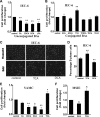
Bile acids (BAs) differentially affect intestinal epithelial cell proliferation. A: IEC-6 cells were serum-starved for 24 h and then treated with unconjugated BAs at nontoxic doses: 200 μM cholic acid (CA), 50 μM chenodeoxycholic acid (CDCA), 50 μM deoxycholic acid (DCA), and 25 μM lithocholic acid (LCA). After 24 h, cells were stained with crystal violet and counted. B: IEC-6 cells were exposed to conjugated BAs [200 μM glycocholic acid (GCA), glycodeoxycholic acid (GDCA), taurine-conjugated CA (TCA), taurochenodeoxycholic acid (TCDCA), taurodeoxycholic acid (TDCA), and taurolithocholic acid (TLCA)] for 24 h, stained with crystal violet, and counted. C and D: IEC-6 cells were labeled with 5-ethynyl-2′-deoxyuridine (EdU) after 24 h of BA exposure, and the percentage of EdU-positive nuclei was determined. E and F: young adult mouse colon (YAMC) and mouse small intestine epithelial (MSIE) cells were exposed to BAs for 24 h after serum starvation, stained with crystal violet, and counted. Values are means ± SE; n > 10 for each (A), n ≥ 4 for each (B), n > 10 for each (D), and n ≥ 3 (E and F). *P < 0.05; **P < 0.01.
Fig. 2.
Fig. 2.
Effects of TCA and DCA on proliferation are dose-dependent. A and B: IEC-6 cells were treated with varying doses of TCA and DCA, stained with crystal violet, and counted. Maximal effects were seen at 250 μM TCA (31.6 ± 9.5%) and 50 μM DCA (22.1 ± 8.0%). Values are means ± SE; n > 5. DCA at 100 μM was toxic as determined by visual inspection and by cell counts. C: lysates from cells treated with 100 or 200 μM DCA for 24 h were assessed by immunoblotting for caspase-3 cleavage. Akt was used as a loading control; 500 μM LCA was used as a positive control. Because of abundant cell death, there was less protein in the positive control condition. *P < 0.05; **P < 0.01.
Fig. 3.
Fig. 3.
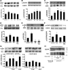
TCA stimulates phosphorylation of Src at Tyr416, epidermal growth factor (EGF) receptor (EGFR), and ERK. IEC-6 cells were exposed to TCA at 5–120 min, and lysates were immunostained by Western blotting. A–C: phosphorylation of Src at Tyr416, EGFR, and ERK was increased, with maximal phosphorylation at 5, 60, and 30 min, respectively. D and E: cells were pretreated with the EGFR inhibitor AG1478 (AG, 150 nM) prior to TCA administration, and phosphorylation of Src at Tyr416 was assessed at 5 min and ERK phosphorylation at 30 min. F: Src inhibition with PP2 (10 μM) eliminated TCA-induced EGFR phosphorylation at 1 h. G: EGFR−/− mouse colon epithelial (MCE) cells expressing wild-type human EGFR (hEGFR) or EGFR−/− MCE cells lacking EGFR were treated with TCA, and Src activation was assessed. H: cells were treated with TCA at 5 or 60 min and immunoprecipitated (IP) with Src, and total EGFR was analyzed. Values are means ± SE; n ≥ 4 (A–G) and n = 3 (H). *P < 0.05; **P < 0.01.
Fig. 4.
Fig. 4.
Effect of TCA on cell proliferation is mediated by EGFR, Src, and ERK. A: after 24 h of serum starvation, cells were pretreated for 1 h with EGFR, Src, or ERK inhibitors (150 nM AG1478, 10 μM PP2, or 10 μM PD98059, respectively). Proliferation was assessed by crystal violet counting and confirmed by EdU incorporation. B: cells were pretreated with the phosphoinositide 3-kinase inhibitor LY294002 (10 μM). C: proliferation was assessed after TCA administration with EdU incorporation in EGFR knockout cells transfected with a plasmid containing human EGFR (−−EGFR +hEGFR), a kinase-dead EGFR (mutant EGFR kinase), or an empty vector (−−EGFR). EGFR was required for TCA-induced proliferation. D: cells were pretreated with the matrix metalloproteinase inhibitor transarterial chemoembolization inhibitor TAPI-1 for 1 h prior to exposure to TCA and assessment of cell proliferation. Values are means ± SE; n > 6 for each (A), n = 8 (B), n = 5 (C), and n = 4 (D). *P < 0.05.
Fig. 5.
Fig. 5.
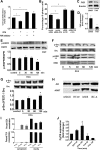
DCA inhibits proliferation through the farnesoid X receptor (FXR). A and B: after 24 h of serum starvation, proliferation was assessed using crystal violet and confirmed by EdU incorporation on cells after 1 h of pretreatment with the FXR inhibitor Z-guggulsterone (10 μM) or transfection with the FXR-specific siRNA (10 nM) or nontargeting control for 48 h prior to assessment of proliferation. C: FXR protein knockdown was assessed by immunoblotting of cell lysates exposed to FXR-specific siRNA. D: reduction of FXR gene expression was assessed by real-time RT-PCR. E–G: IEC-6 cells were treated with DCA for 5–120 min and assessed for EGFR phosphorylation, Src (Tyr416) and ERK phosphorylation, and Src (Tyr527) phosphorylation. H: Madin-Darby canine kidney (MDCK), HT29, MSIE, and IEC-6 cell lysates were probed for apical sodium-dependent BA transporter (ASBT) by immunoblotting. I: IEC-6 cells were treated with DCA or TCA for 30 min or 24 h, media and cell lysates were collected, and BA levels were measured using high-performance liquid chromatography-mass spectroscopy. J: IEC-6 cells were treated for 30 or 120 min with DCA or TCA, and fibroblast growth factor 15 (Fgf15) levels were measured using real-time RT-PCR. Values are means ± SE; n ≥ 5 each (A and B), n = 4 (D and J), n ≥ 3 (E–G), and n = 3 (I). *P < 0.05.
Fig. 6.
Fig. 6.
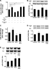
Proliferative effects of TCA and DCA converge on Src kinase. Proliferation was assessed by counting after crystal violet staining and confirmed by EdU incorporation. A: cells were treated with TCA, the FXR agonist GW4064 (GW, 10 μM), and the FXR antagonist Z-guggulsterone (GUG, 10 μM) alone or in combination. B: Western blots on cell lysates were exposed to GW4064 for Src (Tyr416) phosphorylation. C–E: cells were treated with TCA and DCA, alone or in combination, and assessed for proliferation, phosphorylation of Src (Tyr416), and phosphorylation of Src (Tyr527). Values are means ± SE; n ≥ 4 for each (A), n = 5 (B), and n ≥ 3 (C–E). *P < 0.05; **P < 0.01.
Fig. 7.
Fig. 7.
Newborn rats were fed formula with or without TCA or DCA 3 times daily and subjected to hypoxia for 4 days. EdU was injected 6 h prior to euthanasia, and the terminal ileum was harvested. A and B: slides were stained for EdU-positive cells, and proportion of EdU-positive cells was determined. Values are means ± SE; n = 5.
Fig. 8.
Fig. 8.
Proposed mechanism of differential effects of TCA and DCA on intestinal epithelial cell proliferation.

References

    1. Alnouti Y, Csanaky IL, Klaassen CD. Quantitative profiling of bile acids and their conjugates in mouse liver, bile, plasma, and urine using LC-MS/MS. J Chromatogr B Analyt Technol Biomed Life Sci 873: 209–217, 2008. - PMC - PubMed
    1. Aries V, Crowther JS, Drasar BS, Hill MJ. Degradation of bile salts by human intestinal bacteria. Gut 10: 575–576, 1969. - "V体育安卓版" PMC - PubMed
    1. Avissar NE, Toia L, Hu Y, Watson TJ, Jones C, Raymond DP, Matousek A, Peters JH. Bile acid alone, or in combination with acid, induces CDX2 expression through activation of the epidermal growth factor receptor (EGFR). J Gastrointest Surg 13: 212–222, 2009. - PubMed (V体育官网入口)
    1. Bae KB, Jeong YJ, Won HJ, Hong KH, Choi IW, Seo SK, Park SG. A human monoclonal antibody scFv to urokinase plasminogen activator. Hybridoma 29: 147–152, 2010. - "V体育平台登录" PubMed
    1. Bailey AM, Zhan L, Maru D, Shureiqi I, Pickering CR, Kiriakova G, Izzo J, He N, Wei C, Baladandayuthapani V, Liang H, Kopetz S, Powis G, Guo GL. FXR silencing in human colon cancer by DNA methylation and KRAS signaling. Am J Physiol Gastrointest Liver Physiol 306: G48–G58, 2014. - V体育2025版 - PMC - PubMed

Publication types

MeSH terms

LinkOut - more resources