Identification of leukocyte-specific protein 1-positive cells: a clue to the cell of origin and a marker for the diagnosis of dermatofibroma
- PMID: 25834354
- PMCID: PMC4377404
- DOI: 10.5021/ad.2015.27.2.157 (V体育平台登录)
Identification of leukocyte-specific protein 1-positive cells: a clue to the cell of origin and a marker for the diagnosis of dermatofibroma
Abstract (V体育官网)
Background: Dermatofibroma (DF) comprises a heterogeneous group of mesenchymal tumors, with fibroblastic and histiocytic elements present in varying proportions. The cell of origin of DF has been investigated, but remains unclear VSports手机版. .
Objective: The present study attempted to investigate the expression of leukocyte-specific protein 1 (LSP1), a marker of fibrocytes, in DF. Additionally, we evaluated the effectiveness of LSP1 in the differential diagnosis of DF from dermatofibrosarcoma protuberans (DFSP). V体育安卓版.
Methods: Immunohistochemical staining was performed on 20 cases of DF using antibodies against LSP1, CD68, and factor XIIIa (FXIIIa). In addition, the expression of LSP1 and FXIIIa was evaluated in 20 cases of DFSP. V体育ios版.
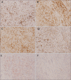
Results: Eighteen of 20 cases (90%) of DF stained positive for LSP1, with variation in the intensity of expression VSports最新版本. CD68 was positive in 10 cases (50%), and FXIIIa was expressed in all cases of DF. There were differences between the regional expression patterns of the three markers in individual tumors. In contrast, only 2 of 20 cases of DFSP expressed LSP1, and none of DFSP cases stained positive for FXIIIa. .
Conclusion: The LSP1-positive cells in DF could potentially be fibrocyte-like cells. FXIIIa and CD68 expression suggests that dermal dendritic cells and histiocytes are constituent cells of DF V体育平台登录. It is known that fibrocytes, dermal dendritic cells and histiocytes are all derived from CD14+ monocytes. Therefore, we suggest that DF may originate from CD14+ monocytes. Additionally, the LSP1 immunohistochemical stain could be useful in distinguishing between DF and DFSP. .
Keywords: Dermatofibroma; Dermatofibrosarcoma; Leukocyte-specific protein 1. VSports注册入口.
Figures


References (V体育ios版)
-
- Song Y, Sakamoto F, Ito M. Characterization of factor XIIIa+ dendritic cells in dermatofibroma: Immunohistochemical, electron and immunoelectron microscopical observations. J Dermatol Sci. 2005;39:89–96. - PubMed
-
- Horenstein MG, Prieto VG, Nuckols JD, Burchette JL, Shea CR. Indeterminate fibrohistiocytic lesions of the skin: is there a spectrum between dermatofibroma and dermatofibrosarcoma protuberans. Am J Surg Pathol. 2000;24:996–1003. - PubMed
-
- Yan X, Takahara M, Xie L, Tu Y, Furue M. Cathepsin K expression: a useful marker for the differential diagnosis of dermatofibroma and dermatofibrosarcoma protuberans. Histopathology. 2010;57:486–488. - PubMed
-
- Goldblum JR, Tuthill RJ. CD34 and factor-XIIIa immunoreactivity in dermatofibrosarcoma protuberans and dermatofibroma. Am J Dermatopathol. 1997;19:147–153. - PubMed
-
- Kuroda K, Tajima S. Proliferation of HSP47-positive skin fibroblasts in dermatofibroma. J Cutan Pathol. 2008;35:21–26. - PubMed
LinkOut - more resources
"V体育平台登录" Full Text Sources
"VSports注册入口" Other Literature Sources
Research Materials
Miscellaneous

