"VSports" Skip to main page content
U.S. flag

An official website of the United States government

Dot gov

The VSports app下载. gov means it’s official. Federal government websites often end in . gov or . mil. Before sharing sensitive information, make sure you’re on a federal government site. .

Https

The site is secure. The https:// ensures that you are connecting to the official website and that any information you provide is encrypted and transmitted securely V体育官网. .

. 2014 Nov;63(11):1769-81.
doi: 10.1136/gutjnl-2013-306271. Epub 2014 Feb 20.

V体育ios版 - Targeted depletion of an MDSC subset unmasks pancreatic ductal adenocarcinoma to adaptive immunity

Affiliations

Targeted depletion of an MDSC subset unmasks pancreatic ductal adenocarcinoma to adaptive immunity (V体育2025版)

Ingunn M Stromnes (V体育平台登录) et al. Gut. 2014 Nov.

Abstract

Background: Pancreatic ductal adenocarcinoma (PDA) is characterised by a robust desmoplasia, including the notable accumulation of immunosuppressive cells that shield neoplastic cells from immune detection VSports手机版. Immune evasion may be further enhanced if the malignant cells fail to express high levels of antigens that are sufficiently immunogenic to engender an effector T cell response. .

Objective: To investigate the predominant subsets of immunosuppressive cancer-conditioned myeloid cells that chronicle and shape the progression of pancreas cancer. We show that selective depletion of one subset of myeloid-derived suppressor cells (MDSC) in an autochthonous, genetically engineered mouse model (GEMM) of PDA unmasks the ability of the adaptive immune response to engage and target tumour epithelial cells V体育安卓版. .

Methods: A combination of in vivo and in vitro studies were performed employing a GEMM that faithfully recapitulates the cardinal features of human PDA V体育ios版. The predominant cancer-conditioned myeloid cell subpopulation was specifically targeted in vivo and the biological outcomes determined. .

Results: PDA orchestrates the induction of distinct subsets of cancer-associated myeloid cells through the production of factors known to influence myelopoiesis VSports最新版本. These immature myeloid cells inhibit the proliferation and induce apoptosis of activated T cells. Targeted depletion of granulocytic MDSC (Gr-MDSC) in autochthonous PDA increases the intratumoral accumulation of activated CD8 T cells and apoptosis of tumour epithelial cells and also remodels the tumour stroma. .

Conclusions: Neoplastic ductal cells of the pancreas induce distinct myeloid cell subsets that promote tumour cell survival and accumulation V体育平台登录. Targeted depletion of a single myeloid subset, the Gr-MDSC, can unmask an endogenous T cell response, disclosing an unexpected latent immunity and invoking targeting of Gr-MDSC as a potential strategy to exploit for treating this highly lethal disease. .

Keywords: Growth Factors; Immune Response; Pancreatic Cancer. VSports注册入口.


Figures

Figure 1
Figure 1
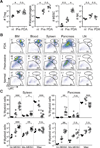
Cancer-conditioned myeloid cells chronicle the evolution of PDA in KPC mice. (A) The number of pancreatic Treg (CD45+CD4+FoxP3+), macrophages (CD45+CD11b+F4/80+), MDSC (CD45+CD11b+RB6-8C5 [Ly6G/Ly6C]+) and NK cells (CD45+NK1.1+) per pancreas were quantified from normal pancreas (nl), 6–8 week old KPC pancreata with confirmed preinvasive disease (Pre), and invasive tumors (PDA). Significant differences were detected in the number of Treg, TAM, and MDSC during disease progression. (B) Evolving profiles of three distinct populations of myeloid cells (gated on CD45+CD11b+) were observed in various organs based on expression patterns of Gr-1 and Ly6C. BM, bone marrow; LN, lymph node. (C) The percentages (top panel) and absolute numbers (bottom panel) of CD45+CD11b+ myeloid populations in the spleen and pancreas in normal (black filled circles) and preinvasive (gray filled circles) and invasive (open circles) disease settings. Data are plotted as mean ± SEM and each data point represents an individual mouse. Gr-MDSC = CD45+CD11b+Gr-1highLy6Cint; Mo-MDSC = CD45+CD11b+Gr-1intLy6Chigh; and macrophage (Mac) = CD45+CD11b+Ly6Cint. (D) Specific immunofluorescence reveals rare Ly6G/Ly6C+ (RB6-8C5) cells in normal pancreas, focal accumulation in pancreata with preinvasive disease, and diffuse infiltration in invasive PDA. Specific Ly6G immunofluorescence demonstrates that Gr-MDSC are absent from normal pancreas, rare in preinvasive disease, and abundant in invasive PDA. The majority of myeloid cells in normal pancreas and surrounding preinvasive lesions appear to be macrophages. Arrowheads, epithelial cells; arrows, myeloid cells; asterisk, Gr-MDSC. Scale bars, 50 µm. (E) Ly6G/Ly6C (RB6-8C5) staining in normal pancreas and KPC salivary gland. Arrowheads, epithelial cells; arrows, myeloid cells. Scale bars, 10 µm. *, p<0.05; **, p<0.005; ***, p<0.0005.
Figure 1
Figure 1
Cancer-conditioned myeloid cells chronicle the evolution of PDA in KPC mice. (A) The number of pancreatic Treg (CD45+CD4+FoxP3+), macrophages (CD45+CD11b+F4/80+), MDSC (CD45+CD11b+RB6-8C5 [Ly6G/Ly6C]+) and NK cells (CD45+NK1.1+) per pancreas were quantified from normal pancreas (nl), 6–8 week old KPC pancreata with confirmed preinvasive disease (Pre), and invasive tumors (PDA). Significant differences were detected in the number of Treg, TAM, and MDSC during disease progression. (B) Evolving profiles of three distinct populations of myeloid cells (gated on CD45+CD11b+) were observed in various organs based on expression patterns of Gr-1 and Ly6C. BM, bone marrow; LN, lymph node. (C) The percentages (top panel) and absolute numbers (bottom panel) of CD45+CD11b+ myeloid populations in the spleen and pancreas in normal (black filled circles) and preinvasive (gray filled circles) and invasive (open circles) disease settings. Data are plotted as mean ± SEM and each data point represents an individual mouse. Gr-MDSC = CD45+CD11b+Gr-1highLy6Cint; Mo-MDSC = CD45+CD11b+Gr-1intLy6Chigh; and macrophage (Mac) = CD45+CD11b+Ly6Cint. (D) Specific immunofluorescence reveals rare Ly6G/Ly6C+ (RB6-8C5) cells in normal pancreas, focal accumulation in pancreata with preinvasive disease, and diffuse infiltration in invasive PDA. Specific Ly6G immunofluorescence demonstrates that Gr-MDSC are absent from normal pancreas, rare in preinvasive disease, and abundant in invasive PDA. The majority of myeloid cells in normal pancreas and surrounding preinvasive lesions appear to be macrophages. Arrowheads, epithelial cells; arrows, myeloid cells; asterisk, Gr-MDSC. Scale bars, 50 µm. (E) Ly6G/Ly6C (RB6-8C5) staining in normal pancreas and KPC salivary gland. Arrowheads, epithelial cells; arrows, myeloid cells. Scale bars, 10 µm. *, p<0.05; **, p<0.005; ***, p<0.0005.
Figure 2
Figure 2
Gr-MDSC isolated from the spleen and PDA are immunosuppressive. (A) Naïve T cell proliferation in response to anti-CD3/CD28 is suppressed by Gr-MDSC (assays performed at 1:1 Gr-MDSC:T cell ratio). Numbers indicate the percentage of CD8 T cells that have undergone ≥ 1 cell division after 48h. Gray filled histograms indicate T cells in the absence of CD3/CD28. (−) indicates no Gr-MDSC added. (B) The suppressive capacity of Gr-MDSC increases at higher Gr-MDSC:T cell ratios. Representative data from 1 of 4 independent experiments are plotted as mean ± SD (n=3 replicates each). (−) = no CD3/CD28 stimulation. (C) Gr-MDSC induce the apoptosis of activated T cells. Representative FACS profiles (of 4 independent experiments) performed at 1:1 Gr-MDSC:T cell ratio and gated on CD8+ Thy1.1+ cells. (−) = no stimulation; (+) = CD3/CD28 stimulation. (D) Gr-MDSC isolated from spleen (Spl) and PDA of KPC mice induce apoptosis of activated CD8 T cells. (−) = T cells only (no MDSC). Data represent mean ± SEM from 4 independent experiments.
Figure 3
Figure 3
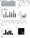
Conditioned media from primary PDA (PDA-CM) or metastatic epithelial cells (Met-CM) promotes the survival of Gr-MDSC. (A) Sorted Gr-MDSC cultured in basic media or PDA-CM for 48h. (B) PDA-CM protects a cultured Gr-MDSC from apoptosis (48h). (C) Relative gene expression of select cytokines and chemokines as determined by quantitative PCR (data represent mean ± SEM of 3 independently derived primary invasive tumor (PDA) and paired metastatic cell preparations and are normalized to expression in preinvasive cells). (D) Impact of conditioned media and cytokines on apoptosis of splenic-derived Gr-MDSC. Bar graphs represent mean ± SEM from 4 independent experiments performed. Treated samples were compared to the basic media (Basic) control and significance was determined by using a one-way ANOVA followed by a Dunnett’s multiple comparison test. (E) Sorted splenic Gr-MDSC from KPC mice with PDA were incubated with the indicated media or cytokines and the percentage of Annexin-V+ Gr-MDSC at 48h determined by FACS (representative results of 4 independent experiments). (F) Sorted splenic Gr-MDSC were incubated with PDA-CM ± the indicated blocking mAb for 48h. The percentage of live cells was determined by FACS analyses of Annexin-Vneg Gr-MDSC. (G) Clusters of PDA cells contain GM-CSF at their stromal surface. CK, cytokeratin. Scale bar, 25 µm. *, p<0.05; **, p<0.005; ***, p<0.0005.
Figure 4
Figure 4
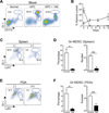
Systemic administration of 1A8 (αLy6G) specifically depletes Gr-MDSC in autochthonous PDA. (A) Representative myeloid cell profiles in PBMC from a normal mouse and untreated (KPC) and 1A8-treated KPC (KPC + 1A8) mice. Numbers indicate the percentage of each subset gated on CD45+ mononuclear cells. We note that the gates for the discrete subpopulations defined in the blood were then applied to the tissue-specific analyses. (B) Percentage of Gr-MDSC (squares) and Mo-MDSC (circles) in blood after 1A8 treatment. Data represent mean ± SD from n=3 independent treated animals. (C) Representative FACS profiles of CD45+ CD11b+ splenocytes from control (−) and 1A8-treated (+) KPC mice (n=4–6 animals per group). (D) Both the percentage and number of splenic Gr-MDSC at endpoint of 1A8 treatment (day 12) are significantly decreased (**, p=0.005). (E) Representative FACS profiles of intratumoral myeloid cells in control (−) and 1A8-treated (+) KPC mice at day 12 of Gr-MDSC depletion. (F) The percentage and number of Gr-MDSC in PDA at endpoint are significantly decreased in 1A8-treated compared to control KPC mice.
Figure 5
Figure 5
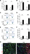
Gr-MDSC depletion increases CD8 T cell number, proliferation, activation and tumor-specific infiltration in autochthonous PDA. The percentage (A) and number (B) of CD8 T cells significantly increased in pancreatic tumors, but not the spleen of 1A8-treated compared with untreated KPC mice. (C) Representative FACS plots of proliferating CD8 T cells in the spleen and pancreatic tumors of control and treated KPC mice as determined by Ki67 expression (n=4–6 mice per cohort). (D) The percentage of Ki67+ CD8 T cells in 1A8-treated (n=4) compared to control KPC mice (n=6 mice) in the spleen and tumors was significantly increased. (E, F) Gr-MDSC depletion increases the percentage of CD8 T cells in both the spleen and PDA that express the activation marker CD69. (G) CD8 T cells (arrow) are rare in control PDA and increase significantly (asterisk) after Gr-MDSC depletion (1A8). Control, open bars; 1A8-treated, filled bars. Scale bars, 50 µm.
Figure 6
Figure 6
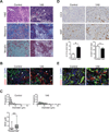
Targeted depletion of Gr-MDSC remodels the stroma and increases tumor epithelial cell apoptosis. (A) Administration of 1A8 increases mononuclear cell infiltrates in autochthonous PDA (outlined) and patency of blood vessels (arrows), and depletes ECM content and integrity (asterisks). Representative fields are shown for histology (H&E), collagen content (Masson’s trichrome) and combined collagen and glycosaminoglycan content (Movat’s pentachrome). Scale bars, 50 µm. (B) CD31 immunofluorescence of PDA reveals a shift toward larger vessel diameters (arrows) after 1A8 treatment (quantified in (C)). Scale bars, 25 µm. (C) Administration of 1A8 significantly increases the mean vessel diameter in PDA. (D) Apoptosis of tumor epithelial cells, as assessed by cleaved caspase-3 (CC3), was significantly increased in 1A8-treated (n=4) compared to control (n=6) KPC mice, while the number of proliferating tumor epithelial cells (Ki67) was not significantly different. (E) CC3 and CK double-positive cells indicate tumor epithelial cells undergoing apoptosis (arrowheads). Scale bars, 25 µm.

References

    1. Pylayeva-Gupta Y, Lee KE, Hajdu CH, et al. Oncogenic Kras-induced GM-CSF production promotes the development of pancreatic neoplasia. Cancer cell. 2012;21:836–847. - PMC - PubMed
    1. Bayne LJ, Beatty GL, Jhala N, et al. Tumor-derived granulocyte-macrophage colony-stimulating factor regulates myeloid inflammation and T cell immunity in pancreatic cancer. Cancer cell. 2012;21:822–835. - "V体育安卓版" PMC - PubMed
    1. Siegel R, Naishadham D, Jemal A. Cancer statistics, 2012. CA: a cancer journal for clinicians. 2012;62:10–29. - "V体育官网" PubMed
    1. Hruban R. In: Tumors of the pancreas. Hruban RH, Pitman MB, Klimstra DS, editors. 2007.
    1. Olive KP, Jacobetz MA, Davidson CJ, et al. Inhibition of Hedgehog signaling enhances delivery of chemotherapy in a mouse model of pancreatic cancer. Science. 2009;324:1457–1461. - PMC - PubMed

Publication types

MeSH terms

Substances