<u date-time="vCy47m"></u><sub date-time="nQtS9k"></sub><sub date-time="aeBQIL"><small dropzone="hPrHv"></small></sub> Skip to main page content
U.S. flag

An official website of the United States government

Dot gov

The . gov means it’s official. Federal government websites often end in . gov or . mil. Before sharing sensitive information, make sure you’re on a federal government site. VSports app下载.

Https

The site is secure. The https:// ensures that you are connecting to the official website and that any information you provide is encrypted and transmitted securely V体育官网. .

. 2013 Nov 19;110(47):19048-53.
doi: 10.1073/pnas.1315336110. Epub 2013 Nov 4.

V体育官网 - Photoswitchable nanoparticles for in vivo cancer chemotherapy

Affiliations

Photoswitchable nanoparticles for in vivo cancer chemotherapy

Rong Tong et al. Proc Natl Acad Sci U S A. .

Abstract

There are many obstacles to effective cancer chemotherapy, including drug penetration and accumulation in tumors and drug systemic toxicity. The penetration of therapies into tumors is limited by the dense tumor matrix and by compression of the tumor vasculature. We have developed spiropyran-based nanoparticles that shrink from 103 to 49 nm upon irradiation at 365 nm. That shrinkage enhanced tissue penetration and drug release. Irradiation of s. c. HT-1080 tumors in nude mice administered i. v. docetaxel-containing nanoparticles was more effective treatment than free docetaxel or encapsulated docetaxel without irradiation. Irradiation at the tumor site also resulted in less systemic toxicity than if the nanoparticles were irradiated before injection, presumably because of less systemically distributed free drug VSports手机版. The enhanced efficacy of nanoparticles in irradiated tumors may have been related to the observed enhanced tumor penetration by nanoparticles and decompression of tumor blood vessels, which may also increase nanoparticle delivery into tumors. .

Keywords: nanomedicine; photoswitching; triggered drug delivery. V体育安卓版.


"VSports在线直播" Conflict of interest statement

The authors declare no conflict of interest.

Figures (V体育ios版)

Fig. 1.
Fig. 1.
In vitro characterization of photoswitching SP NPHCs. (A) Dynamic light-scattering measurement of size changes of SP NPHCs upon alternating UV (35 s) and visible light illumination (500–600 nm, 5 min, 0.5 W/cm2). (B) Release of Dtxl from SP NPHC in the absence of irradiation; with UV irradiation for 10 s at t = 0 or with repeated UV irradiation at t = 0, 12, and 24 h. The times of irradiation are indicated by purple arrows (n = 5). (C) Effect of Dtxl/SP NPHC on HT-1080 cell viability, by MTT assay. Cells were incubated with NPHC for 4 h, washed with NP-free media, irradiated (10 s, 1 W/cm2), and further incubated for a total time of 24 h. Data are means ± SD, n = 6; asterisks indicate P < 0.005. Dtxl, docetaxel; hν, UV irradiation; NPHC, hybrid NPs with cholesterol; SP, spiropyran.
Fig. 2.
Fig. 2.
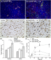
Intratumoral distribution and efficacy of i.t. injected SP NPHCs in animals with s.c. HT-1080 tumors. (A and B) Cy5/SP NPHCs (Cy5 dose, 1 mg/kg) were injected i.t., then the tumors were either (A) not treated or (B) irradiated for 15 s at 365 nm, and excised for immunofluorescence imaging 12 h postinjection. Blue, cell nuclei stained by DAPI; green, Cy5/SP NPHC. (Scale bar, 50 μm.) (C) Quantification of the percentage of tumor area positive for Cy5 (n = 4, with four sections from four tumors). Asterisks indicate P < 0.005. (D) In vivo efficacy of Dtxl/SP NPHC (Dtxl dose: 10 mg/kg, n = 5) given i.t. to s.c. HT-1080 tumors, without or with light triggering (15 s, 1 W/cm2). Two of five mice in the Dtxl/SP NPHC group (green line) were euthanized as their tumor size exceeded 500 mm3 (tumor diameter over 1 cm) on day 18 and 29 (red arrows). Data are medians ± quartiles.
Fig. 3.
Fig. 3.
Efficacy, pharmacokinetics, and biodistribution of i.v.-injected formulations in animals with s.c. HT-1080 tumors. (A) Effect on tumor growth inhibition (Dtxl dose: 40 mg/kg, n = 5). § indicates study termination for the group treated with Dtxl due to significant body weight loss (>20%). Mice with tumor volumes over 500 mm3 were removed from the study (*, from the Dtxl/MC NPHC group; †, from the Dtxl/SP NPHC group with irradiation). (B) Kaplan–Meier plot for the same groups as in A. (C) Plasma concentration of Dtxl over time in mice after i.v. administration of Dtxl, Dtxl/SP NPHC, and Dtxl/MC NPHC (Dtxl dose: 10 mg/kg, n = 5). (D) Biodistribution of Dtxl, free or loaded in NPHCs (with and without light triggering), in various tissues at 24 h after injection (n = 5). Results are expressed as a percentage of the total injected dose per tissue mass (%I.D./g). Data in A, C, and D are medians ± interquartile range. Asterisks indicate P < 0.005. Dtxl, docetaxel; . UV irradiation; NPHC, hybrid NPs with cholesterol; SP, spiropyran.
Fig. 4.
Fig. 4.
Light triggering of SP NPHCs enhanced particle diffusion and induced drug release to decompress vessels, leading to improved intratumoral accumulation of NPHCs. (A and B) Representative images of intratumoral distribution of NPs 24 h after i.v. injection of Cy5/SP NPHCs (red color) (A) without or (B) after light irradiation at tumor site (20 s, 1 W/cm2, 30 min postinjection). Blue, cell nucleus; green, antibody against CD31 staining blood vessel endothelial cells; yellow, colocalization of red and green. (Scale bar, 100 μm.) (C and D) Representative immunohistology (of CD31) of tumor blood vessels 24 h after i.v. injection of (C) Dtxl, or (D) Dtxl/SP NPHCs with irradiation of the tumor site under the same conditions as in B. Dtxl dose was 40 mg/kg in all groups. Vessel diameters were larger (indicated by arrows) in the irradiated group, as quantitated in E. (Scale bar, 200 μm.) In E, “periphery” refers to areas <1 mm away from the tumor edge, and “interior” to areas >1 mm inside tumors (SI Appendix, Fig. S13). Asterisk indicates P < 0.01. Data are means ± SD. (F) Intratumoral Dtxl accumulation over 24 h in animals treated with Dtxl, Dtxl/SP NPHCs, and Dtxl/SP NPHCs irradiated as in D (n = 5). Data are medians ± quartiles.
Fig. 5.
Fig. 5.
Effects of light triggering of Dtxl/SP NPHCs (yellow spheres) in the tumor vasculature. The dense collagen matrix (green lines) and compressed vessels prevent unshrunken NPHCs from delivering drugs within the tumor (gray region). Tumor irradiation shrinks NPHCs (purple spheres), which enhances their penetration through the collagen matrix throughout the tumor. The triggered released of Dtxl (the cyan glow surrounding NPHCs) kills tumor cells, which leads to dilation of compressed intratumoral blood vessels. The decompression of vessels facilitates the transport of NPHCs into the tumor interior.

References

    1. Langer R. Polymer-controlled drug-delivery systems. Acc Chem Res. 1993;26(10):537–542.
    1. Langer R, Folkman J. Polymers for the sustained release of proteins and other macromolecules. Nature. 1976;263(5580):797–800. - PubMed
    1. Gref R, et al. Biodegradable long-circulating polymeric nanospheres. Science. 1994;263(5153):1600–1603. - PubMed
    1. Langer R. Drug delivery and targeting. Nature. 1998;392(6679) Suppl:5–10. - PubMed (VSports手机版)
    1. Matsumura Y, Maeda H. A new concept for macromolecular therapeutics in cancer chemotherapy: Mechanism of tumoritropic accumulation of proteins and the antitumor agent smancs. Cancer Res. 1986;46(12 Pt 1):6387–6392. - PubMed

Publication types