Skip to main page content
U.S. flag

An official website of the United States government

Dot gov

The . gov means it’s official. Federal government websites often end in . gov or . mil. Before sharing sensitive information, make sure you’re on a federal government site. VSports app下载.

Https

The site is secure. The https:// ensures that you are connecting to the official website and that any information you provide is encrypted and transmitted securely. V体育官网.

. 2013 Jul 1;19(13):3404-15.
doi: 10.1158/1078-0432.CCR-13-0525. Epub 2013 May 7.

Inflammatory monocyte mobilization decreases patient survival in pancreatic cancer: a role for targeting the CCL2/CCR2 axis

Affiliations

"V体育2025版" Inflammatory monocyte mobilization decreases patient survival in pancreatic cancer: a role for targeting the CCL2/CCR2 axis

Dominic E Sanford (VSports) et al. Clin Cancer Res. .

Abstract

Purpose: To determine the role of the CCL2/CCR2 axis and inflammatory monocytes (CCR2(+)/CD14(+)) as immunotherapeutic targets in the treatment of pancreatic cancer. VSports手机版.

Experimental design: Survival analysis was conducted to determine if the prevalence of preoperative blood monocytes correlates with survival in patients with pancreatic cancer following tumor resection. Inflammatory monocyte prevalence in the blood and bone marrow of patients with pancreatic cancer and controls was compared. The immunosuppressive properties of inflammatory monocytes and macrophages in the blood and tumors, respectively, of patients with pancreatic cancer were assessed. CCL2 expression by human pancreatic cancer tumors was compared with normal pancreas. A novel CCR2 inhibitor (PF-04136309) was tested in an orthotopic model of murine pancreatic cancer. V体育安卓版.

Results: Monocyte prevalence in the peripheral blood correlates inversely with survival, and low monocyte prevalence is an independent predictor of increased survival in patients with pancreatic cancer with resected tumors. Inflammatory monocytes are increased in the blood and decreased in the bone marrow of patients with pancreatic cancer compared with controls. An increased ratio of inflammatory monocytes in the blood versus the bone marrow is a novel predictor of decreased patient survival following tumor resection. Human pancreatic cancer produces CCL2, and immunosuppressive CCR2(+) macrophages infiltrate these tumors V体育ios版. Patients with tumors that exhibit high CCL2 expression/low CD8 T-cell infiltrate have significantly decreased survival. In mice, CCR2 blockade depletes inflammatory monocytes and macrophages from the primary tumor and premetastatic liver resulting in enhanced antitumor immunity, decreased tumor growth, and reduced metastasis. .

Conclusions: Inflammatory monocyte recruitment is critical to pancreatic cancer progression, and targeting CCR2 may be an effective immunotherapeutic strategy in this disease VSports最新版本. .


Figures

Figure 1
Figure 1. The prevalence of peripheral blood monocytes is prognostic in PC patients
A, patients were stratified into low (< 6% of leukocytes, > 1 SD below mean), mid (≥ 6 to < 11% of leukocytes, within 1 SD of mean), and high (≥ 11% of leukocytes, > 1 SD above mean) peripheral blood monocyte groups. Kaplan Meier survival curves compare PC patients in high (A) and low (B) blood monocyte groups to the rest of the cohort as well as patients in low, mid, and high blood monocyte groups separately (C). p values are by log-rank (Mantel-Cox) test.
Figure 2
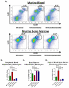
Figure 2. Inflammatory monocyte equilibrium in the peripheral blood and bone marrow is prognostic in pancreatic cancer patients
A, flow cytometry gating strategies used to define IM (CD115+/CD16/CX3CR1low/CD14+/CCR2+) and RM (CD115+/CD16+/CX3CR1hi/CD14/CCR2low) in peripheral blood and bone marrow of PC patients (n=21) and healthy controls (n=11). Graphs compare the prevalence of IM in the blood (B) and bone marrow (C) of PC patients and controls. D, graph compares the blood:bone marrow IM ratios of PC patients with <1 year survival to ≥ 1 year survivors following tumor resection. All graphs show means ± SEM, and p values are by Mann-Whitney test.
Figure 3
Figure 3. Human pancreatic cancers express CCL2 and are infiltrated by immunosuppressive CCR2+ macrophages
A, bar graph compares CCL2 gene expression in human PC (n=11) to normal pancreas (n=10) and representative immunofluorescent confocal images (40×) of human PC stained for cytokeratin 7 (green) and CCL2 (red) with nuclear (Topro) counterstain (blue). B, representative flow cytometry images (gated on CD45+ cells) and immunofluorescent confocal images (40×; CD14 = green, CCR2 = red, and nuclear (Topro) counterstain = blue) of human PC tumors reveal tumor-infiltrating CCR2+ TAM, while bar graph compares tumor-infiltrating TAM to CD8 T cells using flow cytometry. C, representative flow cytometry histogram from T cell suppression assay depicts stimulated, CFSE-labeled CD8 T cells alone (red) or co-cultured at a 1:1 IM (blue) or TAM (green) to lymphocyte ratio after 72 hours, and bar graph compares effect of IM and TAM on T cell division index at various ratios. n=6 experiments. All graphs depict means ± SEM and * denotes p<0.05 by Mann-Whitney test. D, Kaplan-Meier survival curves compare overall survival in PC patients with high CD8 T cell tumor infiltrate/low tumor CCL2 expression to patients with low CD8 T cell infiltrate/high tumor CCL2 expression by tissue microarray (n=60 tumors). p-value is by log-rank (Mantel-Cox) test.
Figure 4
Figure 4. CCR2 mediates inflammatory monocyte mobilization from the bone marrow to the blood in murine pancreatic cancer
A, representative flow cytometry images demonstrate changes in IM prevalence in the blood and bone marrow of control, vehicle-treated, and CCR2i-treated tumor-bearing WT mice after 28 days. Bar graphs show the prevalence of IM in the blood (B) and bone marrow (C) as well as the blood:bone marrow IM ratios (D) of control, vehicle-treated, CCR2−/−, and CCR2i-treated tumor bearing WT mice. All graphs depict means ± SEM and horizontal bars denote statistically significant differences between groups defined as p<0.05 by Mann-Whitney test.
Figure 5
Figure 5. CCR2 blockade promotes anti-tumor immunity and impairs tumor growth
Bar graphs compare tumor myeloid infiltrate (A) as well as the prevalence of tumor infiltrating T lymphocytes (B) and immune gene expression (C) in WT tumor-bearing mice treated with vehicle, GEM, CCR2i, and CCR2i+GEM combination after 28 days. D, tumor growth curves and graph compare effect of CCR2 blockade on subcutaneous and orthotopic (at 28 days) KCKO tumor growth. Note: CCR2−/− tumors were significantly smaller than CCR2i-treated tumors at days 25 and 28 on growth curve, p< 0.05. All graphs depict means +/− SEM. Horizontal bars or * denotes p<0.05 whereas ** denotes p<0.01 by Mann-Whitney test.
Figure 6
Figure 6. CCR2 inhibition reduces inflammatory monocytes and metastasis-associated macrophages in premetastatic livers and impairs hepatic metastasis in murine pancreatic cancer
A, graph compares CCL2 expression using qRT-PCR in baseline (Day 0), premetastatic (Day 6) and metastatic (Day 28) livers of WT mice. B, representative immunofluorescence images (20×) of livers 9 days post-injection from control (matrigel only, MG) mice, vehicle-treated tumor bearing mice, and CCR2i-treated tumor bearing mice for F4/80 (green) expression with a nuclear (Topro) stain (blue). C, graphs depict macrophage and IM prevalence in the livers of MG control or tumor bearing mice treated with vehicle, CCR2i, GEM, or CCR2i+GEM combination by flow cytometry. All flow cytometry and qRT-PCR performed on grossly normal liver (i.e. excluded metastatic liver deposits). All graphs depict means ± SEM and horizontal bars denote statistically significant differences between groups defined as p<0.05 by Mann-Whitney test. D, graph demonstrates the incidence of liver metastasis after 28 days in tumor bearing mice treated with vehicle, GEM, CCR2i, and CCR2i+GEM combination as well as CCR2−/− mice. n = 13-20 mice per group. Horizontal bars denote p<0.05 by Fisher’s exact test.

"VSports手机版" References

    1. Jemal A, Siegel R, Xu J, Ward E. Cancer statistics, 2010. CA Cancer J Clin. 2010;60(5):277–300. Epub 2010/07/09. doi: caac.20073 [pii] 10.3322/caac.20073. PubMed PMID: 20610543. - PubMed
    1. Clark CE, Beatty GL, Vonderheide RH. Immunosurveillance of pancreatic adenocarcinoma: insights from genetically engineered mouse models of cancer. Cancer Lett. 2009;279(1):1–7. Epub 2008/11/18. doi: 10.1016/j.canlet.2008.09.037. PubMed PMID: 19013709. - PubMed
    1. Clark CE, Hingorani SR, Mick R, Combs C, Tuveson DA, Vonderheide RH. Dynamics of the immune reaction to pancreatic cancer from inception to invasion. Cancer Res. 2007;67(19):9518–27. Epub 2007/10/03. doi: 67/19/9518 [pii] 10.1158/0008-5472.CAN-07-0175. PubMed PMID: 17909062. - V体育2025版 - PubMed
    1. Provenzano PP, Cuevas C, Chang AE, Goel VK, Von Hoff DD, Hingorani SR. Enzymatic targeting of the stroma ablates physical barriers to treatment of pancreatic ductal adenocarcinoma. Cancer Cell. 2012;21(3):418–29. Epub 2012/03/24. doi: 10.1016/j.ccr.2012.01.007. PubMed PMID: 22439937; PubMed Central PMCID: PMC3371414. - PMC - PubMed
    1. Kim EJ, Simeone DM. Advances in pancreatic cancer. Current opinion in gastroenterology. 2011;27(5):460–6. Epub 2011/07/23. doi: 10.1097/MOG.0b013e328349e31f. PubMed PMID: 21778878. - PubMed

Publication types

MeSH terms