V体育ios版 - Skip to main page content
U.S. flag

An official website of the United States government

Dot gov

The . gov means it’s official. Federal government websites often end in . gov or . mil VSports app下载. Before sharing sensitive information, make sure you’re on a federal government site. .

Https

The site is secure. The https:// ensures that you are connecting to the official website and that any information you provide is encrypted and transmitted securely. V体育官网.

. 2013 Apr;14(4):356-63.
doi: 10.1038/ni.2547. Epub 2013 Mar 10.

Secondary T cell-T cell synaptic interactions drive the differentiation of protective CD8+ T cells

Affiliations

"V体育2025版" Secondary T cell-T cell synaptic interactions drive the differentiation of protective CD8+ T cells

Audrey Gérard (VSports最新版本) et al. Nat Immunol. 2013 Apr.

Abstract

Immunization results in the differentiation of CD8+ T cells, such that they acquire effector abilities and convert into a memory pool VSports手机版. Priming of T cells takes place via an immunological synapse formed with an antigen-presenting cell (APC). By disrupting synaptic stability at different times, we found that the differentiation of CD8+ T cells required cell interactions beyond those made with APCs. We identified a critical differentiation period that required interactions between primed T cells. We found that T cell-T cell synapses had a major role in the generation of protective CD8+ T cell memory. T cell-T cell synapses allowed T cells to polarize critical secretion of interferon-γ (IFN-γ) toward each other. Collective activation and homotypic clustering drove cytokine sharing and acted as regulatory stimuli for T cell differentiation. .


Figures
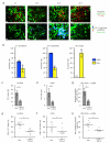
Figure 1
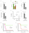
Figure 1. Temporal requirement for CD8 T cell differentiation
(a) CD11c-YFP mice were adoptively transferred with OT-I-RFP cells. Individual “snapshots” of OTI cells (red) and DCs (green) acquired 2, 10, 24 or 72 hours post-immunization with DEC-OVA. Images are displayed as maximum intensity projections along the z axis (top view). Lower panels: Pseudo-colored time projection of a 30 minutes run showing the spatial persistence of OTI cells in clusters 24 hours, but not 2, 10 hours and 72 hours after immunization. Image intensities were scaled to a normalized time projection intensity range of 0–1. Scale bar, 30μm. Data representative of at least three independent experiments (b) T cell priming (CD69 blue label) was quantified by CD69 expression on OTI cells 32 hours post-immunization with DEC-OVA with temporal LFA-1 blockade. T cell differentiation (IFN-γ yellow label) was quantified as the percentage of OTI secreting IFN-γ 6 days after immunization with temporal LFA-1 blockade. Results are expressed as percent of induction compared to immunization without temporal blockade. n=5 - graphs indicate the percentage of induction compared to the control mice – error bars represent SEM (c,d) Influence of temporal LFA-1 blockade on the percentage of OTI among CD8 (c, *P < 0.05) and percentage of OTI secreting IFN-γ (d, *P<0.001) after LM-OVA immunization. Data are from three independent experiments. Each dot is an individual mouse. (e,f) Influence of temporal LFA-1 blockade on the percentage of P14 among CD8 (e, *P<0.05) and percentage of P14 secreting IFN-γ (f, *P<0.05) after LCMV immunization. Data are from three independent experiments. Each dot is an individual mouse. (g–h) Influence of temporal LFA-1 blockade on the endogenous response against DEC-OVA in YETI mice as quantified by the percentage of pentamer positive CD8 cells (g, *P<0.01) and percentage of YFP positive pentamer cells (h, *P<0.01). Data are from four independent experiments. Each dot is an individual mouse.
Figure 2
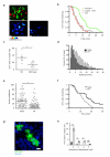
Figure 2. Generation of central memory precursor cells and recall response are dependent on LFA-1-dependent stable interactions during the CDP
(a–c) Mice adoptively transferred with 5×10³ P14 cells were immunized with LCMV and LFA-1 was blocked during the CDP. 15 days post-immunization, the number of P14 cells (a, *P<0.05) and the expression of surface markers KLRGI, IL7R (b, *P<0.05 and **P<0.001) and CD62L (c, *P<0.01) were analyzed. SLEC population was defined as KLRGI high, IL7R low P14 cells, and MPEC population was defined as KLRGI low, IL7R high P14 cells. Data are from three independent experiments - n=6 – error bars correspond to SEM. (d–f) Mice adoptively transferred with 5×10³ OTI cells were immunized with DEC-OVA and LFA-1 was blocked during the CDP. d- Percentage of CD44 and CD62L positive cells among OTI cells was analyzed 8 days post-immunization. *P < 0.001. Data are from three independent experiments. Percentage of OTI secreting IFN-γ (e, *P<0.05) and percentage of OTI among CD8 (f, *P<0.001) were quantified after recall with low dose of DEC-OVA 30 days post-immunization. Data are from three independent experiments. Each dot is an individual mouse. (g–h) Mice adoptively transferred with 5×10³ OTI cells were vaccinated with OVA peptide-pulsed DCs and LFA-1-dependent interactions were blocked during the CDP. (g) Forty to sixty days after vaccination, mice were challenged with a lethal dose of LM-OVA (2–10× LD50). P<0.001 – n=13 (h) Six days post-immunization, OTI cells were isolated and the same number of cells was transferred back in naïve mice. Seventy days after vaccination, mice were challenged with a lethal dose of LM-OVA (2x LD50). P<0.01 - n=10 - Data are from two independent experiments.
Figure 3
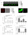
Figure 3. CD8 cell differentiation largely relies on T-T contacts
(a–b) H2bbm1 mice bearing H2bbm1 OTI cells were immunized with OVA peptide-pulsed BMDCs generated from CD11c-DTR mice. APCs were ablated (DT) or LFA-1-dependent interactions were inhibited (CDP blockade) during the CDP. Percentage of OTI secreting IFN-γ (a, *P<0.001) and percentage of OTI among CD8 (b, *P<0.05) were analyzed at the peak of the effector response. Data are from at least three independent experiments. Each dot is an individual mouse. (c) 1×10⁶ fluorescently labelled WT (green) and ICAM-1−/− (red) T cells were ad-mixed in a flat-bottomed well and activated with PMA+Ionomycin. 24h post-activation, the presence of WT vs ICAM-1−/− cells in clusters was evaluated by microscopy. Scale bar, 5μm. (d) The effector response of WT and ICAM-1−/− OTI cells after DEC-OVA immunization was quantified as the percentage of OTI secreting IFN-γ 6 days post-immunization. *P < 0.05 and **P < 0.001. Data are from three independent experiments. (e) The effector response of WT and ICAM-1−/− OTI cells after LM-OVA immunization was quantified as the percentage of OTI secreting IFN-γ 8 days post-immunization. *P < 0.01. Data are from three independent experiments. (f) The effector response of WT and ICAM-1−/− P14 cells after LCMV immunization was quantified as the percentage of P14 secreting IFN-γ 8 days post-immunization. *P < 0.05. Data are from three independent experiments.
Figure 4
Figure 4. Characterization of T-T contacts
(a–b) Fluorescently labelled WT and ICAM-1−/− OTI cells (2×10⁶) were transferred in WT recipients. Mice were immunized with DEC-OVA. a- Individual “snapshot” showing WT (green) and ICAM-1−/− (red) OTI cells during the CDP, i.e 24h after immunization in vivo. Images are displayed as maximum intensity projections along the z axis (top view). Image intensities were scaled to a normalized time projection intensity range of 0–1. Lower panels: Pseudo-colored time projection of a 30 minute run showing the spatial persistence of WT (left panel) and ICAM-1−/− (right panel) OTI cells. Scale bar, 15μm. b- Graph shows the fraction of OTI cells remaining in cluster over time in vivo after DEC-OVA immunization. P < 0.001. Data are from two independent experiments. c- CD11c-YFP mice bearing OTI-RFP cells were immunized with DEC-OVA. When indicated, mice were treated with LFA-1 Ab 22h post-immunization. Explanted lymph nodes were subjected to 2-photon imaging 2h after LFA-1 Ab treatment. Graph shows the percentage of OTI cells engaged in homotypic interaction during a 30 minute run. Every dot corresponds to an individual field. *P < 0.001. Data are from four independent experiments. (d–f) H2bbm1 mice bearing CFSE-labelled OTI H2bbm1 were immunized with CMTMR-labelled and OVA peptide-pulsed BMDCs generated from Cd11c-DTR mice. When indicated, mice were treated with DT 8h post-immunization (DT). Explanted lymph nodes were subjected to 2-photon imaging 24 to 30h post-immunization. Instantaneous speed (d), Arrest Coefficient (defined as the percentage of time a given cell has an instantaneous speed < 2μm/min, every dot correspond to an individual cell) (e, *P<0.001), and the fraction of OTI cells remaining in cluster over time (f) were analyzed during the CDP. g- Example of a cluster composed of 4 OTI cells found during the CDP after immunization with DEC-OVA when low precursor frequency (104 OTI cells transferred) is used. Image is displayed as maximum intensity projection along the z axis (top view). Cells are numbered from 0 to 4. Cell #0 is isolated, whereas cells #1 to 4 are clustered. h- Quantification of the percentage of OTI cell clusters found in a whole popliteal LN of naïve or immunized animals when low precursor frequency is used (n=338 for naïve, n=320 for DEC-OVA condition, over 3 independent experiments). *P < 0.05 and **P < 0.001.
Figure 5
Figure 5. OTI cell differentiation is regulated by cytokine secretion at T-T contacts
(a–b) OTI blasts were transduced with a plasmid encoding IFN-γ fused to GFP (IFN-γ-GFP). Cells were stimulated with PMA and Ionomycin to induce cell clustering and imaged for 15 minutes. Time-lapse images in (a) show an IFN-γ-GFP expressing OTI cell at the periphery of a cluster. Upper panels: Overlay between IFN-γ-GFP and contrast. Scale bar, 10 μm. Lower panel: magnification showing IFN-γ-GFP localization within a cell (cell contour is represented by a dashed gray line) relative to cell-cell contact (depicted in red). Scale bar, 5 μm. Graph in (b) shows the percentage of IFN-γ-GFP expressing OTI cells at the periphery of a cell cluster showing localization of IFN-γ-GFP at T-T contact, or not specifically localized (n=20). *P<0.01. (c–d) OTI cells were stimulated with PMA+Ionomycin for 24 hours. Representative images of the sites of IFN-γ secretion in clustered cells (c, Scale bar, 10 μm) and quantification of percentage of cells at the periphery of a cluster showing captured IFN-γ facing another T cell or facing away (n=50) (d, *P<0.05). Data are from three independent experiments. e- Picture shows an example of 2 OTI clustered cells with polarized IFN-γ localization in vivo during the CDP after DEC-OVA immunization. Cells are shown in the xyz dimensions. OTI cells are green and IFN-γ is depicted in red. Scale bar, 5 μm. f- Time-line of the experimental protocol. (f–i) OTI cells were activated in vitro with PMA and Ionomycin, treated with LFA-1 Ab, IFN-γ Ab (f–g) or IFN-γ (h–i) and transferred in WT mice 3 days post-activation. Percentage of OTI secreting IFN-γ (f and h, * P<0.01 **P<0.001) and percentage of OTI among CD8 (h and j, *P<0.05 and **P<0.001) were analyzed after in vivo recall. Data are from three independent experiments.

Comment in

References

    1. Lefrancois L, Obar JJ. Once a killer, always a killer: from cytotoxic T cell to memory cell. Immunological Reviews. 2010;235:206–218. - PMC - PubMed
    1. Pipkin ME, et al. Interleukin-2 and inflammation induce distinct transcriptional programs that promote the differentiation of effector cytolytic T cells. Immunity. 2010;32:79–90. - "V体育2025版" PMC - PubMed
    1. Prlic M, Williams MA, Bevan MJ. Requirements for CD8 T-cell priming, memory generation and maintenance. Current opinion in immunology. 2007;19:315–319. - "V体育官网" PubMed
    1. Mescher MF, et al. Signals required for programming effector and memory development by CD8+ T cells. Immunological reviews. 2006;211:81–92. - PubMed
    1. Obar JJ, Lefrancois L. Early events governing memory CD8+ T-cell differentiation. International immunology. 2010;22:619–625. - "VSports手机版" PMC - PubMed

Publication types

MeSH terms (VSports注册入口)