Skip to main page content
U.S. flag

An official website of the United States government

Dot gov

The . gov means it’s official. Federal government websites often end in . gov or . mil. Before sharing sensitive information, make sure you’re on a federal government site. VSports app下载.

Https

The site is secure V体育官网. The https:// ensures that you are connecting to the official website and that any information you provide is encrypted and transmitted securely. .

. 2012 Oct 22;209(11):1985-2000.
doi: 10.1084/jem.20111665. Epub 2012 Oct 1.

Disrupting galectin-1 interactions with N-glycans suppresses hypoxia-driven angiogenesis and tumorigenesis in Kaposi's sarcoma (V体育安卓版)

Affiliations

"V体育官网" Disrupting galectin-1 interactions with N-glycans suppresses hypoxia-driven angiogenesis and tumorigenesis in Kaposi's sarcoma

Diego O Croci et al. J Exp Med. .

V体育2025版 - Abstract

Kaposi's sarcoma (KS), a multifocal vascular neoplasm linked to human herpesvirus-8 (HHV-8/KS-associated herpesvirus [KSHV]) infection, is the most common AIDS-associated malignancy. Clinical management of KS has proven to be challenging because of its prevalence in immunosuppressed patients and its unique vascular and inflammatory nature that is sustained by viral and host-derived paracrine-acting factors primarily released under hypoxic conditions. We show that interactions between the regulatory lectin galectin-1 (Gal-1) and specific target N-glycans link tumor hypoxia to neovascularization as part of the pathogenesis of KS VSports手机版. Expression of Gal-1 is found to be a hallmark of human KS but not other vascular pathologies and is directly induced by both KSHV and hypoxia. Interestingly, hypoxia induced Gal-1 through mechanisms that are independent of hypoxia-inducible factor (HIF) 1α and HIF-2α but involved reactive oxygen species-dependent activation of the transcription factor nuclear factor κB. Targeted disruption of Gal-1-N-glycan interactions eliminated hypoxia-driven angiogenesis and suppressed tumorigenesis in vivo. Therapeutic administration of a Gal-1-specific neutralizing mAb attenuated abnormal angiogenesis and promoted tumor regression in mice bearing established KS tumors. Given the active search for HIF-independent mechanisms that serve to couple tumor hypoxia to pathological angiogenesis, our findings provide novel opportunities not only for treating KS patients but also for understanding and managing a variety of solid tumors. .


Figures
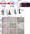
Figure 1.
Figure 1.
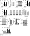
Gal-1 expression is a hallmark of KS. (A and B) Gal-1 transcript profile of mouse mECK36 KS tumors and human AIDS-KS compared with normal skin (Array data were obtained from Mutlu et al., 2007 and from Wang et al., 2004). (C) Left, qRT-PCR analysis of Gal-1 mRNA in mECK36 KS tumors and normal skin. Results are the mean ± SEM of three independent experiments. Right, confocal microscopy of mECK36 tumors stained for Gal-1 and LANA. Data are representative of three independent experiments. (D) Left, immunoblot of Gal-1 in KS-Imm cells. Right, flow cytometry of Gal-1 in permeabilized KS cells. Data are representative of six experiments. (E) ELISA of Gal-1 secretion by KS cells and HUVEC. Data are the mean ± SEM of three independent experiments. (F and G) Immunohistochemical analysis of human benign vascular lesions (n = 26) and primary KS tumors (n = 15) stained with anti–Gal-1 polyclonal Ab or with H&E. Data are the mean ± SEM (F) .Representative micrographs are shown (G). (C, E, and F) **, P < 0.01; ***, P < 0.001.
Figure 2.
Figure 2.
KSHV controls Gal-1 expression. (A) qRT-PCR analysis of Gal-1 mRNA in uninfected 293 and 293rKSHV.219 cells upon stimulation with 3 mM sodium butyrate to induce lytic gene expression. Data indicate fold increase of mRNA as measured by triplicates of two independent experiments. (B) KSHV lytic gene expression (RTA, vGPCR, gB, K8.1) in 293rKSHV cells upon stimulation with sodium butyrate for 24 h. Data indicate fold increase of mRNA as measured by triplicates of two independent experiments. (C) qRT-PCR analysis of Gal-1 mRNA in iSLK.219 cells upon stimulation with doxycycline to induce RTA-driven lytic gene expression. Data indicate fold increase of mRNA as measured by triplicates of two independent experiments. (D) KSHV lytic gene expression (RTA, vGPCR, gB) in iSLK.219 cells upon stimulation with doxycycline. Data indicate fold increase of mRNA as measured by triplicates of two independent experiments. (A–D) Error bars represent SEM. (A and C) **, P < 0.01.
Figure 3.
Figure 3.
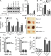
Hypoxia controls Gal-1 expression in KS through HIF-independent, NF-κB–dependent mechanisms. (A–D) Expression of Gal-1 in KS cells transfected with or without HIF-1α siRNA or a super-repressor form of IκB-α (IκB-α-SR) and incubated under hypoxia or normoxia. (A) Promoter activity. (B) qRT-PCR of Gal-1 mRNA relative to RN18S1. AU, arbitrary units. Data are the mean ± SEM of five (A) or three (B) independent experiments. (C) Immunoblot of Gal-1, IκB-α, HIF-1α, and actin. Data are representative of four independent experiments. (D) ELISA of Gal-1 secretion. Data are the mean ± SEM of three independent experiments. (E) ELISA of Gal-1 secretion by KS cells cultured under hypoxic or normoxic conditions in the presence or absence of HIF-1α or NF-κB inhibitors. Data are the mean ± SEM of three independent experiments. (F and G) Immunoblot (F) and qRT-PCR (G) of Gal-1 expression induced by hypoxia (Hyp) in human and mouse melanoma (A375 and B16-F0), mouse breast carcinoma (4T1), and human prostate carcinoma (LNCaP) cell lines. Data are representative (F) or are the mean ± SEM (G) of three independent experiments. (H) Western blot of HIF-1α, IκB-α, Gal-1, and actin upon treatment of KS cells with CoCl2 (chemical activator of HIF-1α). Data are representative of four experiments. (I) Gal-1 promoter activity upon treatment of KS cells with CoCl2. Modulation of pGL3–Gal-1–Luciferase activity relative to Renilla expression is shown. Data are the mean ± SEM of three independent experiments. (J) ELISA of VEGF secretion by KS cells transfected with HIF-1α or scr siRNA cultured under hypoxic or normoxic conditions. Data are the mean ± SEM of three independent experiments. (K) Immunoblot of Gal-1, HIF-2α, c/EBPα, and actin in KS cells transfected with or without siRNA for HIF-2α, C/EBPα, or scrambled (scr) and incubated under hypoxia or normoxia. Data are representative of three independent experiments. (L) ELISA of Gal-1 secretion by KS cells transfected with or without siRNA for HIF-1α, HIF-2α, or C/EBPα or incubated with an NF-κB inhibitor (BAY 11–7082; 1 µM) and cultured under hypoxic or normoxic conditions. Data are the mean ± SEM of three independent experiments. (A, B, D, E, J, and L) *, P < 0.05; **, P < 0.01.
Figure 4.
Figure 4.
Hypoxia controls Gal-1 expression in KS through mechanisms involving ROS-dependent NF-κB activation. (A) Immunoblot of Gal-1, IκB-α, and actin expression in KS cells cultured under hypoxic or normoxic conditions in the presence or absence of 5 mM of the ROS scavenger NAC. Data are representative of three independent experiments. (B) ELISA of Gal-1 secretion by KS cells cultured under hypoxic or normoxic conditions in the presence of increasing concentrations of NAC. Data are the mean ± SEM of three independent experiments. (C) ELISA of Gal-1 secretion by KS cells cultured with increasing concentrations of H2O2. Data are the mean ± SEM of three independent experiments. (D) ELISA of Gal-1 secretion by KS cells exposed to 0.5 mM H2O2 in the presence or absence of 1 µM BAY 11–7082. Data are the mean ± SEM of three independent experiments. (E) Immunostaining of Gal-1 and Hypoxyprobe-1 in nonhypoxic and hypoxic areas of KS xenografts. Right, chart shows quantification of pixel intensity of red fluorescence (Gal-1) along the dashed line. Images are representative of three independent experiments. (B and D) **, P < 0.01.
Figure 5.
Figure 5.
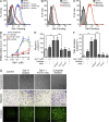
Gal-1–N-glycan interactions link tumor hypoxia to angiogenesis in KS. (A) Immunoblot (left) and ELISA (right) of Gal-1 in KS cells expressing shRNA constructs that target different sequences of human Gal-1 mRNA (sh-Gal-1.1, sh-Gal-1.2 and sh-Gal-1.3) or scrambled shRNA (sh-scr) compared with nontransfected KS cells (KS). Data are representative (left) or are the mean ± SEM (right) of five independent experiments. (B) Tube formation by HUVEC incubated with SFCM from normoxic or hypoxic KS cells transfected or not with scr or Gal-1 shRNA. Data are the mean ± SEM of four independent experiments. (C) Hemoglobin content of Matrigel plugs containing SFCM of KS cells transfected or not with Gal-1 or scr shRNA, cultured under hypoxic or normoxic conditions and inoculated into B6 WT or Lgals1−/− mice. Data are the mean ± SEM of three independent experiments. (D) In vivo vascularization of Matrigel plugs containing SFCM of KS cells transfected or not with Gal-1 shRNA or scr shRNA. Data are representative of three independent experiments with three animals per group. (E and F) qRT-PCR analysis of C2GnT-1 (E) or GnT5 (F) mRNA of HUVEC transfected with different concentrations of specific siRNA relative to RN18S1 mRNA (AU: arbitrary units). Data are the mean ± SEM of three independent experiments. (G) Tube formation by HUVEC transfected with GnT5, C2GnT-1, or scr siRNA incubated with SFCM from normoxic or hypoxic KS cells. Data are the mean ± SEM of four independent experiments. (B, C, and E–G) **, P < 0.01.
Figure 6.
Figure 6.
Silencing of Gal-1 expression does not alter the secretion of KS-derived proangiogenic cytokines. (A–C) ELISA of VEGF (A), ANGPTL4 (B), and Oncostatin M (C) secretion by KS cells transfected or not with Gal-1 or scr shRNA and cultured under hypoxic or normoxic conditions. Data are the mean ± SEM of three independent experiments.
Figure 7.
Figure 7.
Targeting Gal-1–glycan interactions prevents the angiogenic switch in KS. (A) Tumor growth in nude mice inoculated with 5 × 10⁶ knockdown KS cells expressing Gal-1 shRNA (sh-Gal-1.1 and sh-Gal-1.2), KS cells expressing scr shRNA (sh-scr), or nontransfected KS cells (KS). Data are the mean ± SEM of four independent experiments with five animals per group. (B) In vitro cell growth of KS clones expressing Gal-1 shRNA or scr shRNA (sh-scr) or nontransfected KS cells (KS). Data are the mean ± SEM of three independent experiments. (C) Flow cytometry of tumor-associated CD34+ ECs. Dot plots are representative of four independent experiments. Right, data are the mean ± SEM of four independent experiments. (D) Microvessel density. Data are the mean ± SEM of four independent experiments. (E) Tumor hemoglobin content. Data are the mean ± SEM of four independent experiments. (A and C–E) **, P < 0.01.
Figure 8.
Figure 8.
A Gal-1–specific neutralizing mAb prevents Gal-1–induced EC proliferation, migration, invasion, and tube formation. (A) Binding of 20 µg/ml 488-Gal-1 to HUVEC in the presence or absence of 0.5 µM F8.G7 anti–Gal-1 mAb, 0.5 µM isotype control, or 30 mM lactose. Data are representative of three independent experiments. (B and C) Binding of 20 µg/ml 488-Gal-3 (B) or 20 µg/ml 488-Gal-8 (C) to HUVEC in the presence or absence of 0.5 µM F8.G7 anti–Gal-1 mAb. Filled histogram, nonspecific binding determined with unlabeled galectins. Data are representative of three independent experiments. (D–G) Functional activity of F8.G7 mAb in vitro. Proliferation (D), migration (E), and tube formation (F) of HUVEC incubated with or without increasing concentrations of Gal-1 in the presence or absence of 0.5 µM F8.G7 mAb, 0.5 µM isotype control, or 30 mM lactose. (G) Representative micrographs of tube formation (top), migration (middle), and invasion (bottom) of HUVEC exposed to different treatments. Data are the mean ± SEM (D–F) or are representative (G) of three independent experiments. (D–F) **, P < 0.01.
Figure 9.
Figure 9.
Therapeutic administration of a Gal-1–specific neutralizing mAb promotes tumor regression in established KS. (A–E) Nude mice were inoculated with KS cells and treated in vivo with the indicated doses of F8.G7 mAb or with isotype control when tumors reached 100 mm3. (A) Kinetics of tumor growth. Data are the mean ± SEM of four independent experiments. (B) Microvessel density. Left, representative confocal micrographs of three experiments are shown (green, CD31; red, propidium iodide). Right, results are the mean ± SEM of three independent experiments. (C) Tumor hemoglobin content. Data are the mean ± SEM of three independent experiments. (D) Top, in vivo proliferation rate of KS xenografts from mice receiving F8.G7 mAb or isotype control, determined by incorporation of EdU injected into mice 2 h before sacrifice. Bottom, apoptosis of tumor sections from mice receiving F8.G7 mAb or isotype control (green, TUNEL; red, propidium iodide). Dashed lines indicate the peripheral borders of the tumor. Data are representative (left) or are the mean ± SEM (right) of three independent experiments. (E) Number of infiltrating B cells (B220+), macrophages (F4/80+), and NK cells (NK1.1+) in tumor sections from mice receiving F8.G7 mAb or isotype control. Data are representative (left) or are the mean ± SEM (right) of three independent experiments with five animals per group. (A–E) *, P < 0.05; **, P < 0.01.

References

    1. Albini A., Morini M., D’Agostini F., Ferrari N., Campelli F., Arena G., Noonan D.M., Pesce C., De Flora S. 2001. Inhibition of angiogenesis-driven Kaposi’s sarcoma tumor growth in nude mice by oral N-acetylcysteine. Cancer Res. 61:8171–8178 - PubMed
    1. Alcendor D.J., Knobel S.M., Desai P., Zhu W.Q., Vigil H.E., Hayward G.S. 2010. KSHV downregulation of galectin-3 in Kaposi’s sarcoma. Glycobiology. 20:521–532 10.1093/glycob/cwp204 - DOI (VSports手机版) - PMC - PubMed
    1. Banh A., Zhang J., Cao H., Bouley D.M., Kwok S., Kong C., Giaccia A.J., Koong A.C., Le Q.T. 2011. Tumor galectin-1 mediates tumor growth and metastasis through regulation of T-cell apoptosis. Cancer Res. 71:4423–4431 10.1158/0008-5472.CAN-10-4157 - DOI (V体育安卓版) - PMC - PubMed
    1. Casper C. 2011. The increasing burden of HIV-associated malignancies in resource-limited regions. Annu. Rev. Med. 62:157–170 10.1146/annurev-med-050409-103711 - DOI - PubMed
    1. Cedeno-Laurent F., Watanabe R., Teague J.E., Kupper T.S., Clark R.A., Dimitroff C.J. 2012. Galectin-1 inhibits the viability, proliferation, and Th1 cytokine production of nonmalignant T cells in patients with leukemic cutaneous T-cell lymphoma. Blood. 119:3534–3538 10.1182/blood-2011-12-396457 - DOI - PMC - PubMed

V体育官网 - MeSH terms