Skip to main page content
U.S. flag

An official website of the United States government

Dot gov

The . gov means it’s official. Federal government websites often end in . gov or VSports app下载. mil. Before sharing sensitive information, make sure you’re on a federal government site. .

Https

The site is secure. The https:// ensures that you are connecting to the official website and that any information you provide is encrypted and transmitted securely. V体育官网.

. 2012 Feb 1;188(3):1255-65.
doi: 10.4049/jimmunol.1101579. Epub 2011 Dec 28.

"V体育2025版" Population dynamics of naive and memory CD8 T cell responses after antigen stimulations in vivo

Affiliations

Population dynamics of naive and memory CD8 T cell responses after antigen stimulations in vivo

Matthew D Martin et al. J Immunol. .

Abstract

The extent to which the progeny of one primary memory CD8 T cell differs from the progeny of one naive CD8 T cell of the same specificity remains an unresolved question. To explore cell-autonomous functional differences between naive and memory CD8 T cells that are not influenced by differences in the priming environment, an experimental model has been developed in which physiological numbers of both populations of cells were cotransferred into naive hosts before Ag stimulation. Interestingly, naive CD8 T cells undergo greater expansion in numbers than do primary memory CD8 T cells after various infections or immunizations VSports手机版. The intrinsic ability of one naive CD8 T cell to give rise to more effector CD8 T cells than one memory CD8 T cell is independent of the number and quality of primary memory CD8 T cells present in vivo. The sustained proliferation of newly activated naive CD8 T cells contributed to their greater magnitude of expansion. Additionally, longitudinal analyses of primary and secondary CD8 T cell responses revealed that on a per-cell basis naive CD8 T cells generate higher numbers of long-lived memory cells than do primary memory CD8 T cells. This enhanced "memory generation potential" of responding naive CD8 T cells occurred despite the delayed contraction of secondary CD8 T cell responses. Taken together, the data in this study revealed previously unappreciated differences between naive and memory CD8 T cells and will help further define the functional potential for both cell types. .


Conflict of interest statement

Disclosures

The authors have no financial conflict of interest.

Figures
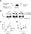
Figure 1
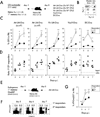
Figure 1. Naïve CD8 T cells undergo a higher magnitude of expansion than memory CD8 T cells after LM infection
A) Experimental design. Naïve Thy1.1/1.1 OT-I (1x10³) were mixed with an equal number of primary memory Thy1.1/1.2 OT-I (day 200+ after primary VacV-OVA infection) and injected into naïve B6 Thy1.2/1.2 recipients. Mice were challenged 24 hrs later with Att LM-OVA (5x10⁶ CFU per mouse; i.v.). B) Representative plots showing the primary (Thy1.1/1.1) and secondary (Thy1.1/1.2) OT-I responses at the indicated days after Att LM-OVA infection. Numbers inside the plots indicate the percentage of primary or secondary OT-I CD8 T cells in peripheral blood, and numbers below the plots indicate the ratio of primary to secondary OT-I cells at the indicated days post infection (p.i.). C) Kinetic analysis of OT-I in peripheral blood. Data are presented as the percentage of primary or secondary OT-I cells in peripheral blood (mean +/− SEM for 5 mice per group). D) The ratio of primary to secondary OT-I cells at the indicated days after infection. Dots represent individual mice, solid lines indicate the mean, and the dashed line indicates the starting ratio of naïve and primary memory OT-I cells in the mix used for adoptive transfer before infection. The data are representative of at least three independent experiments. * indicates a p value between 0.01 and 0.05, and ** indicates a p value less than 0.01.
Figure 2
Figure 2. Naïve CD8 T cells undergo a greater magnitude of expansion than memory CD8 T cells in response to infections or non-infectious immunization
A) Experimental design. Naïve Thy1.1/1.1 OT-I (1x10³) were mixed with an equal number of primary memory Thy1.1/1.2 OT-I (day 200+ after primary VacV-OVA infection) and injected into naïve B6 Thy 1.2/1.2 recipients. Mice were challenged 24 hrs later with Att LM-OVA (5x10⁶ CFU or 5x10⁴ CFU per mouse; i.v.), Vir LM-OVA (5x10⁴ CFU per mouse; i.v.), VacV-OVA (3x10⁶ PFU per mouse; i.p.), or DC-OVA (1x10⁶ cells per mouse; i.v.). B) Dot plot showing the mix of naïve and memory OT-I cells used for adoptive transfer. Numbers inside the plot indicate the percentage of naïve OT-I (Thy1.1/1.1) or primary memory OT-I (Thy1.1/1.2). C) Kinetic analysis of OT-I in peripheral blood at the indicated days after infection or immunization. Data are presented as the percentage of primary or secondary OT-I cells in peripheral blood (mean +/− SEM for 5 mice per group). D) The ratio of primary to secondary OT-I at the indicated days after infection or immunization. Dots represent individual mice, solid lines indicate the mean, and the dashed line indicates the starting ratio of naïve and memory OT-I cells before infection. E) Experimental design. Endogenous primary memory Ova257-specific CD8 T cells (day 113 after primary VacV-OVA infection) were transferred (5x10³ cells per mouse i.v.) into naïve B6 CD45.1/CD45.1 recipients one day before Att LM-OVA (5x10⁶ CFU; i.v.) challenge. F) Representative plots showing the primary (CD45.2 negative) and secondary (CD45.2 positive) KbOva257 endogenous CD8 T cell responses at the indicated days after Att LM-OVA infection. Numbers inside the plots indicate the percentage of primary or secondary Ova257-specific CD8 T cells in peripheral blood (PBL). G) Kinetic analysis of Ova257-specific CD8 T cells in peripheral blood at the indicated days after infection. Data are presented as the percentage of primary or secondary cells in peripheral blood (mean +/− SEM for 5 mice per group). These data are representative of two to three similar and independent experiments. * indicates a p value between 0.01 and 0.05, and ** indicates a p value less than 0.01.
Figure 3
Figure 3. Systemic inflammation influences the expansion potential of primary memory CD8 T cells
A) Experimental design. Naïve Thy1.1/1.1 OT-I cells (1x10³) were mixed with an equal number of primary memory Thy1.1/1.2 OT-I cells (d350+ after primary infection) and injected into naïve B6 Thy1.2/1.2 recipients. Mice were immunized 24 hrs later with Ova257-coated dendritic cells (DC-OVA; 2x10⁵ cells per recipient) in the presence or absence of Att LM co-infection (5x10⁶ CFU per mouse). B) Graph showing the ratio of primary to secondary OT-I cells in PBL at the indicated days after DC-OVA immunization in the presence or absence of infection. C) The ratio of primary to secondary OT-I cells in the indicated organs at day 7 after immunization. The dotted lines in B and C indicate the ratio of naïve to primary memory OT-I cells used in the adoptive transfer mix. Representative experiment out of three similar and independent experiments is shown.
Figure 4
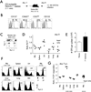
Figure 4. Differences in trafficking before infection or in tissue localization after infection do not explain the differences in expansion potential of naïve and memory CD8 T cells
A) Experimental design. The indicated numbers of naïve Thy1.1/1.1 OT-I were mixed with an equal number of primary memory Thy1.1/1.2 OT-I (day 300+ after primary VacV-OVA infection) and injected into naïve B6 Thy1.2/1.2 recipients. Mice were challenged 24 hrs later with Att LM-OVA (5x10⁶ CFU per mouse). B) The expression of the indicated markers was evaluated on primary memory OT-I cells. Shaded histograms represent isotype control staining and open histograms represent specific staining of gated primary memory OT-I CD8 T cells. Numbers indicate the percentage of cells positive for the indicated molecules. C) Dot plot showing the mix of naïve and memory OT-I cells used for adoptive transfer. Numbers inside the plot indicate the percentage of naïve OT-I (Thy1.1/1.1) or primary memory OT-I (Thy1.1/1.2) cells. D) The ratio of primary to secondary OT-I in the indicated organs 24 hrs after adoptive transfer of naïve OT-I and primary memory OT-I (3x10⁵ of each cell types per recipient). Dots represent individual mice, solid lines indicate the mean, and the dashed line indicates the starting ratio of naïve and memory OT-I cells before infection. E) Total numbers of naïve or primary memory OT-I in the spleens one day after adoptive transfer. Data are presented as mean + SEM for 4 mice per group. F) Representative histograms showing the percentage of secondary among all gated OT-I cells in the indicated organs at day 7 after Att LM-OVA infection (5x10⁶ CFU per mouse; i.v.). The total number of naïve and primary OT-I cells initially transferred is indicated. G) The ratio of primary to secondary OT-I cells in the indicated organs at day 7 p.i. Dots represent individual mice, solid lines indicate the mean, and the dashed line indicates the starting ratio of naïve and memory OT-I cells before infection. * indicates a p value between 0.01 and 0.05.
Figure 5
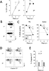
Figure 5. Sustained proliferation of primary effectors rather than increased death of secondary effectors leads to the greater magnitude of expansion of the naïve CD8 T cells
A) Kinetic analysis of primary or secondary OT-I in peripheral blood. Naïve OT-I or primary memory OT-I cells were transferred into separate mice, and mice were challenged 24 hrs later with Att LM-OVA (5x10⁶ CFU per recipient; i.v.). Data are presented as the percentage of primary or secondary OT-I cells in peripheral blood (mean +/− SEM for 4–10 mice per group per time point). B) Representative dot plots of BrdU staining of OT-I cells in the spleen at day 7 after Att LM-OVA infection. Numbers inside the plots indicate the percentage of OT-I cells positive for BrdU. C) Kinetic analysis of BrdU incorporation. Data are presented as the percentage of primary or secondary OT-I cells in peripheral blood or the spleen positive for BrdU (mean +/− SEM for 3–4 mice per group per time point). D) Representative dot plots of caspase-3/7 staining of primary or secondary OT-I cells in the spleens at the indicated time points after Att LM-OVA infection. Numbers indicate the percentage of OT-I cells positive for caspase-3/7. E) The percentage of primary or secondary OT-I cells positive for caspase-3/7 at the indicated days after Att LM-OVA infection (mean + SEM for 3–4 mice per group per time point). Data are representative of two independent experiments. ** indicates a p value less than 0.01; ns- not significant.
Figure 6
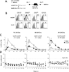
Figure 6. Longitudinal analysis of primary and secondary CD8 T cell responses in vivo
A) Experimental Design. Naïve Thy1.1/1.1 OT-I (1x10³) were mixed with primary memory Thy1.1/1.2 OT-I (day 200+ after primary VacV-OVA infection) at the indicated ratios and injected into naïve B6 Thy1.2/1.2 recipients. Mice were challenged 24 hrs later with Att LM-OVA (5x10⁶ CFU per recipient; i.v.). B) Kinetic analysis of OT-I in peripheral blood at the indicated days after infection or immunization. Data are presented as the mean percentage of primary or secondary OT-I cells in PBL +/− SEM for 4 or 5 mice per group. C) Kinetic analysis of primary and secondary OT-I CD8 T cell responses presented as the ratio of primary to secondary OT-I cells in peripheral blood (mean +/− SEM for 4 or 5 mice per group). The dashed line indicates the starting ratio of naïve and memory OT-I cells transferred before infection. The data are representative of at least three similar and independent experiments.
Figure 7
Figure 7. Tissue distribution and homeostatic proliferation of primary and secondary memory CD8 T cells analyzed in the same host
A) The percentage of primary or secondary OT-I per indicated organ on day 227 after Att LM-OVA infection. Data are presented as mean + SEM for 3 mice per organ. B) The ratio of primary to secondary OT-I in the indicated organs at day 227 after Att LM-OVA infection. C) Representative dot plots of BrdU staining of primary or secondary OT-I cells in the spleen or peripheral blood (PBL) on day 216 after infection (the ratio of naïve to primary memory OT-I cells used for adoptive transfer was 1:10). Numbers inside the plots indicate the percentage of OT-I cells positive for BrdU. D) The percentage of BrdU positive primary or secondary OT-I cells in spleen or PBL at day 216 after Att LM-OVA infection. Dots represent individual mice, solid lines indicate the mean, and the dashed line indicates the starting ratio of naïve and memory OT-I cells transferred before infection. * indicates a p value between 0.01 and 0.05, and ** indicates a p value less than 0.01.
Figure 8
Figure 8. Higher proliferation potential but indistinguishable kinetics of secondary CD8 T cell responses generated from late versus early primary memory CD8 T cells
A) Experimental Design. Day 33 old primary memory (earlyM; Thy1.1/1.1 OT-I - 1x10³ or 1x10⁴) were mixed with day 250+ old primary memory (lateM Thy1.1/1.2 OT-I) at the indicated ratios and injected into naïve B6 Thy1.2/1.2 recipients. Mice were challenged 24 hrs later with Att LM-OVA or Vir LM-OVA (5x10⁶ and 5x10⁴ CFU per mouse, respectively). B) The expression of the indicated markers was evaluated for earlyM and lateM OT-I donors. Shaded histograms represent isotype control staining and open histograms represent staining of earlyM or lateM OT-I CD8 T cells. Numbers indicate the percentage of cells positive for the indicated molecules. C) Kinetic analysis of the earlyM or lateM OT-I response in PBL after the indicated infection. Data are presented as the mean percentage of earlyM or lateM OT-I cells in PBL +/− SEM for five mice per group. D) The ratio of earlyM or lateM OT-I in the PBL at the indicated days after infection. Dots represent individual mice, solid lines indicate the mean, and the dashed line indicates the starting ratio of earlyM and lateM OT-I cells transferred before infection. The data are representative of at least two similar and independent experiments.
Figure 9
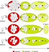
Figure 9. Population dynamics of primary and multiple stimulated CD8 T cell responses – a model
After antigen encounter, naïve and memory CD8 T cells (primary, secondary, and tertiary) undergo proliferative expansion. The magnitude of the expansion, duration of contraction, and ability to generate long-lived progeny (‘memory generation potential’) of naïve and/or memory CD8 T cells is dependent on the Ag-stimulation history.

V体育官网入口 - References

    1. Harty JT, Badovinac VP. Shaping and reshaping CD8+ T-cell memory. Nat Rev Immunol. 2008;8:107–119. - V体育官网 - PubMed
    1. Jameson SC, Masopust D. Diversity in T cell memory: an embarrassment of riches. Immunity. 2009;31:859–871. - "VSports在线直播" PMC - PubMed
    1. Kaech SM, Wherry EJ, Ahmed R. Effector and memory T-cell differentiation: implications for vaccine development. Nat Rev Immunol. 2002;2:251–262. - PubMed
    1. Lefrancois L. Development, trafficking, function of memory T-cell subsets. Immunol Rev. 2006;211:93–103. - PubMed (V体育官网)
    1. Sallusto F, Lanzavecchia A, Araki K, Ahmed R. From vaccines to memory and back. Immunity. 2010;33:451–463. - PMC - PubMed

Publication types

Substances